Default-mode network streams for coupling to language and control systems
- PMID: 32632019
- PMCID: PMC7382234
- DOI: 10.1073/pnas.2005238117
Default-mode network streams for coupling to language and control systems
Abstract
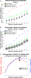
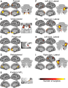
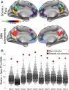
The human brain is organized into large-scale networks identifiable using resting-state functional connectivity (RSFC). These functional networks correspond with broad cognitive domains; for example, the Default-mode network (DMN) is engaged during internally oriented cognition. However, functional networks may contain hierarchical substructures corresponding with more specific cognitive functions. Here, we used individual-specific precision RSFC to test whether network substructures could be identified in 10 healthy human brains. Across all subjects and networks, individualized network subdivisions were more valid-more internally homogeneous and better matching spatial patterns of task activation-than canonical networks. These measures of validity were maximized at a hierarchical scale that contained ∼83 subnetworks across the brain. At this scale, nine DMN subnetworks exhibited topographical similarity across subjects, suggesting that this approach identifies homologous neurobiological circuits across individuals. Some DMN subnetworks matched known features of brain organization corresponding with cognitive functions. Other subnetworks represented separate streams by which DMN couples with other canonical large-scale networks, including language and control networks. Together, this work provides a detailed organizational framework for studying the DMN in individual humans.
Keywords: Default network; brain networks; fMRI; functional connectivity; individual variability.
Conflict of interest statement
Competing interest statement: N.U.F.D. is cofounder of Nous Imaging.
Figures


Similar articles
-
A robust dissociation among the language, multiple demand, and default mode networks: Evidence from inter-region correlations in effect size.Neuropsychologia. 2018 Oct;119:501-511. doi: 10.1016/j.neuropsychologia.2018.09.011. Epub 2018 Sep 20. Neuropsychologia. 2018. PMID: 30243926 Free PMC article.
-
Task-based co-activation patterns reliably predict resting state canonical network engagement during development.Dev Cogn Neurosci. 2022 Dec;58:101160. doi: 10.1016/j.dcn.2022.101160. Epub 2022 Oct 8. Dev Cogn Neurosci. 2022. PMID: 36270101 Free PMC article.
-
Risk seeking for losses modulates the functional connectivity of the default mode and left frontoparietal networks in young males.Cogn Affect Behav Neurosci. 2018 Jun;18(3):536-549. doi: 10.3758/s13415-018-0586-4. Cogn Affect Behav Neurosci. 2018. PMID: 29616472
-
Mapping cognitive and emotional networks in neurosurgical patients using resting-state functional magnetic resonance imaging.Neurosurg Focus. 2020 Feb 1;48(2):E9. doi: 10.3171/2019.11.FOCUS19773. Neurosurg Focus. 2020. PMID: 32006946 Free PMC article. Review.
-
Default mode network as a potential biomarker of chemotherapy-related brain injury.Neurobiol Aging. 2014 Sep;35 Suppl 2:S11-9. doi: 10.1016/j.neurobiolaging.2014.03.036. Epub 2014 May 15. Neurobiol Aging. 2014. PMID: 24913897 Free PMC article. Review.
Cited by
-
Effects of a new speech support application on intensive speech therapy and changes in functional brain connectivity in patients with post-stroke aphasia.Front Hum Neurosci. 2022 Sep 22;16:870733. doi: 10.3389/fnhum.2022.870733. eCollection 2022. Front Hum Neurosci. 2022. PMID: 36211132 Free PMC article.
-
The human brainstem's red nucleus was upgraded to support goal-directed action.Nat Commun. 2025 Apr 10;16(1):3398. doi: 10.1038/s41467-025-58172-z. Nat Commun. 2025. PMID: 40210909 Free PMC article.
-
Functional specialization within the inferior parietal lobes across cognitive domains.Elife. 2021 Mar 2;10:e63591. doi: 10.7554/eLife.63591. Elife. 2021. PMID: 33650486 Free PMC article.
-
Tasks activating the default mode network map multiple functional systems.Brain Struct Funct. 2022 Jun;227(5):1711-1734. doi: 10.1007/s00429-022-02467-0. Epub 2022 Feb 18. Brain Struct Funct. 2022. PMID: 35179638 Free PMC article.
-
Cognitive Implications of Correlated Structural Network Changes in Schizophrenia.Front Integr Neurosci. 2022 Jan 20;15:755069. doi: 10.3389/fnint.2021.755069. eCollection 2021. Front Integr Neurosci. 2022. PMID: 35126065 Free PMC article.
References
Publication types
MeSH terms
Grants and funding
LinkOut - more resources
Full Text Sources
Medical

