. 2020 Jul 6;11(7):506.
doi: 10.1038/s41419-020-2701-z.

PD-L1 promotes tumor growth and progression by activating WIP and β-catenin signaling pathways and predicts poor prognosis in lung cancer

Affiliations

PD-L1 promotes tumor growth and progression by activating WIP and β-catenin signaling pathways and predicts poor prognosis in lung cancer

Wendan Yu et al. Cell Death Dis. .

Abstract

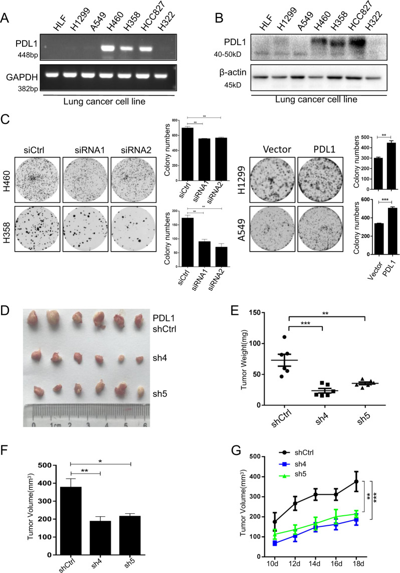
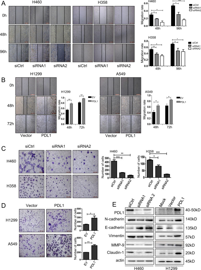
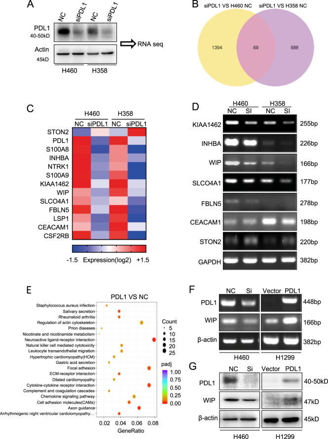
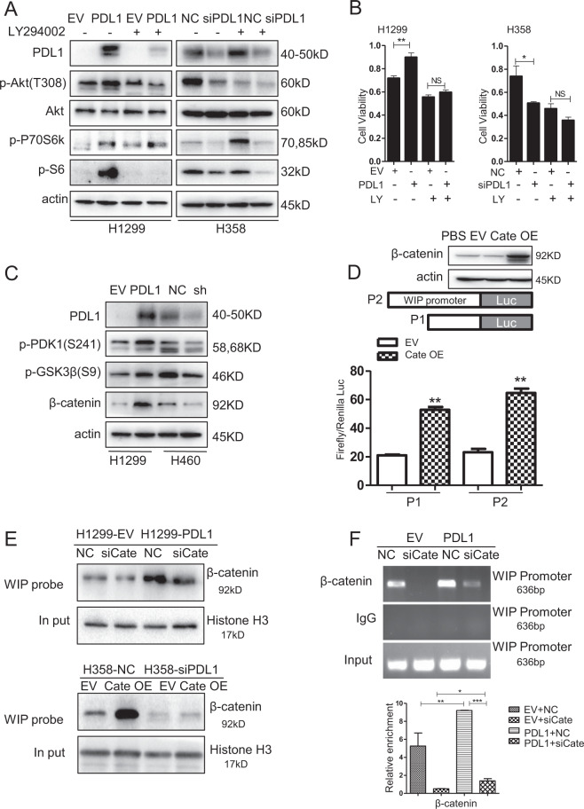
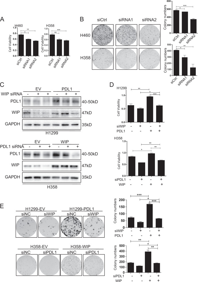
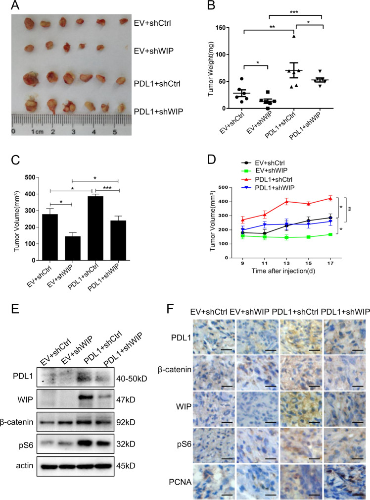
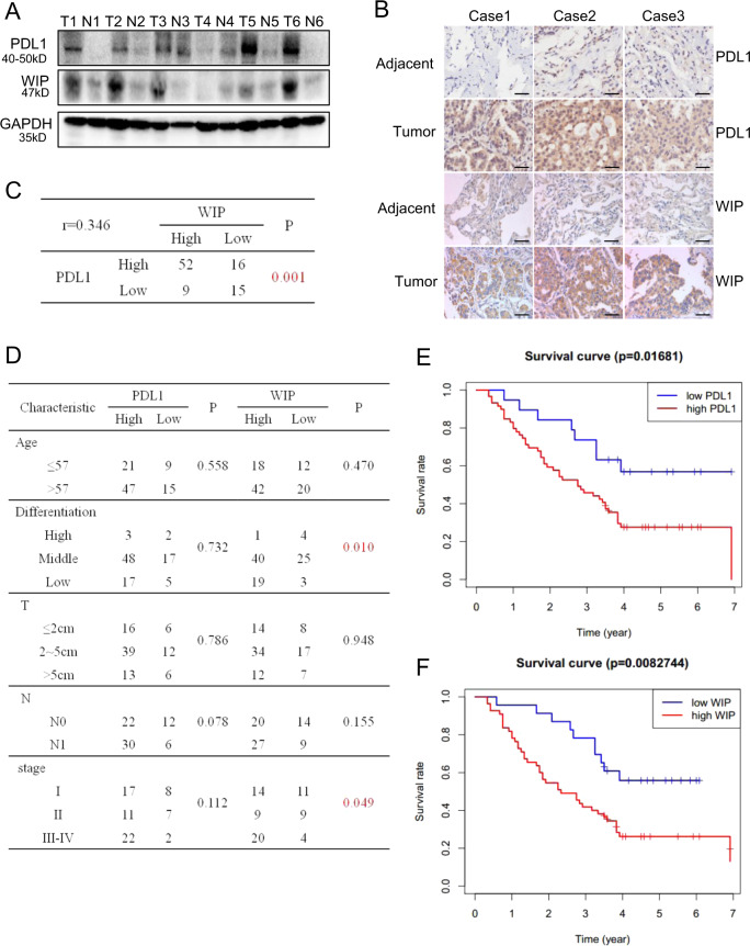
PD-L1 is overexpressed in tumor cells and contributes to cancer immunoevasion. However, the role of the tumor cell-intrinsic PD-L1 in cancers remains unknown. Here we show that PD-L1 regulates lung cancer growth and progression by targeting the WIP and β-catenin signaling. Overexpression of PD-L1 promotes tumor cell growth, migration and invasion in lung cancer cells, whereas PD-L1 knockdown has the opposite effects. We have also identified WIP as a new downstream target of PD-L1 in lung cancer. PD-L1 positively modulates the expression of WIP. Knockdown of WIP also inhibits cell viability and colony formation, whereas PD-L1 overexpression can reverse this inhibition effects. In addition, PD-L1 can upregulate β-catenin by inhibiting its degradation through PI3K/Akt signaling pathway. Moreover, we show that in lung cancer cells β-catenin can bind to the WIP promoter and activate its transcription, which can be promoted by PD-L1 overexpression. The in vivo experiments in a human lung cancer mouse model have also confirmed the PD-L1-mediated promotion of tumor growth and progression through activating the WIP and β-catenin pathways. Furthermore, we demonstrate that PD-L1 expression is positively correlated with WIP in tumor tissues of human adenocarcinoma patients and the high expression of PD-L1 and WIP predicts poor prognosis. Collectively, our results provide new insights into understanding the pro-tumorigenic role of PD-L1 and its regulatory mechanism on WIP in lung cancer, and suggest that the PD-L1/Akt/β-catenin/WIP signaling axis may be a potential therapeutic target for lung cancers.


Conflict of interest statement

The authors declare that they have no conflict of interest.

Figures

Fig. 1
Fig. 1. PD-L1 promotes lung cancer cell growth in vitro and in vivo.
a PD-L1 mRNA expression in different human lung cancer cell lines was analyzed by RT-PCR. b PD-L1 expression in human lung cancer cells was detected by western blot. c Colony formation assay in human lung cancer cells with PD-L1 knockdown or overexpression. d The xenografts were harvested at 18 days after injection and the morphology of tumor xenografts from each nude mouse was photographed. e The tumor weight of each nude mouse was measured. f The tumor volume of each mouse from different groups was measured and calculated as Volume = (Width2 × length)/2. g Growth kinetics (mean ± SD) of PD-L1-sh4, PD-L1-sh5 versus the negative control in BALB/c-nude mice (n = 6). The data are presented as mean ± SD of three independent tests. *P < 0.05, **P < 0.01, ***P < 0.001.
Fig. 2
Fig. 2. PD-L1 promotes lung cancer cell migration and invasion via EMT signaling.
H460 and H358 cells were transfected with PD-L1 siRNAs, and H1299 and A549 cells were transfected with PD-L1-OE plasmid. After 48 h, a, b Cell migration was analyzed by cell wound-healing assay and the migration rate was calculated; c, d Cell invasion assay was performed, and the number of invasion cells was counted. The data are presented as mean ± SD of three independent tests. *P < 0.05, **P < 0.01, ***P < 0.001; e The expression levels of PD-L1, N-cadherin, E-cadherin, Vimentin, MMP-9, Caludin-1 and β-actin were analyzed by western blot.
Fig. 3
Fig. 3. WIP is a downstream molecule of PD-L1.
a PD-L1 was knocked down by siRNA in H460 and H358 cells. (b) Overlap of changed genes affected by PD-L1 siRNA silencing in H460 and H358 lung cancer cells. c Heatmap showing the subset of 12 genes identified in H460 and H358 NSCLC cells followed by PD-L1 silencing. d The regulation of gene expression by PD-L1 was analyzed by RT-PCR assay in H460 and H358 cells. e KEGG analysis showed that genes were enriched in the actin cytoskeleton pathway. f, g The expression of WIP was detected by RT-PCR and western blot.
Fig. 4
Fig. 4. PD-L1 activates PI3K/Akt pathway to promote the β-catenin mediated transcription of WIP.
a H1299 and H358 cells were pretreated with 10 µM LY294002 for 4 h, and then cells were transfected with PD-L1-OE plasmid or siRNA for 48 h. The expression of p-Akt, Akt, p-P70S6K, p-S6, and β-actin were detected by western blot. b H1299 and H358 cells were pretreated with 10 µM LY294002 for 4 h and then transfected with PD-L1-OE plasmid or siRNA for 48 h, and cell viability was measured by MTT assay. c Expression of PD-L1, p-PDK1, p-GSK3β, β-catenin were detected by western blotting after 48 h transfection of PD-L1-OE plasmid or siRNA in H1299 and H460 cells. d H1299 cells were co-transfected with β-catenin-OE plasmid or empty vector and WIP promoter reporter plasmid. At 48 h after transfection, promoter activity was analyzed using dual-luciferase assay. e Binding of β-catenin to the WIP promoter was detected by DNA pulldown assay after PD-L1 overexpressed alone or PD-L1 overexpressed and β-catenin silenced. f Binding of β-catenin to the WIP promoter was analyzed by ChIP analysis. For b, d, f, the experiments were repeated three times, and the results were shown as mean ± SD. *P < 0.05, **P < 0.01, ***P < 0.001. NS means no significant differences between two groups. LY means LY294002.
Fig. 5
Fig. 5. The PD-L1 mediated cell growth promotion is partially dependent on WIP.
a, b H460 and H358 cells were transfected with WIP siRNAs. After 48 h, cell viability and colony numbers were analyzed by MTT and colony formation assays. c H1299 cells were transfected with PD-L1-OE and WIP siRNAs, and H358 cells were transfected with PD-L1 siRNAs and WIP-OE plasmid. After 48 h, the expression of PD-L1, WIP, and GAPDH were detected by western blot. d, e H1299 cells were treated with PD-L1-OE and WIP siRNAs, and H358 cells were treated with PD-L1 siRNA and WIP-OE plasmid. After 48 h, cell viability and colony numbers were analyzed by MTT and colony formation assays. For a, b, d, e, the data represent the mean ± SD of three independent experiments, *P < 0.05, **P < 0.01, ***P < 0.001.
Fig. 6
Fig. 6. PD-L1 promotes tumor growth by upregulating WIP in human lung cancer mouse model.
a The morphology of tumor xenografts from each mouse was photographed. b, c The mouse of each group was sacrificed, the tumor weight and tumor volume were measured. d Tumor diameters of each nude mouse from different group were measured at a regular interval of 2 days after 9 days of injection. Tumor volume = (Width2 × length)/2. e The expression level of PD-L1, β-catenin, WIP, and p-S6 within tumor xenografts in each group of nude mice were detected. f The expression of PD-L1, β-catenin, WIP, p-S6, and Ki67 in tumor tissues was detected by immunohistochemistry staining. N = 6 mice/group. Scale bars = 50 μm. Original magnification: ×40. The level of significance was indicated by *P < 0.05, **P < 0.01, ***P < 0.001.
Fig. 7
Fig. 7. PD-L1 is positively correlated with WIP expression in lung adenocarcinoma tissues and predicts poor prognosis in patients.
a PD-L1 and WIP expression of six human patient tissues and paired adjacent tissues was detected by Western blot. b PD-L1 and WIP expression from human lung adenocarcinoma tissue microarray of three cases with tumor and paired adjacent tissues was analyzed by immunostaining analysis. The representative images are shown. Scale bars = 100 μm. Original magnification: ×40. c The correlation between the expression of PD-L1 and WIP in human lung cancer tissues from 92 patients. d The relationship between the level of PD-L1 or WIP expression and clinicopathologic characteristics. e, f The relation between overall survival and expression of PD-L1 (p = 0.017) or WIP (p = 0.008) was analyzed by Kaplan–Meier analysis.
Fig. 8
Fig. 8. Schematic diagram illustrating the mechanism of PD-L1 involves in regulating lung cancer growth and progression.
PD-L1 promotes p-S6 expression through PI3K/AKT/mTOR pathway, and also upregulates β-catenin expression by increasing phosphorylation of GSK3β (inactive form Ser9). β-Catenin binds to the WIP promoter and activates its expression in lung cancer cells.

References

    1. Siegel, R. L., Miller, K. D. & Jemal, A. Cancer statistics, 2019. CA: a cancer J. clinicians69, 7–34 (2019). - PubMed
    1. Ohe, Y. et al. Long-term follow-up of patients with unresectable locally advanced non-small cell lung cancer treated with chemoradiotherapy: a retrospective analysis of the data from the Japan Clinical Oncology Group trials (JCOG0003A). Cancer Sci.94, 729–734 (2003). - DOI - PMC - PubMed
    1. Yasumoto, K., Hanagiri, T. & Takenoyama, M. Lung cancer-associated tumor antigens and the present status of immunotherapy against non-small-cell lung cancer. Gen. Thorac. Cardiovasc. Surg.57, 449–457 (2009). - DOI - PubMed
    1. Hirsch, F. R. et al. Lung cancer: current therapies and new targeted treatments. Lancet389, 299–311 (2017). - DOI - PubMed
    1. Song, X., Liu, J., Lu, Y., Jin, H. & Huang, D. Overexpression of B7-H1 correlates with malignant cell proliferation in pancreatic cancer. Oncol. Rep.31, 1191–1198 (2014). - DOI - PubMed

Publication types

MeSH terms