. 2020 Jul 6;11(1):3377.
doi: 10.1038/s41467-020-17102-x.

Uncovering mutation-specific morphogenic phenotypes and paracrine-mediated vessel dysfunction in a biomimetic vascularized mammary duct platform

Affiliations

Uncovering mutation-specific morphogenic phenotypes and paracrine-mediated vessel dysfunction in a biomimetic vascularized mammary duct platform

Matthew L Kutys et al. Nat Commun. .

Abstract

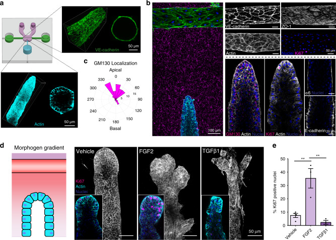
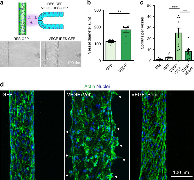
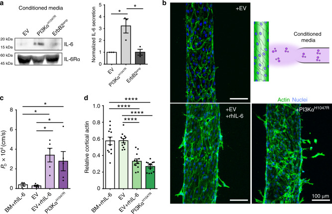
The mammary gland is a highly vascularized tissue capable of expansion and regression during development and disease. To enable mechanistic insight into the coordinated morphogenic crosstalk between the epithelium and vasculature, we introduce a 3D microfluidic platform that juxtaposes a human mammary duct in proximity to a perfused endothelial vessel. Both compartments recapitulate stable architectural features of native tissue and the ability to undergo distinct forms of branching morphogenesis. Modeling HER2/ERBB2 amplification or activating PIK3CA(H1047R) mutation each produces ductal changes observed in invasive progression, yet with striking morphogenic and behavioral differences. Interestingly, PI3KαH1047R ducts also elicit increased permeability and structural disorganization of the endothelium, and we identify the distinct secretion of IL-6 as the paracrine cause of PI3KαH1047R-associated vascular dysfunction. These results demonstrate the functionality of a model system that facilitates the dissection of 3D morphogenic behaviors and bidirectional signaling between mammary epithelium and endothelium during homeostasis and pathogenesis.


Conflict of interest statement

The authors declare no competing interests.

Figures

Fig. 1
Fig. 1. A vascularized mammary duct platform models native 3D tissue architectures and morphogenesis.
Tissues are at one week of co-culture unless otherwise indicated. a Organotypic microfluidic device consisting of an engineered 3D endothelial vessel (green) and mammary epithelial duct (cyan) in physiologic ECM (magenta), enabling long-term culture and paracrine signaling. Inset: 3D reconstruction of endothelial vessel (green—VE-cadherin) and epithelial duct (cyan—phalloidin). b Left: Composite stitched maximum intensity projection micrograph of tissue base to medial section of a mammary epithelial duct (cyan, phalloidin) and proximal vasculature (green, phalloidin); Alexa Fluor-labeled collagen I (magenta), DAPI (blue). Top right: Representative maximum intensity micrographs of mature endothelial vessels immunostained for VE-cadherin, ZO-1, actin, and Ki67, as indicated. Bottom right: Individual confocal slice micrographs from 3D epithelial duct midsections immunostained for GM130, Ki67, α6 integrin, E-cadherin, actin, and DAPI, as is indicated in each panel. c Quantification of golgi localization as measured by the nuclear-GM130 axis (n = 52 cells examined from three independent ducts). 0 corresponds to a nuclear-GM130 axis apically oriented toward the lumen, while 180 corresponds to a basal orientation. d Cartoon depicting morphogen gradients established by perfusing growth factors through an acellular vascular channel. Immunographs of quiescent MCF10A ducts exposed to vehicle (DMSO), FGF2 (3 nM), or TGFβ1 (5 ng/ml) gradients for three days. Inset: high magnification slice micrograph of duct terminus immunostained with phalloidin (cyan), DAPI (blue) and Ki67 (magenta). e Quantification of percentage of Ki67 positive nuclei in ducts treated with each morphogen gradient (n = 4, 3, 3 ducts examined across three independent experiments; one-way ANOVA with Bonferroni post test Vehicle vs FGF2 **p = 0.0036, FGF2 vs TGFβ1 **p = 0.002, mean ± s.e.m). All images are representative of at least three independent experiments. Source Data are provided as a Source Data file.
Fig. 2
Fig. 2. Vascular morphogenesis driven by mammary paracrine signaling.
a Cartoon of co-culture paracrine experimental setup. MCF10A cells expressing IRES-GFP or VEGF-IRES-GFP were seeded into the duct channels adjacent to an endothelialized vessel. Phase-contrast images of vessels with control (left) and VEGF expressing ducts (right) after two days in co-culture. b Quantification of endothelial vessel diameter after three days of culture with either GFP or VEGF expressing MCF10A cells (n = 6, 9 vessels examined across three independent experiments; two-tailed, unpaired Student’s t test **p = 0.0019). c Quantification of sprout number per endothelial vessel after three days of culture with acellular basal assay medium (BM), GFP expressing ducts (GFP), or VEGF expressing ducts in which DMSO (VEGF + Veh) or 10 µM Semaxanib (VEGF + Sem) was delivered to the vasculature. (n = 9, 6, 9, 8 vessels examined across three independent experiments; one-way ANOVA with Bonferroni post test ***P = 0.000014, **P = 0.0011). d Maximum intensity projection micrographs of tissue base to medial section of endothelial vessels co-cultured with GFP or VEGF ducts and treated with DMSO or 10 µM Semaxanib; phalloidin (green) and DAPI (blue). Arrows indicate nascent angiogenic sprouts. For all plots, values mean ± s.e.m and all images are representative of at least three independent experiments. Source Data are provided as a Source Data file.
Fig. 3
Fig. 3. Ductal and vascular consequences resulting from specific breast cancer mutations.
a Left: Cartoon of co-culture paracrine experimental setup with mosaic mutant/wild-type ducts. Ducts were generated from MCF10A cells stably expressing empty vector (EV), wild-type hemagglutinin (HA)-tagged ErbB2 (ErbB2amp), or constitutively active HA-tagged PI3Kα H1047R (PI3KαH1047R) along with each associated vascular compartment. Right: Western blot of lysates from EV, ErbB2amp, or PI3KαH1047R MCF10A. b Composite stitched maximum intensity projection micrographs of endothelial vessels co-cultured for five days along with their corresponding modified ducts. Endothelial vessels: phalloidin (green), DAPI (blue), epithelial ducts: phalloidin (cyan), DAPI (blue). c Diffusive permeability (PD) of 70 kDa dextran across the endothelial barrier (n = 8, 8, 6 vessels examined across three independent experiments; one-way ANOVA with Bonferroni post test, **P = 0.0049, *P = 0.012) and (d) endothelial vessel diameter in each co-culture setting (n = 8, 8, 6 vessels examined across three independent experiments; one-way ANOVA with Bonferroni post test). e Left: Quantification of cortical actin measured from phalloidin labeled micrographs (n = 12, 13, 13 cells taken from three independent vessel experiments one-way ANOVA with Bonferroni post test, ****P = 7.6845e−06, ***P = 0.0003). Right: Averaged, cross-sectional actin distribution in endothelial vessels co-cultured with specific mutant ducts (n = 11 cell actin profiles taken from three independent vessel experiments). For all plots, values mean ± s.e.m. and all images are representative of at least three independent experiments. Source Data are provided as a Source Data file.
Fig. 4
Fig. 4. PI3KαH1047R increases IL-6 secretion to drive vascular dysfunction and remodeling.
a Representative western blot and associated quantification of conditioned media from EV, ErbB2amp, and PI3KαH1047R ducts immunoblotted for human IL-6 and IL-6Rα (n = 3 western blots from three independent experiments). b Top right: Experimental schematic of endothelial vessel-conditioned media setup. Maximum intensity projection micrographs of endothelial vessels treated overnight with EV, EV+ recombinant human IL-6 (rhIL-6) (200 ng/ml), or PI3KαH1047R conditioned media; phalloidin (green), DAPI (blue). c Diffusive permeability PD (n = 4, 5, 5, 5 vessels examined across three independent experiments; one-way ANOVA with Bonferroni post test, BM + rhIL-6 vs EV + rhIL-6 *P = 0.019, BM + rhIL-6 vs PI3KαH1047R *P = 0.035, EV vs EV + rhIL-6 *P = 0.0104, EV vs PI3KαH1047R *P = 0.0205) and (d) relative cortical actin measured from vessels treated overnight with basal assay medium supplemented with 200 ng/ml rhIL-6 (BM + rhIL-6) or EV, EV + rhIL-6, or PI3KαH1047R conditioned media (n = 12, 12, 12, 12 cells examined from three vessel experiments; one-way ANOVA with Bonferroni post test, BM + rhIL-6 vs EV + rhIL-6 ***P = 1.32402e−06, BM+rhIL-6 vs PI3KαH1047R ****P = 1.08309e−08, EV vs EV + rhIL-6 ****P = 1.0819e−06, EV vs PI3KαH1047R ****P = 8.84689e−09). For all plots, values mean ± s.e.m. and all images are representative of at least three independent experiments. Source Data are provided as a Source Data file.

References

    1. Hannezo E, Heisenberg CP. Mechanochemical feedback loops in development and disease. Cell. 2019;178:12–25. - PubMed
    1. Hebner C, Weaver VM, Debnath J. Modeling morphogenesis and oncogenesis in three-dimensional breast epithelial cultures. Annu Rev. Pathol. 2008;3:313–339. - PubMed
    1. Khademhosseini A, Langer R. A decade of progress in tissue engineering. Nat. Protoc. 2016;11:1775–1781. - PubMed
    1. Jain A, et al. Primary human lung alveolus-on-a-chip model of intravascular thrombosis for assessment of therapeutics. Clin. Pharm. Ther. 2018;103:332–340. - PMC - PubMed
    1. Park SE, Georgescu A, Huh D. Organoids-on-a-chip. Science. 2019;364:960–965. - PMC - PubMed

Publication types

MeSH terms

Substances