

Impact of histological diagnosis on the treatment of atypical brainstem lesions
- PMID: 32632139
- PMCID: PMC7338439
- DOI: 10.1038/s41598-020-68063-6
Impact of histological diagnosis on the treatment of atypical brainstem lesions
Abstract
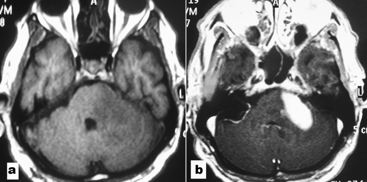
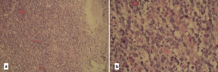
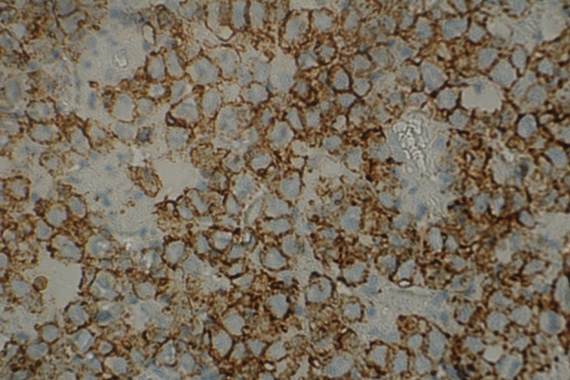
For atypical brainstem lesions, histological diagnosis can have an impact on treatment, especially in cases where diffuse glioma is not found. Since radiotherapy is the only therapeutic modality that has shown clinical and radiographic improvement in patients with diffuse glioma, the misdiagnosis of diffuse glioma can have drastic consequences, particularly in patients with nontumorous lesions. Thus, the purpose of this study was to evaluate the impact of histological diagnosis on the treatment of atypical brainstem lesions. This was a retrospective study of 31 patients who underwent biopsy of atypical brainstem lesions. The procedures were performed between January 2008 and December 2018 at the Life Center Hospital and Santa Casa de Belo Horizonte, MG, Brazil. A diagnosis was obtained in 26 (83.9%) cases. Three patients presented complications: one presented bleeding with no clinical repercussions and two showed worsening of neurological deficit, only one of which was definitive. No mortality occurred due to the procedure. The histological diagnosis was diffuse glioma in seven cases (22.6%) and not diffuse glioma in 19 cases (61.3%). Thus, the histological diagnosis had an impact on the treatment of 19 patients (treatment impact rate: 61.3%). The histological diagnosis of intrinsic brainstem lesions is a safe, efficient procedure with a high diagnosis rate, and as such, it should be considered in the management of atypical lesions.
Conflict of interest statement
The authors declare that they have no conflicts of interest.
Figures
References
-
- Albright AL, et al. Magnetic resonance scans should replace biopsies for the diagnosis of diffuse brainstem gliomas. Neurosurgery. 1993;33(6):1026–1030. - PubMed
-
- Frazier J, et al. Treatment of diffuse brainstem gliomas: Failed approaches and futures strategies. J. Neurosurg. 2009;3(4):259–269. - PubMed
Publication types
MeSH terms
LinkOut - more resources
Full Text Sources
