Review
. 2021 Mar;22(3):183-195.
doi: 10.1038/s41580-020-0264-6. Epub 2020 Jul 6.

RNA contributions to the form and function of biomolecular condensates

Affiliations
Review

RNA contributions to the form and function of biomolecular condensates

Christine Roden et al. Nat Rev Mol Cell Biol. 2021 Mar.

Abstract

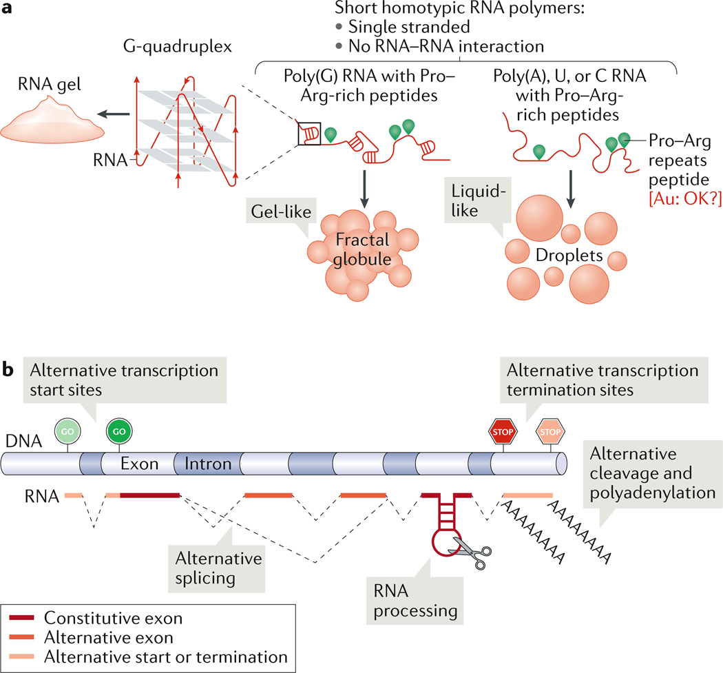
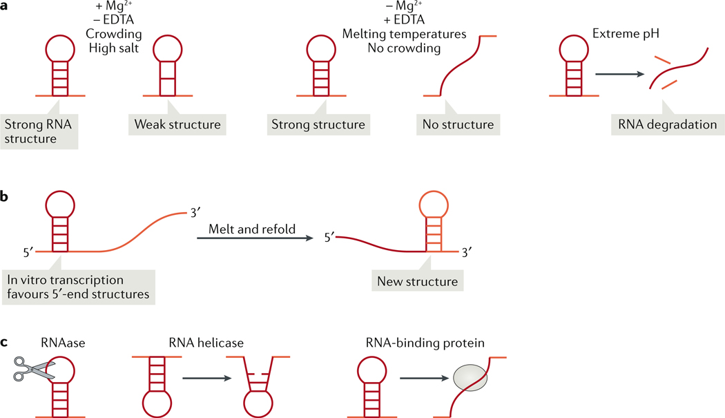
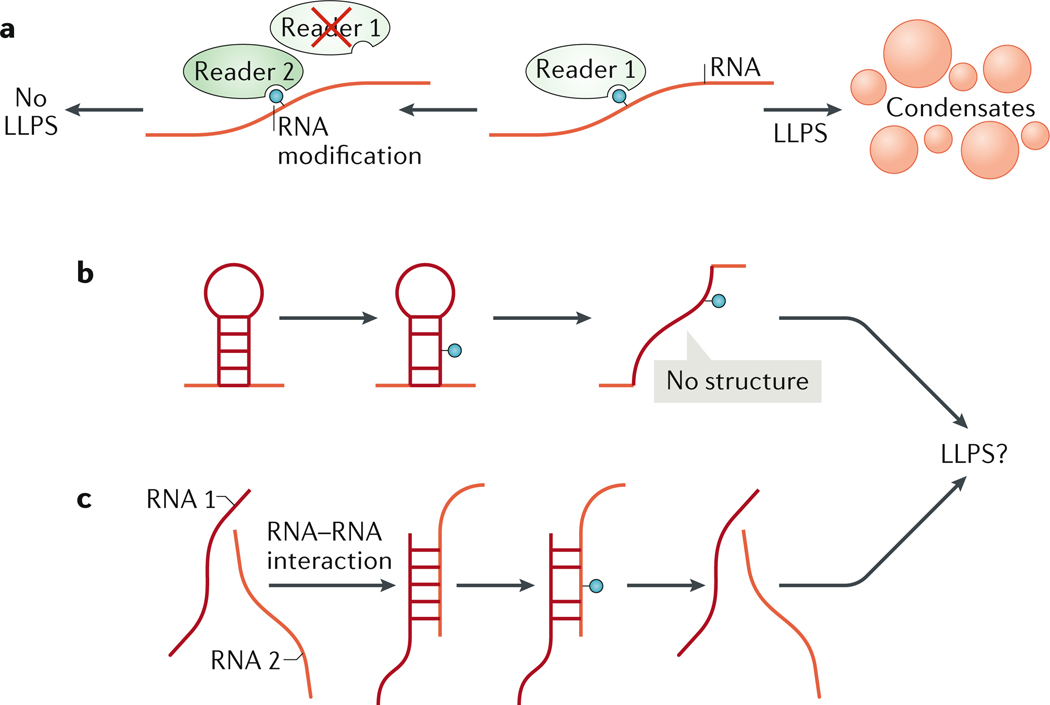
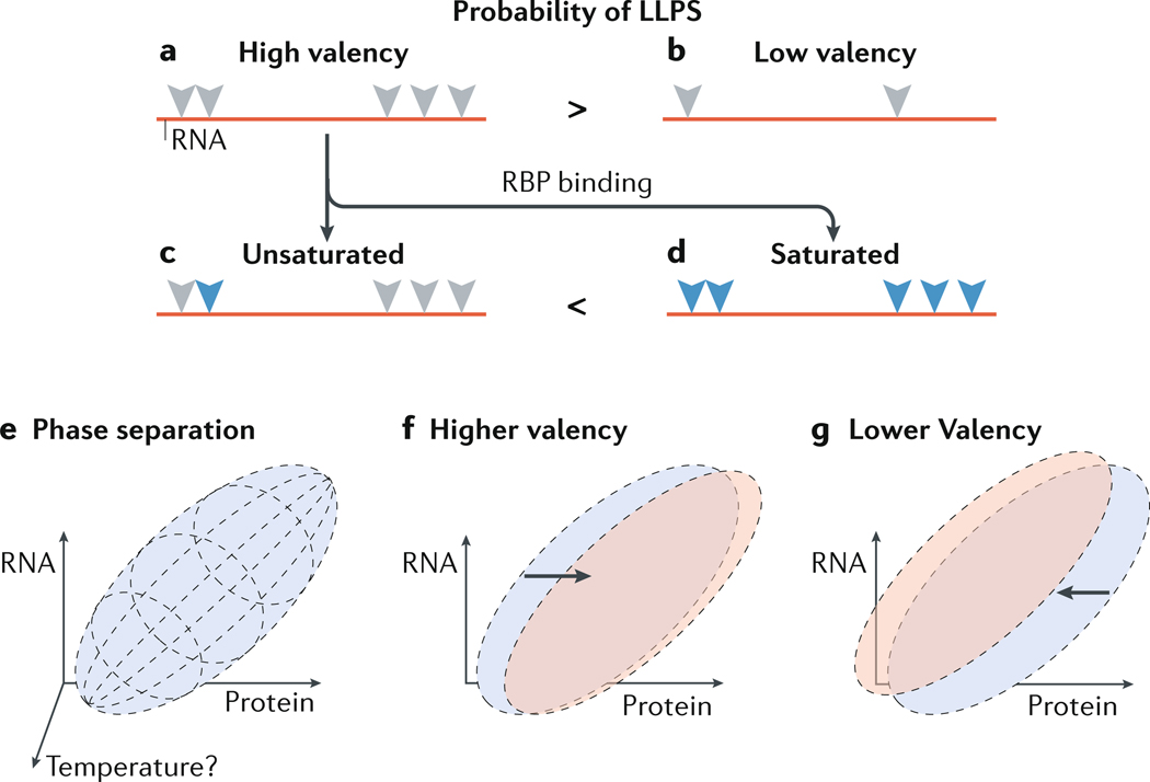
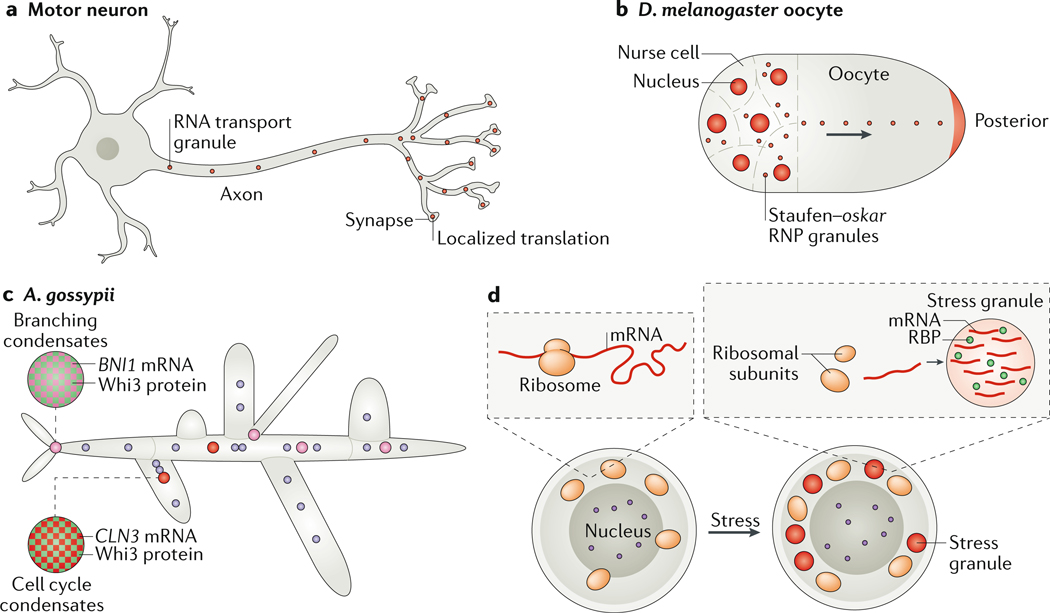
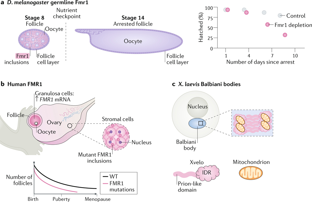
Biomolecular condensation partitions cellular contents and has important roles in stress responses, maintaining homeostasis, development and disease. Many nuclear and cytoplasmic condensates are rich in RNA and RNA-binding proteins (RBPs), which undergo liquid-liquid phase separation (LLPS). Whereas the role of RBPs in condensates has been well studied, less attention has been paid to the contribution of RNA to LLPS. In this Review, we discuss the role of RNA in biomolecular condensation and highlight considerations for designing condensate reconstitution experiments. We focus on RNA properties such as composition, length, structure, modifications and expression level. These properties can modulate the biophysical features of native condensates, including their size, shape, viscosity, liquidity, surface tension and composition. We also discuss the role of RNA-protein condensates in development, disease and homeostasis, emphasizing how their properties and function can be determined by RNA. Finally, we discuss the multifaceted cellular functions of biomolecular condensates, including cell compartmentalization through RNA transport and localization, supporting catalytic processes, storage and inheritance of specific molecules, and buffering noise and responding to stress.


Conflict of interest statement

Conflict of Interest:

The authors declare no competing financial interests

Figures

Figure 1:
Figure 1:. Regulation of condensate properties through RNA sequence and length.
(A) Homotypic RNA polymers mixed with proline–arginine-repeats peptides yield either liquid-like (poly(A), U or C) or gel-like (poly(G)) condensates. At high-enough concentrations, poly(G) RNAs can form G-quadruplexes and RNA gels. (B) Generating RNA sequence (chemical) complexity. RNA sequence length can be regulated by choice of transcription start or termination sites, alternative splicing, RNA processing or alternate cleavage and polyadenylation.
Figure 2:
Figure 2:. Tuning condensate properties through RNA modifications.
(A) RNA modifications are recognized by ‘reader’ proteins. Interaction with a reader can enhance or inhibit RNA undergoing liquid–liquid phase separation (LLPS). (B and C) RNA modifications can tune RNA structure by blocking or promoting RNA–RNA interactions in cis, within the same RNA molecule (B) or in trans between different RNA molecules(C). Modification of RNA sequence and structure could alter phase separation of target RNA.
Figure 3:
Figure 3:. Modifying RNA structure in vitro and in vivo.
(A) In vitro, buffers that include magnesium ions (Mg2+) and a crowding agent and exclude the chelating agent EDTA, will support the formation of both strong RNA structures (structures with many base pairs) and weak RNA structures (fewer base pairs) at lower temperatures (left), whereas in the opposite buffer conditions or in higher temperature, the weaker RNA structures will unfold (middle). Extreme pH will denature and cause the degradation of even strong RNA structures (right). (B) In vitro transcription favours interactions at the 5’-end region, as this region is transcribed first. Melting and refolding of the in vitro transcribed RNA will allow and the formation of interactions between the 5’ and 3’ regions of the molecule. (C) In vivo, RNA structures are modulated by cellular RNAses, which actively remove particular RNA sequences and structures; by RNA helicases, which unwind double-stranded RNA; and by RNA-binding proteins, which can shield RNA sequences from RNA–RNA interaction or block structure formation. The cellular environment helps to explain the discrepancy between RNA-structure probing data obtained in vitro or from cells. This discrepancy may be explained also by RNA modifications (Figure 2).
Figure 4:
Figure 4:. Balancing RNA and protein ratios in condensates.
(A and B) RNA with high valency (A) refers to an RNA molecule that contains multiple sequences and/or structures (grey triangles) that can be recognized by an RNA-binding protein (RBP). Low valency (B) refers to an RNA with few such sequences or structures. High valency may favor liquid–liquid phase separation (LLPS) over low valency. A fraction, or all of the potential binding sites for RBPs could be actually bound by RBPs (blue triangles), resulting in unsaturated (C) or saturated (D) RNA to protein ratios, respectively. Saturation may favor LLPS. (E) A possible phase diagram of a mixture of RNA and protein at a particular temperature. Phase separation between the RNA and protein occurs within the parameters of the purple ellipsoid and is a factor of the concentrations of RNA, of protein, and of buffer (not shown). (F) Increasing the valency of the RNA target may shift the phase diagram to the right (red ellipse), indicating that a lower RNA concentration is required to induce LLPS. (G) Reducing RNA valency may have an opposite effect.
Figure 5:
Figure 5:. Examples of cytoplasmic RNA–protein condensates.
Compartmentalization of cytoplasmic RNA is prevalent in large (A and B) or multinucleate cells (C and D). (A) In motor neurons, RNA transport granules move mRNAs on microtubules (not shown) towards synapses, to allow their localized translation away from the cell body. (B) Oocytes compartmentalize and store maternal RNA. For example, in Drosophila melanogaster oocytes, the oskar mRNA is transported along microtubules to the posterior pole of the embryo by the RNA-binding protein (RBP) Staufen. (C) Ashbya gossypii is a multinucleate filamentous fungus that utilizes condensates to compartmentalize its cytoplasm. The RBP Whi3 is used to define sites of branching, by binding to the SPA2 and BNI1 mRNAs, and to control nuclear cell cycle asynchrony by binding G1 cyclin RNA (CLN3). CLN3 and BNI1–SPA2 condensates are immiscible; condensate immiscibility is controlled by RNA sequence and structure. Note that the nuclei are not drawn to scale — condensates are roughly 1/10–1/5 the size of nuclei. (D)Cells in conditions of acute stress undergo translation shutdown and release mRNAs from the translation machinery. The mRNAs form stress granules with RBPs. The purple circle is the nucleus.
Figure 6:
Figure 6:. Oocytes may use condensates for long-term RNA storage.
(A) Loss of fragile X mental retardation protein 1 (Fmr1) in Drosophila melanogaster oocytes accelerates the reduction in hatch rate in arrested oocytes over the fly life time. Fmr1 is expressed and forms condensates in the cytoplasm of stage 8 oocytes. Starvation of female flies leads to arrest of oocyte development at stage 14. Fmr1 depletion leads to a reduction in hatch rate over time. (B) In humans, mutations in FMR1 that cause fragile X syndrome are associated with female infertility. The FMR1 protein forms inclusions in ovarian stromal cells. The number of follicles in women with fragile X syndrome decreases overtime more than in healthy women, leading to premature ovarian failure. (C) In Xenopus laevis oocytes, the protein Xvelo, which contains an intrinsically disordered region (IDR) and a prion-like domain is required for the formation of Balbiani bodies through assembly of an amyloid–like network with RNA and mitochondria.

References

    1. Pak CW et al. Sequence Determinants of Intracellular Phase Separation by Complex Coacervation of a Disordered Protein. Mol Cell 63, 72–85 (2016). - PMC - PubMed
    1. Li P. et al. Phase transitions in the assembly of multivalent signalling proteins. Nature 483, 336–40 (2012). - PMC - PubMed
    1. Martin EW et al. Valence and patterning of aromatic residues determine the phase behavior of prion-like domains. Science 367, 694–699 (2020). - PMC - PubMed
    1. Harmon TS, Holehouse AS, Rosen MK & Pappu RV Intrinsically disordered linkers determine the interplay between phase separation and gelation in multivalent proteins. Elife 6 (2017). - PMC - PubMed
    1. Langdon EM et al. mRNA structure determines specificity of a polyQ-driven phase separation. Science 360, 922–927 (2018). - PMC - PubMed

Publication types

MeSH terms