Review
. 2020 Feb;3(1):e1156.
doi: 10.1002/cnr2.1156. Epub 2019 Jan 29.

Tumor dormancy in bone

Affiliations
Review

Tumor dormancy in bone

Vera Mayhew et al. Cancer Rep (Hoboken). 2020 Feb.

Abstract

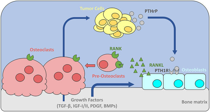
Background: Bone marrow is a common site of metastasis for a number of tumor types, including breast, prostate, and lung cancer, but the mechanisms controlling tumor dormancy in bone are poorly understood. In breast cancer, while advances in drug development, screening practices, and surgical techniques have dramatically improved survival rates in recent decades, metastatic recurrence in the bone remains common and can develop years or decades after elimination of the primary tumor.

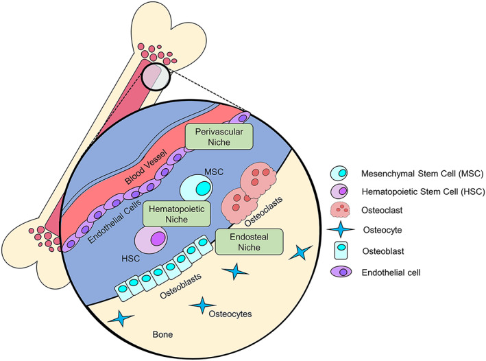
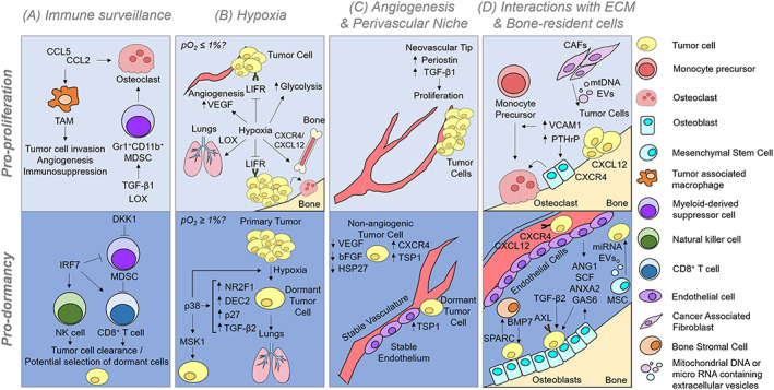
Recent findings: It is now understood that tumor cells disseminate to distant metastatic sites at early stages of tumor progression, leaving cancer survivors at a high risk of recurrence. This review will discuss mechanisms of bone lesion development and current theories of how dormant cancer cells behave in bone, as well as a number of processes suspected to be involved in the maintenance of and exit from dormancy in the bone microenvironment.

Conclusions: The bone is a complex microenvironment with a multitude of cell types and processes. Many of these factors, including angiogenesis, immune surveillance, and hypoxia, are thought to regulate tumor cell entry and exit from dormancy in different bone marrow niches.

Keywords: angiogenesis; bone marrow; dormancy; hypoxia; immune surveillance; metastasis.


Conflict of interest statement

CONFLICT OF INTEREST The authors have no conflicts of interest to declare.

Figures

Figure 1
Figure 1
Mechanisms of tumor‐induced bone destruction. Tumor cells secrete parathyroid hormone related protein (PTHrP), which signals through parathyroid hormone receptor type 1 (PTH1R) on the surface of osteoblasts to promote their activity and differentiation. Increased osteoblast activity results in increased secretion of the receptor activator of nuclear factor kappa‐B ligand (RANKL), which binds to RANK on pre‐osteoclasts to promote osteoclastogenesis and on mature osteoclasts to stimulate their activity. Increased number and activity of osteoclasts results in more bone resorption and release of growth factors from the bone matrix, including transforming growth factor β (TGF‐β), insulin‐like growth factor I and II (IGF‐I/II), platelet derived growth factor (PDGF), and bone morphogenetic proteins (BMPs). These growth factors normally function to couple osteoclast and osteoblast activity, but can also stimulate further tumor cell proliferation and PTHrP production by the tumor cells to feed this cycle of bone destruction
Figure 2
Figure 2
The bone is composed of three distinct niches that can exert pro‐ or anti‐dormancy effects. The perivascular niche contains blood vessel lining endothelial cells that can secrete factors to push cell into, or out of, dormancy, and signaling between endothelial cells and hypoxic tumor cells can drive tumor vascularization and escape from dormancy. Hematopoietic stem cells (HSC) and mesenchymal stem cells (MSC) that reside in the hematopoietic niche secrete signaling factors or produce extracellular vesicles that can influence a cell's dormancy status. The endosteal niche contains osteoclasts and osteoblasts that play a role in the vicious cycle, but can also secrete factors to promote dormancy. Since these niches are in close proximity to one another, tumor cells will often seed in the bone marrow and experience the effects of multiple niches
Figure 3
Figure 3
Tumor cells enter or exit dormancy depending on the micro‐environmental signals they encounter. A, Immune surveillance mechanisms can stimulate angiogenesis, immunosuppression, and osteoclastogenesis to stimulate tumor growth, but immune cells can also clear tumor cells and potentially select for non‐immunogenic or, potentially, dormant clones. B, Hypoxia promotes dissemination of tumor cells to distant sites and their ability to colonize the bone marrow. Hypoxia has also been shown to stimulate the expression of several dormancy‐associated genes. We speculate that severe hypoxia (pO2  0.5%) may stimulate tumor cell exit from dormancy while moderate hypoxia (pO2 < 4% and  0.5%) may favor dormancy. C, According to the angiogenic dormancy model, micrometastases that cannot induce angiogenesis will remain dormant. It is now clear that the spatial relationship of tumor cells in relation to endothelial derived factors can control the proliferation or dormancy of disseminated tumor cells, in that cells located along neovascular tips are stimulated to proliferate, while cells residing along the stable vasculature will remain dormant. D, the bone is an organ packed with many cell types. Tumor cells in bone can induce the vicious cycle of bone destruction due to PTHrP expression, as described in more detail in Figure 1. Additionally, if tumor cells have upregulated VCAM1 expression, they can stimulate osteoclastogenesis by recruiting monocyte precursors that can differentiate into osteoclasts. Endothelial cells and osteoblasts in particular secrete many factors that are critical for bone homeostasis and hematopoietic stem cell quiescence and maintenance, which can be coopted by some tumor cells. Thus, once tumor cells home to the bone marrow, they are exposed to many signals that promote their survival and quiescence. CCL2 = C‐C motif chemokine ligand 2 (monocyte chemotactic protein 1); CCL5 = C‐C motif chemokine ligand 5; TAM = tumor associated macrophage; MDSC = monocyte‐derived suppressor cell; TGF‐B1/2 = transforming growth factor Beta 1/2; LOX = lysyl oxidase; DKK1 = dikkopf‐1; IRF7 = interferon regulatory factor 7; NK cell = natural killer cell; LIFR = leukemia inhibitory factor receptor; VEGF = vascular endothelial growth factor; NR2F1 = nuclear receptor subfamily 2 group F member 1; DEC2 = differentially expressed in chondrocytes 2; MSK1 = mitogen‐ and stress‐activated protein kinase 1; TSP1 = thrombospondin 1; HSP27 = heat shock protein 27; bFGF = basic fibroblast growth factor; VCAM1 = vascular cell adhesion molecule 1; PTHrP = parathyroid hormone related protein; mtDNA = mitochondrial DNA; EV = extracellular vesicle; CXCR4 = C‐X‐C motif chemokine receptor 4; CXCL12 = C‐X‐C motif chemokine ligand 12 (stromal cell‐derived factor 1); miRNA = micro RNA; ANG1 = angiopoietin 1; SCF = stem cell factor; ANXA2 = Annexin II; GAS6 = growth arrest‐specific 6; BMP7 = bone morphogenetic protein 7; SPARC = secreted protein rich in cysteine

References

    1. Suva LJ, Washam C, Nicholas RW, Griffin RJ. Bone metastasis: mechanisms and therapeutic opportunities. Nat Rev Endocrinol. 2011;7(4):208‐218. - PMC - PubMed
    1. Coleman RE. Clinical features of metastatic bone disease and risk of skeletal morbidity. Clin Cancer Res. 2006;12(20 Pt 2):6243s‐6249s. - PubMed
    1. Riihimaki M, Hemminki A, Fallah M, et al. Metastatic sites and survival in lung cancer. Lung Cancer. 2014;86(1):78‐84. - PubMed
    1. Katakami N, Kunikane H, Takeda K, et al. Prospective study on the incidence of bone metastasis (BM) and skeletal‐related events (SREs) in patients (pts) with stage IIIB and IV lung cancer‐CSP‐HOR 13. J Thorac Oncol. 2014;9(2):231‐238. - PMC - PubMed
    1. Gomis RR, Gawrzak S. Tumor cell dormancy. Mol Oncol. 2016;11:62. - PMC - PubMed

Publication types

MeSH terms

Substances