Review
. 2020 Dec;19(6):833-868.
doi: 10.1007/s12311-020-01155-1.

Consensus Paper: Cerebellum and Social Cognition

Affiliations
Review

Consensus Paper: Cerebellum and Social Cognition

Frank Van Overwalle et al. Cerebellum. 2020 Dec.

Abstract

The traditional view on the cerebellum is that it controls motor behavior. Although recent work has revealed that the cerebellum supports also nonmotor functions such as cognition and affect, only during the last 5 years it has become evident that the cerebellum also plays an important social role. This role is evident in social cognition based on interpreting goal-directed actions through the movements of individuals (social "mirroring") which is very close to its original role in motor learning, as well as in social understanding of other individuals' mental state, such as their intentions, beliefs, past behaviors, future aspirations, and personality traits (social "mentalizing"). Most of this mentalizing role is supported by the posterior cerebellum (e.g., Crus I and II). The most dominant hypothesis is that the cerebellum assists in learning and understanding social action sequences, and so facilitates social cognition by supporting optimal predictions about imminent or future social interaction and cooperation. This consensus paper brings together experts from different fields to discuss recent efforts in understanding the role of the cerebellum in social cognition, and the understanding of social behaviors and mental states by others, its effect on clinical impairments such as cerebellar ataxia and autism spectrum disorder, and how the cerebellum can become a potential target for noninvasive brain stimulation as a therapeutic intervention. We report on the most recent empirical findings and techniques for understanding and manipulating cerebellar circuits in humans. Cerebellar circuitry appears now as a key structure to elucidate social interactions.

Keywords: Body language reading; Cerebellar stimulation; Crus I/II; Innate hand-tool overlap; Mind reading; Posterior cerebellum; Social action sequences; Social cognition; Social mentalizing; Social mirroring; Stone-tool making.


Conflict of interest statement

The authors declare that they have no conflict of interest.

Figures

Fig. 1
Fig. 1
Transversal view of the inferior and superior cerebellum at MNI z-coordinates − 50 and – 32, respectively. [Left] The most active areas in the cerebellum from the automated meta-analyses of NeuroSynth (50 topics) [right] overlaid on the 7-network structure of Buckner et al. [12] with coordinates denoted by white crosses. Three green “mirror” areas associated with “action” and “mirror” keywords in NeuroSynth (#19) are part of the green somatomotor integration and purple ventral attention networks; two red “mentalizing” areas associated with the “mentalizing” keyword in NeuroSynth (#8) are part of the red mentalizing network
Fig. 2
Fig. 2
a Cerebellar task activation maps [84] (top) and resting-state networks [12] (bottom). 1 = indication of emotion processing activation in lobule IX (for clarity). 2 = indication of area of language/social overlap (for clarity). Asterisk (left lobule IX) = indication of region of working memory task activation if a lower effect size threshold is used, as shown in the supplementary material of [84]. b Cerebellar functional gradients [85]. Atlas indicates the position of each motor and nonmotor representation [84]. c Relationship of functional gradients 1 and 2 with task activation maps (top) and resting-state networks (bottom). Each dot corresponds to one cerebellar voxel; vertical/horizontal position of each dot corresponds to gradient 1/gradient 2 values for that voxel; the color of each dot indicates whether each voxel belongs to a particular task activation (top) or resting-state network (bottom) map [85]
Fig. 3
Fig. 3
Results of the meta-analysis for distinct mentalizing task subcategories in proportion to 100% of all identified studies. All other nonmentalizing functions are denoted by white. ROIs 1 and 2 with MNI coordinates ± 25, − 75, − 40 are superimposed on the 7-network parcellation from Bruckner et al. [12], where the white area reflects the mentalizing/default network; ROIs 3 and 4 with MNI coordinates ± 26, − 84, − 34 are from the mentalizing meta-analysis in NeuroSynth
Fig. 4
Fig. 4
An example of a social false belief sequence in the picture sequencing task ([76]; the correct order is 2–1–4–3; the numbers are not shown to the participants but given here for display purposes). Participants had to select, in the correct order, the first picture on the screen, then the second picture, and so on
Fig. 5
Fig. 5
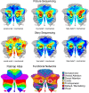
Top: Activation in the posterior cerebellum in the Picture and Story sequencing tasks for social scripts, true and false belief > mechanical comparisons shown on a SUIT flatmap [124] without threshold. True and false beliefs strongly activate Crus II in the default mode/mentalizing network, while social scripts activate this area in Crus II less so. Bottom: SUIT flatmap atlas showing the cerebellar lobules from [124] and functional networks from Fig. 2 of this consensus paper
Fig. 6
Fig. 6
Left: Experimental procedure (abridged). Participants were instructed to learn the given temporal order of a set of six sentences involving a single person or object, and had to infer from these six sentences a common trait of the person or feature of the object. Right: A preliminary analysis comparing social (person) and nonsocial (object) conditions during this study phase revealed activation in the bilateral posterior cerebellum (MNI coordinates 20, – 76, − 36; n = 19)
Fig. 7
Fig. 7
Left: The serial belief reaction time task. In this design, on each trial, participants had to report how many green flowers were received among green clovers according to one of two smurfs (i.e., Papa Smurf or Smurfette). On true trials, the smurf was turned to the screen and participants should report what the smurf could observe (the number of flowers); on false trials, the smurf was turned away from the screen and participants should report what they believed that the smurf saw last. Participants implicitly learned the fixed (but unknown) sequences embedded in the task, in particular the sequence of true and false beliefs. Implicit learning was attested by interspersing blocks with random instead of fixed standard sequences (e.g., random true–false beliefs), and observing significantly increasing response times as a consequence. Right: In a follow-up fMRI study [135], a parallel increasing pattern of posterior cerebellar activation during true–false belief randomization was observed (MNI coordinates − 36, − 64, − 42; n = 18)

References

    1. Schmahmann JD, Sherman JC. The cerebellar cognitive affective syndrome. Brain. 1998;141(1998):561–579. - PubMed
    1. Van Overwalle F, Baetens K, Mariën P, Vandekerckhove M. Social cognition and the cerebellum: a meta-analysis of over 350 fMRI studies. Neuroimage. 2014;86(2014):554–572. - PubMed
    1. Van Overwalle F, De Coninck S, Heleven E, Perrotta G, Taib NO. Ben, Manto M, et al. The role of the cerebellum in reconstructing social action sequences: a pilot study. Soc Cogn Affect Neurosci. 2019;14(5):549–558. - PMC - PubMed
    1. Schurz M, Radua J, Aichhorn M, Richlan F, Perner J. Fractionating theory of mind: a meta-analysis of functional brain imaging studies. Neurosci Biobehav Rev. 2014;42:9–34. - PubMed
    1. Van Overwalle F. Social cognition and the brain: a meta-analysis. Hum Brain Mapp. 2009;30(3):829–858. - PMC - PubMed