. 2020 Oct;17(4):1907-1918.
doi: 10.1007/s13311-020-00884-9.

Intranasal Dexamethasone Reduces Mortality and Brain Damage in a Mouse Experimental Ischemic Stroke Model

Affiliations

Intranasal Dexamethasone Reduces Mortality and Brain Damage in a Mouse Experimental Ischemic Stroke Model

Alejandro Espinosa et al. Neurotherapeutics. 2020 Oct.

Abstract

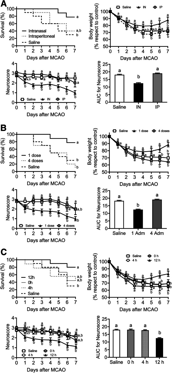
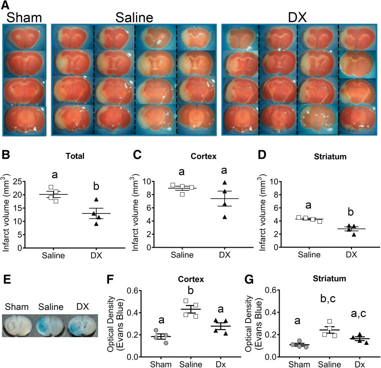
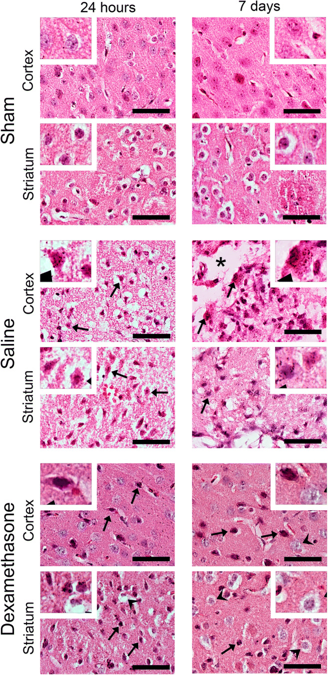
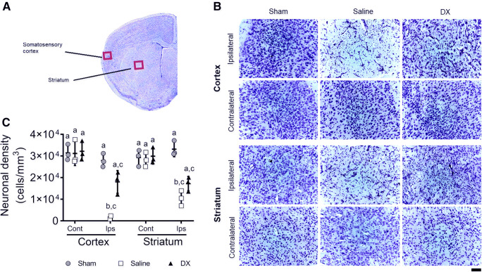
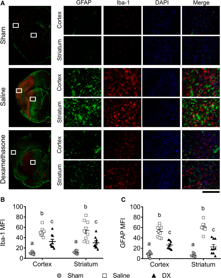
Neuroinflammation triggered by the expression of damaged-associated molecular patterns released from dying cells plays a critical role in the pathogenesis of ischemic stroke. However, the benefits from the control of neuroinflammation in the clinical outcome have not been established. In this study, the effectiveness of intranasal, a highly efficient route to reach the central nervous system, and intraperitoneal dexamethasone administration in the treatment of neuroinflammation was evaluated in a 60-min middle cerebral artery occlusion (MCAO) model in C57BL/6 male mice. We performed a side-by-side comparison using intranasal versus intraperitoneal dexamethasone, a timecourse including immediate (0 h) or 4 or 12 h poststroke intranasal administration, as well as 4 intranasal doses of dexamethasone beginning 12 h after the MCAO versus a single dose at 12 h to identify the most effective conditions to treat neuroinflammation in MCAO mice. The best results were obtained 12 h after MCAO and when mice received a single dose of dexamethasone (0.25 mg/kg) intranasally. This treatment significantly reduced mortality, neurological deficits, infarct volume size, blood-brain barrier permeability in the somatosensory cortex, inflammatory cell infiltration, and glial activation. Our results demonstrate that a single low dose of intranasal dexamethasone has neuroprotective therapeutic effects in the MCAO model, showing a better clinical outcome than the intraperitoneal administration. Based on these results, we propose a new therapeutic approach for the treatment of the damage process that accompanies ischemic stroke.

Keywords: Dexamethasone; MCAO; inflammation; intranasal administration; ischemic stroke.


Conflict of interest statement

The authors declare that they have no conflict of interest.

Figures

Fig. 1
Fig. 1
One single dose of dexamethasone (DX) intranasally administered 12 h after stroke improves survival and neurological performance in MCAO mice. Intranasal (IN) versus intraperitoneal (IP) DX routes of administration were compared at 12 h after MCAO. Each mouse was treated with 0.25 mg/kg of DX by the IN or IP route or received 20 μl IN saline solution (A). A single dose of 0.25 mg/kg of DX administered 12 h after MCAO was compared with 4 repeated administrations every 24 h starting at 12 h (B), and with single administrations at 0 and 4 h after the insult (C) (n = 10 per group). The percentage of survival, body weight, and the area under the curve (AUC) for neuroscore were determined daily for 7 days after MCAO. Data represent the mean ± SEM. Survival was analyzed by the Mantel–Cox log–rank test; the percentage of body weight loss was analyzed by 2-way ANOVA, followed by Bonferroni’s multiple-comparisons test. The AUC was calculated averaging neuroscore scale for 7 days and peaks were compared by 1-way ANOVA, followed by Tukey’s multiple-comparisons test. Different letters (a and b) indicate significant differences between groups (p < 0.05)
Fig. 2
Fig. 2
Dexamethasone (DX) treatment decreased mortality and improved the neuroscore and weight at 7 days and promotes long-term recovery after MCAO. Experimental design line (A). Twelve hours after MCAO, each mouse was treated with 0.25 mg/kg DX or 20 μl saline solution by the intranasal route. The percentage of survival (B), body weight (C), and area under the curve (AUC) for neuroscore (D) were determined daily for 7 days after MCAO. From mice that survived 7 days after the MCAO, a group of animals (n = 10 per group) were evaluated for an extra period of 42 days (6 weeks) (A, B, and C). Data represent the mean ± SEM. Survival was analyzed by the Mantel–Cox log–rank test; the percentage of body weight loss was analyzed by 2-way ANOVA, followed by Bonferroni’s multiple-comparisons test. The AUC was calculated averaging the neuroscore scale for 7 days, and peaks were compared by 1-way ANOVA, followed by Tukey’s multiple-comparisons test. Different letters (a, b, and c) indicate significant differences in the survival, body weight, and neuroscore between sham, treated, and nontreated mice (p < 0.05)
Fig. 3
Fig. 3
Intranasal dexamethasone (DX) reduced blood–brain barrier permeability and infarct volume 7 days after the MCAO. Coronal brain sections of the sham-, saline-, and DX-treated groups are shown (A). Images correspond to the 4 saline- and DX-treated mice presented in vertical columns separated by pointed lines. The total infarction volume (B), and infarct volume in the cortex (C) and striatum (D) were quantified using the Image J software. Representative coronal brain slices of Evans blue extravasation evaluated in the cortex and striatum (marked with dotted lines) of the sham, saline, and DX groups are shown (E). Quantification of Evans blue is shown as relative units of optical density in the cortex (F) and striatum (G) from sham-, saline-, or DX-treated mice. Each point represents the data of an individual mouse (n = 4 per group). Data represent the mean ± SEM. Data were analyzed by a 2-tailed unpaired t test (infarct volume) and 1-way ANOVA, followed by Tukey’s multiple-comparisons test (BBB permeability). Different letters (a, b, and c) indicate significant differences in the infarct volume and BBD permeability between treated and nontreated mice (p < 0.05)
Fig. 4
Fig. 4
Intranasal dexamethasone reduces markedly the ischemic brain tissue damage in an MCAO model. Representative images of a brain section (ipsilateral hemisphere) stained with hematoxylin and eosin (H&E) of the sham-, saline-, or DX-treated groups are shown. Coronal sections of whole brains of sham mice and mice subjected to MCAO for 24 h and 7 days are observed. One control group was treated by the intranasal route with saline solution, and the other group received 0.25 mg/kg intranasal dexamethasone. For histologic examination, low (× 60)-magnification H&E-stained micrographs of the somatosensory cortex and striatum were examined for each animal (n = 3 per group). High-magnification images are shown in white boxes. Scale bar, 50 μm
Fig. 5
Fig. 5
Increased neuronal density in the somatosensory cortex and the striatum after intranasal dexamethasone (DX) treatment in the MCAO model. Representative image of a brain section stained with Nissl to illustrate where neuronal quantifications were analyzed (A). Representative images of Nissl staining of the somatosensory cortex and striatum from the sham-, saline-, and DX-treated mice after 24 h of performing MCAO (B). Quantification of the neuronal density of 4 adjacent histological sections of 20 μm (800 μm total distance) stained with Nissl in the cortex and striatum from the ipsilateral (Ips) and contralateral (Cont) hemisphere of the sham, saline, or DX animals (C). Each point represents the data of an individual mouse (n = 3 per group). Data represent the mean ± SEM. Data were analyzed by a Kruskal–Wallis, followed by Dunn’s multiple-comparisons test. Different letters (a, b, and c) indicate significant differences in the neuronal density between groups (p < 0.05). Scale bar, 50 μm
Fig. 6
Fig. 6
Analysis of brain GFAP (green) and Iba-1 (red) expression in treated and nontreated mice 7 days after the MCAO. Nuclei were stained with DAPI (blue). Representative mouse brain sections of the sham-, saline-, and dexamethasone (DX)-treated mice (A). Left images show a low-magnification reconstruction of the ipsilateral hemisphere. The mean fluorescence intensity (MFI) of Iba-1 (B) and GFAP (C) were quantified using the Image J software. Each point represents the data from 1 of 3 photographs analyzed in both the cortex and striatum (n = 3 animals per group). Data represent the mean ± SEM. Data were analyzed with 2-way ANOVA followed by Bonferroni’s test. Different letters (a, b, and c) indicate significant differences in the MFI between groups (p < 0.05). Scale bar, 200 μm

Similar articles

Cited by

References

    1. Institute for Health Metrics and Evaluation (IHME) Findings from the Global Burden of Disease Study 2017. Seattle, WA: IHME; 2018.
    1. López-Espuela F, Pedrera-Zamorano JD, Jiménez-Caballero PE, et al. Functional status and disability in patients after acute stroke: a longitudinal study. Am J Crit Care. 2016;25:144–151. - PubMed
    1. Noe-Sebastian E, Balasch-Bernat M, Colomer-Font C, et al. Disability after stroke: a longitudinal study in moderate and severe stroke patients included in a multidisciplinary rehabilitation program. Rev Neurol. 2017;64:385–392. - PubMed
    1. Gülke E, Gelderblom M, Magnus T. Danger signals in stroke and their role on microglia activation after ischemia. Ther Adv Neurol Disord. 2018;11:1756286418774254. - PMC - PubMed
    1. Patel AR, Ritzel R, McCullough LD, Liu F. Microglia and ischemic stroke: a double-edged sword. Int J Physiol Pathophysiol Pharmacol. 2013;5:73–90. - PMC - PubMed

Publication types

MeSH terms