. 2021 Mar;529(4):786-801.
doi: 10.1002/cne.24978. Epub 2020 Aug 3.

Estrogen receptor alpha, G-protein coupled estrogen receptor 1, and aromatase: Developmental, sex, and region-specific differences across the rat caudate-putamen, nucleus accumbens core and shell

Affiliations

Estrogen receptor alpha, G-protein coupled estrogen receptor 1, and aromatase: Developmental, sex, and region-specific differences across the rat caudate-putamen, nucleus accumbens core and shell

Amanda A Krentzel et al. J Comp Neurol. 2021 Mar.

Abstract

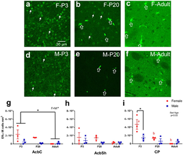
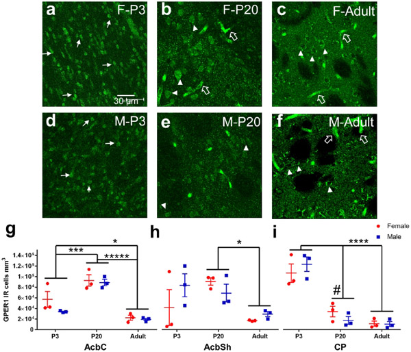
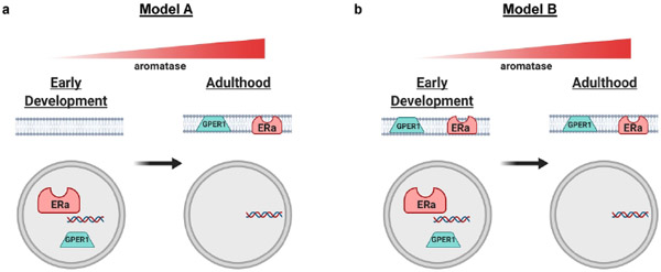
Sex steroid hormones such as 17β-estradiol (estradiol) regulate neuronal function by binding to estrogen receptors (ERs), including ERα and GPER1, and through differential production via the enzyme aromatase. ERs and aromatase are expressed across the nervous system, including in the striatal brain regions. These regions, comprising the nucleus accumbens core, shell, and caudate-putamen, are instrumental for a wide-range of functions and disorders that show sex differences in phenotype and/or incidence. Sex-specific estrogen action is an integral component for generating these sex differences. A distinctive feature of the striatal regions is that in adulthood neurons exclusively express membrane but not nuclear ERs. This long-standing finding dominates models of estrogen action in striatal regions. However, the developmental etiology of ER and aromatase cellular expression in female and male striatum is unknown. This omission in knowledge is important to address, as developmental stage influences cellular estrogenic mechanisms. Thus, ERα, GPER1, and aromatase cellular immunoreactivity was assessed in perinatal, prepubertal, and adult female and male rats. We tested the hypothesis that ERα, GPER1, and aromatase exhibits sex, region, and age-specific differences, including nuclear expression. ERα exhibits nuclear expression in all three striatal regions before adulthood and disappears in a region- and sex-specific time-course. Cellular GPER1 expression decreases during development in a region- but not sex-specific time-course, resulting in extranuclear expression by adulthood. Somatic aromatase expression presents at prepuberty and increases by adulthood in a region- but not sex-specific time-course. These data indicate that developmental period exerts critical sex-specific influences on striatal cellular estrogenic mechanisms.

Keywords: RRID AB_1141090; RRID AB_310305; RRID AB_566942; aromatase; estrogen receptor; rat; sex differences; striatum.


Conflict of interest statement

Conflict of Interest: The authors declare that the research was conducted in the absence of any commercial or financial relationships that could be construed as a potential conflict of interest.

Figures

Figure 1:
Figure 1:. Anatomical locations.
Depiction of locations where subregions were imaged. Imaged areas are 1) caudate-putamen (CP), 2) nucleus accumbens core (AcbC), 3) nucleus accumbens shell (AcbSh), and 4) cingulate cortex (Cg). Adapted from Rat Brain in Stereotaxic Coordinates.
Figure 2:
Figure 2:. Nuclear ERα present in early-life and disappears by adulthood in the striatal subregions.
a-f) Representative images of caudate-putamen depicting ERα-IR cells in females P3 (a), P20 (b), and adult (c) and males P3 (d), P20 (e), and adult (f). Examples of ERα-IR nuclei are marked by small, closed, white arrows. Examples of blood vessel/staining artifact around marked by large, open, white arrows. g) Cell counts of ERα-IR nuclei from each male (blue) and female (red) subject of the nucleus accumbens core (AcbC). Lines and error bars depict means and standard errors. h) Cell counts of ERα-IR nuclei from nucleus accumbens shell (AcbSh). i) Cell counts of ERα-IR nuclei from caudate-putamen (CP). * p<0.05 and red letters depict significant posthoc age comparisons for females.
Figure 3:
Figure 3:. Nuclear ERα present in adulthood in the arcuate nucleus.
Representative image of ERα-IR nuclei staining in an adult female arcuate nucleus to demonstrate that not all brain regions eliminate nuclear ERα in adulthood. Small, closed, white arrows point to examples of ERα-IR nuclei.
Figure 4:
Figure 4:. GPER1 changes cellular compartments and decreases in density with age.
a-f) Representative images of caudate-putamen depicting GPER1-IR cells in females at P3 (a), P20 (b), and adults (c) and males at P3 (d), P20 (e), and adults (f). Examples of GPER1-IR nuclei or soma+nuclei are marked by white triangles. Examples of cytoplasm only GPER1-IR are marked by white triangles. Examples of blood vessel/staining artifact around marked by large, open, white arrows. g) Cell counts of GPER1-IR cells from each male (blue) and female (red) subject of the nucleus accumbens core (AcbC). Lines and error bars depict means and standard errors. h) Cell counts of GPER1-IR cells from nucleus accumbens shell (AcbSh). i) Cell counts of GPER1-IR cells from caudate-putamen (CP).# p<0.10, * p<0.05, *** p<0.001, **** p<0.0001, ***** p<0.00001.
Figure 5:
Figure 5:. GPER1 does not have similar age dependent changes in cingulate cortex.
a) Representative image of cingulate cortex depicting GPER1-IR cells in an adult female. Examples of cytoplasm only GPER1-IR are marked by white triangles. Examples of blood vessel/staining artifact around marked by large, open, white arrows. b) Cell counts of GPER1-IR cells from each male (blue) and female (red) subject of the cingulate cortex. Ns=non-significant.
Figure 6:
Figure 6:. Somatic aromatase-IR cells increases in density with age.
a-f) Representative images of caudate-putamen depicting aromatase-IR cells in females at P3 (a), P20 (b), and adults (c) and males at P3 (d), P20 (e), and adults (f). Examples of cytoplasm only aromatase-IR are marked by white triangles. Examples of blood vessel/staining artifact around marked by large, open, white arrows. g) Cell counts of aromatase-IR cells from each male (blue) and female (red) subject of the nucleus accumbens core (AcbC). Lines and error bars depict means and standard errors. h) Cell counts of aromatase-IR cells from nucleus accumbens shell (AcbSh). i) Cell counts of aromatase-IR cells from caudate-putamen (CP). * p<0.05, ** p<0.01, *** p<0.001, **** p<0.0001.
Figure 7:
Figure 7:. Somatic aromatase-IR cell density also increases with age in cingulate cortex.
a) Representative image from cingulate cortex depicting aromatase-IR cells in females. Examples of cytoplasm only aromatase-IR are marked by white triangles. Examples of blood vessel/staining artifact around marked by large, open, white arrows. b) Cell counts of aromatase-IR cells from each male (blue) and female (red) subject of the cingulate cortex. Lines and error bars depict means and standard errors. ** p<0.01
Figure 8:
Figure 8:. Somatic aromatase-IR in the medial amygdala.
Representative image from medial amygdala depicting aromatase-IR cells in females. Examples of cytoplasm only aromatase-IR are marked by white triangles.
Figure 9:
Figure 9:. Hypothetical models for estrogen receptor compartmentalization across development in the striatum.
These depict two hypotheses of how estrogen receptors ERα and GPER1 may exist in neuron cellular compartments from early development. Both models are based on this study’s current findings of transition from nuclear compartment to extranuclear compartments. Both models show a rise of aromatase expression with age. a) Model A depicts only nuclear expression of ERα and GPER1 in early development that switches to exclusively extranuclear expression by adulthood. b) Model B depicts both nuclear and extranuclear expression of ERα and GPER1 in early development, with nuclear expression disappearing by adulthood. Figure made using Biorender.

References

    1. Acharya KD, & Veney SL (2012). Characterization of the G-protein-coupled membrane-bound estrogen receptor GPR30 in the zebra finch brain reveals a sex difference in gene and protein expression. Dev Neurobiol, 72(11), 1433–1446. doi:10.1002/dneu.22004 - DOI - PubMed
    1. Adlanmerini M, Solinhac R, Abot A, Fabre A, Raymond-Letron I, Guihot AL, … Lenfant F (2014). Mutation of the palmitoylation site of estrogen receptor alpha in vivo reveals tissue-specific roles for membrane versus nuclear actions. Proc Natl Acad Sci U S A, 111(2), E283–290. doi:10.1073/pnas.1322057111 - DOI - PMC - PubMed
    1. Agarwal VR, Sinton CM, Liang C, Fisher C, German DC, & Simpson ER (2000). Upregulation of estrogen receptors in the forebrain of aromatase knockout (ArKO) mice. Mol Cell Endocrinol, 162(1-2), 9–16. doi:10.1016/s0303-7207(00)00227-6 - DOI - PubMed
    1. Almey A, Cannell E, Bertram K, Filardo E, Milner TA, & Brake WG (2014). Medial prefrontal cortical estradiol rapidly alters memory system bias in female rats: ultrastructural analysis reveals membrane-associated estrogen receptors as potential mediators. Endocrinology, 155(11), 4422–4432. doi:10.1210/en.2014-1463 - DOI - PMC - PubMed
    1. Almey A, Filardo EJ, Milner TA, & Brake WG (2012). Estrogen receptors are found in glia and at extranuclear neuronal sites in the dorsal striatum of female rats: evidence for cholinergic but not dopaminergic colocalization. Endocrinology, 153(11), 5373–5383. doi:10.1210/en.2012-1458 - DOI - PMC - PubMed

Publication types

MeSH terms

Substances