

Evaluating Biochemically Recurrent Prostate Cancer: Histologic Validation of 18F-DCFPyL PET/CT with Comparison to Multiparametric MRI
- PMID: 32633674
- PMCID: PMC7457947
- DOI: 10.1148/radiol.2020192018
Evaluating Biochemically Recurrent Prostate Cancer: Histologic Validation of 18F-DCFPyL PET/CT with Comparison to Multiparametric MRI
Abstract
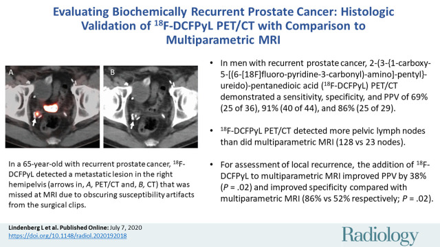
Background Prostate cancer recurrence is found in up to 40% of men with prior definitive (total prostatectomy or whole-prostate radiation) treatment. Prostate-specific membrane antigen PET agents such as 2-(3-{1-carboxy-5-[(6-[18F]fluoro-pyridine 3-carbonyl)-amino]-pentyl}-ureido)-pentanedioic acid (18F-DCFPyL) may improve detection of recurrence compared with multiparametric MRI; however, histopathologic validation is lacking. Purpose To determine the sensitivity, specificity, and positive predictive value (PPV) of 18F-DCFPyL PET/CT based on histologic analysis and to compare with pelvic multiparametric MRI in men with biochemically recurrent prostate cancer. Materials and Methods Men were prospectively recruited after prostatectomy and/or radiation therapy with rising prostate-specific antigen level (median, 2.27 ng/mL; range, 0.2-27.45 ng/mL) and a negative result at conventional imaging (bone scan and/or CT). Participants underwent 18F-DCFPyL PET/CT imaging and 3.0-T pelvic multiparametric MRI. Statistical analysis included Wald and modified χ2 tests. Results A total of 323 lesions were visualized in 77 men by using 18F-DCFPyL or multiparametric MRI, with imaging detection concordance of 25% (82 of 323) when including all lesions in the MRI field of view and 53% (52 of 99) when only assessing prostate bed lesions. 18F-DCFPyL depicted more pelvic lymph nodes than did MRI (128 vs 23 nodes). Histologic validation was obtained in 80 locations with sensitivity, specificity, and PPV of 69% (25 of 36; 95% confidence interval [CI]: 51%, 88%), 91% (40 of 44; 95% CI: 74%, 98%), and 86% (25 of 29; 95% CI: 73%, 97%) for 18F-DCFPyL and 69% (24 of 35; 95% CI: 50%, 86%), 74% (31 of 42; 95% CI: 42%, 89%), and 69% (24 of 35; 95% CI: 50%, 88%) for multiparametric MRI (P = .95, P = .14, and P = .07, respectively). In the prostate bed, sensitivity, specificity, and PPV were 57% (13 of 23; 95% CI: 32%, 81%), 86% (18 of 21; 95% CI: 73%, 100%), and 81% (13 of 16; 95% CI: 59%, 100%) for 18F-DCFPyL and 83% (19 of 23; 95% CI: 59%, 100%), 52% (11 of 21; 95% CI: 29%, 74%), and 66% (19 of 29; 95% CI: 44%, 86%) for multiparametric MRI (P = .19, P = .02, and P = .17, respectively). The addition of 18F-DCFPyL to multiparametric MRI improved PPV by 38% overall (P = .02) and by 30% (P = .09) in the prostate bed. Conclusion Findings with 2-(3-{1-carboxy-5-[(6-[18F]fluoro-pyridine 3-carbonyl)-amino]-pentyl}-ureido)-pentanedioic acid (18F-DCFPyL) were histologically validated and demonstrated high specificity and positive predictive value. In the pelvis, 18F-DCFPyL depicted more lymph nodes and improved positive predictive value and specificity when added to multiparametric MRI. © RSNA, 2020 Online supplemental material is available for this article. See also the editorial by Zukotynski and Rowe in this issue.
Figures
![Participant flowchart. 18F-DCFPyL = 2-(3-{1-carboxy-5-[(6-[18F]fluoro-pyridine 3-carbonyl)-amino]-pentyl}-ureido)-pentanedioic acid.](https://cdn.ncbi.nlm.nih.gov/pmc/blobs/568c/7457947/bfe63a86ec9c/radiol.2020192018.fig1.jpg)
![Images show 2-(3-{1-carboxy-5-[(6-[18F]fluoro-pyridine 3-carbonyl)-amino]-pentyl}-ureido)-pentanedioic acid (18F-DCFPyL) PET/CT–positive, multiparametric MRI–negative, and biopsy-positive lesion in a 65-year-old man (prostate-specific antigen level of 1.85 ng/mL) with history of prostate cancer and prior radical prostatectomy, now with biochemically recurrent prostate cancer. 18F-DCFPyL depicted pathologically proven metastatic lesion in right hemipelvis (arrow in [a] PET, [b] PET/CT, and [c] CT) that was missed at multiparametric MRI review due to obscuring susceptibility artifacts from surgical clips at (d) T2-weighted MRI, (e) b = 2000 sec/mm2 diffusion-weighted MRI, (f) apparent diffusion coefficient map, and (g) dynamic contrast material–enhanced MRI.](https://cdn.ncbi.nlm.nih.gov/pmc/blobs/568c/7457947/df3c62dfd262/radiol.2020192018.fig2a.jpg)
![Images show 2-(3-{1-carboxy-5-[(6-[18F]fluoro-pyridine 3-carbonyl)-amino]-pentyl}-ureido)-pentanedioic acid (18F-DCFPyL) PET/CT–positive, multiparametric MRI–negative, and biopsy-positive lesion in a 65-year-old man (prostate-specific antigen level of 1.85 ng/mL) with history of prostate cancer and prior radical prostatectomy, now with biochemically recurrent prostate cancer. 18F-DCFPyL depicted pathologically proven metastatic lesion in right hemipelvis (arrow in [a] PET, [b] PET/CT, and [c] CT) that was missed at multiparametric MRI review due to obscuring susceptibility artifacts from surgical clips at (d) T2-weighted MRI, (e) b = 2000 sec/mm2 diffusion-weighted MRI, (f) apparent diffusion coefficient map, and (g) dynamic contrast material–enhanced MRI.](https://cdn.ncbi.nlm.nih.gov/pmc/blobs/568c/7457947/4a6e88e98d8a/radiol.2020192018.fig2b.jpg)
![Images show 2-(3-{1-carboxy-5-[(6-[18F]fluoro-pyridine 3-carbonyl)-amino]-pentyl}-ureido)-pentanedioic acid (18F-DCFPyL) PET/CT–positive, multiparametric MRI–negative, and biopsy-positive lesion in a 65-year-old man (prostate-specific antigen level of 1.85 ng/mL) with history of prostate cancer and prior radical prostatectomy, now with biochemically recurrent prostate cancer. 18F-DCFPyL depicted pathologically proven metastatic lesion in right hemipelvis (arrow in [a] PET, [b] PET/CT, and [c] CT) that was missed at multiparametric MRI review due to obscuring susceptibility artifacts from surgical clips at (d) T2-weighted MRI, (e) b = 2000 sec/mm2 diffusion-weighted MRI, (f) apparent diffusion coefficient map, and (g) dynamic contrast material–enhanced MRI.](https://cdn.ncbi.nlm.nih.gov/pmc/blobs/568c/7457947/74b498bd3600/radiol.2020192018.fig2c.jpg)
![Images show 2-(3-{1-carboxy-5-[(6-[18F]fluoro-pyridine 3-carbonyl)-amino]-pentyl}-ureido)-pentanedioic acid (18F-DCFPyL) PET/CT–positive, multiparametric MRI–negative, and biopsy-positive lesion in a 65-year-old man (prostate-specific antigen level of 1.85 ng/mL) with history of prostate cancer and prior radical prostatectomy, now with biochemically recurrent prostate cancer. 18F-DCFPyL depicted pathologically proven metastatic lesion in right hemipelvis (arrow in [a] PET, [b] PET/CT, and [c] CT) that was missed at multiparametric MRI review due to obscuring susceptibility artifacts from surgical clips at (d) T2-weighted MRI, (e) b = 2000 sec/mm2 diffusion-weighted MRI, (f) apparent diffusion coefficient map, and (g) dynamic contrast material–enhanced MRI.](https://cdn.ncbi.nlm.nih.gov/pmc/blobs/568c/7457947/51eeccf851a7/radiol.2020192018.fig2d.jpg)
![Images show 2-(3-{1-carboxy-5-[(6-[18F]fluoro-pyridine 3-carbonyl)-amino]-pentyl}-ureido)-pentanedioic acid (18F-DCFPyL) PET/CT–positive, multiparametric MRI–negative, and biopsy-positive lesion in a 65-year-old man (prostate-specific antigen level of 1.85 ng/mL) with history of prostate cancer and prior radical prostatectomy, now with biochemically recurrent prostate cancer. 18F-DCFPyL depicted pathologically proven metastatic lesion in right hemipelvis (arrow in [a] PET, [b] PET/CT, and [c] CT) that was missed at multiparametric MRI review due to obscuring susceptibility artifacts from surgical clips at (d) T2-weighted MRI, (e) b = 2000 sec/mm2 diffusion-weighted MRI, (f) apparent diffusion coefficient map, and (g) dynamic contrast material–enhanced MRI.](https://cdn.ncbi.nlm.nih.gov/pmc/blobs/568c/7457947/5c783205f90d/radiol.2020192018.fig2e.jpg)
![Images show 2-(3-{1-carboxy-5-[(6-[18F]fluoro-pyridine 3-carbonyl)-amino]-pentyl}-ureido)-pentanedioic acid (18F-DCFPyL) PET/CT–positive, multiparametric MRI–negative, and biopsy-positive lesion in a 65-year-old man (prostate-specific antigen level of 1.85 ng/mL) with history of prostate cancer and prior radical prostatectomy, now with biochemically recurrent prostate cancer. 18F-DCFPyL depicted pathologically proven metastatic lesion in right hemipelvis (arrow in [a] PET, [b] PET/CT, and [c] CT) that was missed at multiparametric MRI review due to obscuring susceptibility artifacts from surgical clips at (d) T2-weighted MRI, (e) b = 2000 sec/mm2 diffusion-weighted MRI, (f) apparent diffusion coefficient map, and (g) dynamic contrast material–enhanced MRI.](https://cdn.ncbi.nlm.nih.gov/pmc/blobs/568c/7457947/bf4146bea914/radiol.2020192018.fig2f.jpg)
![Images show 2-(3-{1-carboxy-5-[(6-[18F]fluoro-pyridine 3-carbonyl)-amino]-pentyl}-ureido)-pentanedioic acid (18F-DCFPyL) PET/CT–positive, multiparametric MRI–negative, and biopsy-positive lesion in a 65-year-old man (prostate-specific antigen level of 1.85 ng/mL) with history of prostate cancer and prior radical prostatectomy, now with biochemically recurrent prostate cancer. 18F-DCFPyL depicted pathologically proven metastatic lesion in right hemipelvis (arrow in [a] PET, [b] PET/CT, and [c] CT) that was missed at multiparametric MRI review due to obscuring susceptibility artifacts from surgical clips at (d) T2-weighted MRI, (e) b = 2000 sec/mm2 diffusion-weighted MRI, (f) apparent diffusion coefficient map, and (g) dynamic contrast material–enhanced MRI.](https://cdn.ncbi.nlm.nih.gov/pmc/blobs/568c/7457947/be65cdb3a77f/radiol.2020192018.fig2g.jpg)
![Images show 2-(3-{1-carboxy-5-[(6-[18F]fluoro-pyridine 3-carbonyl)-amino]-pentyl}-ureido)-pentanedioic acid (18F-DCFPyL) PET/CT–negative, multiparametric MRI–positive, and biopsy-positive lesion in a 67-year-old man (prostate-specific antigen level of 0.42 ng/mL) with history of prostate cancer and prior prostatectomy, now with biochemically recurrent prostate cancer. 18F-DCFPyL images ([a] PET and [b] PET/CT) did not show focal findings. (c) Axial T2-weighted MRI, (d) b = 2000 sec/mm2 diffusion-weighted MRI, (e) apparent diffusion coefficient map, and (f) dynamic contrast material–enhanced MRI show grade 5 (moderate-high likelihood for recurrent prostate cancer) left-sided focal abnormality at prostatectomy bed, which was biopsy proven as recurrent prostate cancer.](https://cdn.ncbi.nlm.nih.gov/pmc/blobs/568c/7457947/369c72c02170/radiol.2020192018.fig3a.jpg)
![Images show 2-(3-{1-carboxy-5-[(6-[18F]fluoro-pyridine 3-carbonyl)-amino]-pentyl}-ureido)-pentanedioic acid (18F-DCFPyL) PET/CT–negative, multiparametric MRI–positive, and biopsy-positive lesion in a 67-year-old man (prostate-specific antigen level of 0.42 ng/mL) with history of prostate cancer and prior prostatectomy, now with biochemically recurrent prostate cancer. 18F-DCFPyL images ([a] PET and [b] PET/CT) did not show focal findings. (c) Axial T2-weighted MRI, (d) b = 2000 sec/mm2 diffusion-weighted MRI, (e) apparent diffusion coefficient map, and (f) dynamic contrast material–enhanced MRI show grade 5 (moderate-high likelihood for recurrent prostate cancer) left-sided focal abnormality at prostatectomy bed, which was biopsy proven as recurrent prostate cancer.](https://cdn.ncbi.nlm.nih.gov/pmc/blobs/568c/7457947/3dfeb2a262a5/radiol.2020192018.fig3b.jpg)
![Images show 2-(3-{1-carboxy-5-[(6-[18F]fluoro-pyridine 3-carbonyl)-amino]-pentyl}-ureido)-pentanedioic acid (18F-DCFPyL) PET/CT–negative, multiparametric MRI–positive, and biopsy-positive lesion in a 67-year-old man (prostate-specific antigen level of 0.42 ng/mL) with history of prostate cancer and prior prostatectomy, now with biochemically recurrent prostate cancer. 18F-DCFPyL images ([a] PET and [b] PET/CT) did not show focal findings. (c) Axial T2-weighted MRI, (d) b = 2000 sec/mm2 diffusion-weighted MRI, (e) apparent diffusion coefficient map, and (f) dynamic contrast material–enhanced MRI show grade 5 (moderate-high likelihood for recurrent prostate cancer) left-sided focal abnormality at prostatectomy bed, which was biopsy proven as recurrent prostate cancer.](https://cdn.ncbi.nlm.nih.gov/pmc/blobs/568c/7457947/690847c1d70f/radiol.2020192018.fig3c.jpg)
![Images show 2-(3-{1-carboxy-5-[(6-[18F]fluoro-pyridine 3-carbonyl)-amino]-pentyl}-ureido)-pentanedioic acid (18F-DCFPyL) PET/CT–negative, multiparametric MRI–positive, and biopsy-positive lesion in a 67-year-old man (prostate-specific antigen level of 0.42 ng/mL) with history of prostate cancer and prior prostatectomy, now with biochemically recurrent prostate cancer. 18F-DCFPyL images ([a] PET and [b] PET/CT) did not show focal findings. (c) Axial T2-weighted MRI, (d) b = 2000 sec/mm2 diffusion-weighted MRI, (e) apparent diffusion coefficient map, and (f) dynamic contrast material–enhanced MRI show grade 5 (moderate-high likelihood for recurrent prostate cancer) left-sided focal abnormality at prostatectomy bed, which was biopsy proven as recurrent prostate cancer.](https://cdn.ncbi.nlm.nih.gov/pmc/blobs/568c/7457947/596ff8cee99f/radiol.2020192018.fig3d.jpg)
![Images show 2-(3-{1-carboxy-5-[(6-[18F]fluoro-pyridine 3-carbonyl)-amino]-pentyl}-ureido)-pentanedioic acid (18F-DCFPyL) PET/CT–negative, multiparametric MRI–positive, and biopsy-positive lesion in a 67-year-old man (prostate-specific antigen level of 0.42 ng/mL) with history of prostate cancer and prior prostatectomy, now with biochemically recurrent prostate cancer. 18F-DCFPyL images ([a] PET and [b] PET/CT) did not show focal findings. (c) Axial T2-weighted MRI, (d) b = 2000 sec/mm2 diffusion-weighted MRI, (e) apparent diffusion coefficient map, and (f) dynamic contrast material–enhanced MRI show grade 5 (moderate-high likelihood for recurrent prostate cancer) left-sided focal abnormality at prostatectomy bed, which was biopsy proven as recurrent prostate cancer.](https://cdn.ncbi.nlm.nih.gov/pmc/blobs/568c/7457947/45910c140a0e/radiol.2020192018.fig3e.jpg)
![Images show 2-(3-{1-carboxy-5-[(6-[18F]fluoro-pyridine 3-carbonyl)-amino]-pentyl}-ureido)-pentanedioic acid (18F-DCFPyL) PET/CT–negative, multiparametric MRI–positive, and biopsy-positive lesion in a 67-year-old man (prostate-specific antigen level of 0.42 ng/mL) with history of prostate cancer and prior prostatectomy, now with biochemically recurrent prostate cancer. 18F-DCFPyL images ([a] PET and [b] PET/CT) did not show focal findings. (c) Axial T2-weighted MRI, (d) b = 2000 sec/mm2 diffusion-weighted MRI, (e) apparent diffusion coefficient map, and (f) dynamic contrast material–enhanced MRI show grade 5 (moderate-high likelihood for recurrent prostate cancer) left-sided focal abnormality at prostatectomy bed, which was biopsy proven as recurrent prostate cancer.](https://cdn.ncbi.nlm.nih.gov/pmc/blobs/568c/7457947/700e99ad49cd/radiol.2020192018.fig3f.jpg)
![Images show 2-(3-(5)-ureido)-pentanedioic acid (18F-DCFPyL) PET/CT–positive, multiparametric MRI–positive, and biopsy-positive lesion in a 62-year-old man (prostate-specific antigen level of 1.48 ng/mL) with history of prostate cancer, treated with external beam radiation therapy, now with biochemically recurrent prostate cancer. Histologically proven lesion in left midbase depicted with 18F-DCFPyL (arrows) ([a] PET and [b] PET/CT) and multiparametric MRI ([c] T2-weighted MRI, [d] b = 2000 sec/mm2 diffusion-weighted MRI, [e] apparent diffusion coefficient map, [f] dynamic contrast material–enhanced MRI) show grade 6 (high likelihood for recurrent prostate cancer).](https://cdn.ncbi.nlm.nih.gov/pmc/blobs/568c/7457947/ce7958da524d/radiol.2020192018.fig4a.jpg)
![Images show 2-(3-(5)-ureido)-pentanedioic acid (18F-DCFPyL) PET/CT–positive, multiparametric MRI–positive, and biopsy-positive lesion in a 62-year-old man (prostate-specific antigen level of 1.48 ng/mL) with history of prostate cancer, treated with external beam radiation therapy, now with biochemically recurrent prostate cancer. Histologically proven lesion in left midbase depicted with 18F-DCFPyL (arrows) ([a] PET and [b] PET/CT) and multiparametric MRI ([c] T2-weighted MRI, [d] b = 2000 sec/mm2 diffusion-weighted MRI, [e] apparent diffusion coefficient map, [f] dynamic contrast material–enhanced MRI) show grade 6 (high likelihood for recurrent prostate cancer).](https://cdn.ncbi.nlm.nih.gov/pmc/blobs/568c/7457947/bb948e03e916/radiol.2020192018.fig4b.jpg)
![Images show 2-(3-(5)-ureido)-pentanedioic acid (18F-DCFPyL) PET/CT–positive, multiparametric MRI–positive, and biopsy-positive lesion in a 62-year-old man (prostate-specific antigen level of 1.48 ng/mL) with history of prostate cancer, treated with external beam radiation therapy, now with biochemically recurrent prostate cancer. Histologically proven lesion in left midbase depicted with 18F-DCFPyL (arrows) ([a] PET and [b] PET/CT) and multiparametric MRI ([c] T2-weighted MRI, [d] b = 2000 sec/mm2 diffusion-weighted MRI, [e] apparent diffusion coefficient map, [f] dynamic contrast material–enhanced MRI) show grade 6 (high likelihood for recurrent prostate cancer).](https://cdn.ncbi.nlm.nih.gov/pmc/blobs/568c/7457947/320a4f5a5f2c/radiol.2020192018.fig4c.jpg)
![Images show 2-(3-(5)-ureido)-pentanedioic acid (18F-DCFPyL) PET/CT–positive, multiparametric MRI–positive, and biopsy-positive lesion in a 62-year-old man (prostate-specific antigen level of 1.48 ng/mL) with history of prostate cancer, treated with external beam radiation therapy, now with biochemically recurrent prostate cancer. Histologically proven lesion in left midbase depicted with 18F-DCFPyL (arrows) ([a] PET and [b] PET/CT) and multiparametric MRI ([c] T2-weighted MRI, [d] b = 2000 sec/mm2 diffusion-weighted MRI, [e] apparent diffusion coefficient map, [f] dynamic contrast material–enhanced MRI) show grade 6 (high likelihood for recurrent prostate cancer).](https://cdn.ncbi.nlm.nih.gov/pmc/blobs/568c/7457947/4b69fd33e154/radiol.2020192018.fig4d.jpg)
![Images show 2-(3-(5)-ureido)-pentanedioic acid (18F-DCFPyL) PET/CT–positive, multiparametric MRI–positive, and biopsy-positive lesion in a 62-year-old man (prostate-specific antigen level of 1.48 ng/mL) with history of prostate cancer, treated with external beam radiation therapy, now with biochemically recurrent prostate cancer. Histologically proven lesion in left midbase depicted with 18F-DCFPyL (arrows) ([a] PET and [b] PET/CT) and multiparametric MRI ([c] T2-weighted MRI, [d] b = 2000 sec/mm2 diffusion-weighted MRI, [e] apparent diffusion coefficient map, [f] dynamic contrast material–enhanced MRI) show grade 6 (high likelihood for recurrent prostate cancer).](https://cdn.ncbi.nlm.nih.gov/pmc/blobs/568c/7457947/2c98f78eb378/radiol.2020192018.fig4e.jpg)
![Images show 2-(3-(5)-ureido)-pentanedioic acid (18F-DCFPyL) PET/CT–positive, multiparametric MRI–positive, and biopsy-positive lesion in a 62-year-old man (prostate-specific antigen level of 1.48 ng/mL) with history of prostate cancer, treated with external beam radiation therapy, now with biochemically recurrent prostate cancer. Histologically proven lesion in left midbase depicted with 18F-DCFPyL (arrows) ([a] PET and [b] PET/CT) and multiparametric MRI ([c] T2-weighted MRI, [d] b = 2000 sec/mm2 diffusion-weighted MRI, [e] apparent diffusion coefficient map, [f] dynamic contrast material–enhanced MRI) show grade 6 (high likelihood for recurrent prostate cancer).](https://cdn.ncbi.nlm.nih.gov/pmc/blobs/568c/7457947/635bd8cb6d85/radiol.2020192018.fig4f.jpg)
Comment in
-
Histologic Validation of 18F-DCFPyL PET/CT with Comparison to Multiparametric MRI in Biochemically Recurrent Prostate Cancer.Radiology. 2020 Sep;296(3):573-574. doi: 10.1148/radiol.2020202098. Epub 2020 Jul 7. Radiology. 2020. PMID: 32639193 No abstract available.
References
-
- Bray F, Ferlay J, Soerjomataram I, Siegel RL, Torre LA, Jemal A. Global cancer statistics 2018: GLOBOCAN estimates of incidence and mortality worldwide for 36 cancers in 185 countries. CA Cancer J Clin 2018;68(6):394–424. - PubMed
-
- Babaian RJ, Troncoso P, Bhadkamkar VA, Johnston DA. Analysis of clinicopathologic factors predicting outcome after radical prostatectomy. Cancer 2001;91(8):1414–1422. - PubMed
-
- Hoskin P, Sartor O, O’Sullivan JM, et al. Efficacy and safety of radium-223 dichloride in patients with castration-resistant prostate cancer and symptomatic bone metastases, with or without previous docetaxel use: a prespecified subgroup analysis from the randomised, double-blind, phase 3 ALSYMPCA trial. Lancet Oncol 2014;15(12):1397–1406. - PubMed
Publication types
MeSH terms
Substances
Grants and funding
LinkOut - more resources
Full Text Sources
Medical
