. 2021 Mar;147(3):955-966.e16.
doi: 10.1016/j.jaci.2020.06.024. Epub 2020 Jul 4.

Staphylococcus epidermidis protease EcpA can be a deleterious component of the skin microbiome in atopic dermatitis

Affiliations

Staphylococcus epidermidis protease EcpA can be a deleterious component of the skin microbiome in atopic dermatitis

Laura Cau et al. J Allergy Clin Immunol. 2021 Mar.

Abstract

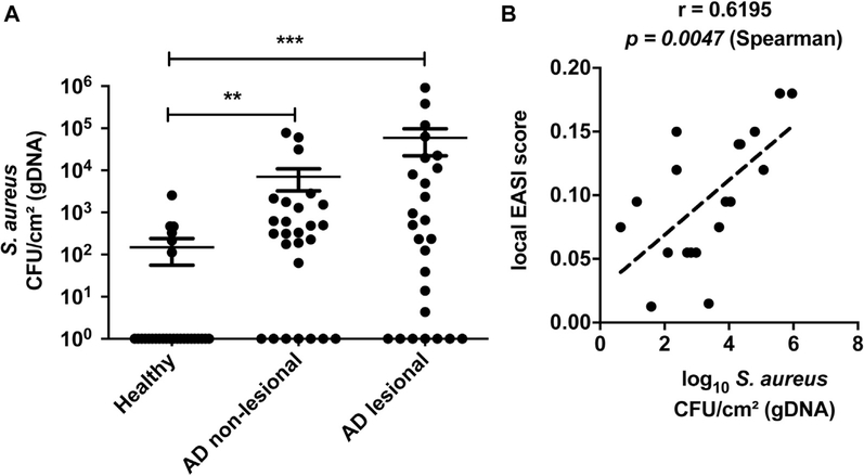
Background: Staphylococcus aureus and Staphylococcus epidermidis are the most abundant bacteria found on the skin of patients with atopic dermatitis (AD). S aureus is known to exacerbate AD, whereas S epidermidis has been considered a beneficial commensal organism.

Objective: In this study, we hypothesized that S epidermidis could promote skin damage in AD by the production of a protease that damages the epidermal barrier.

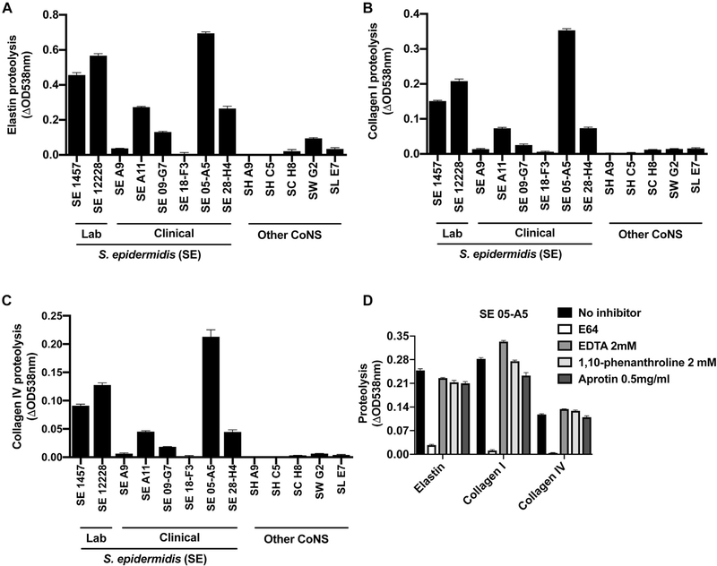
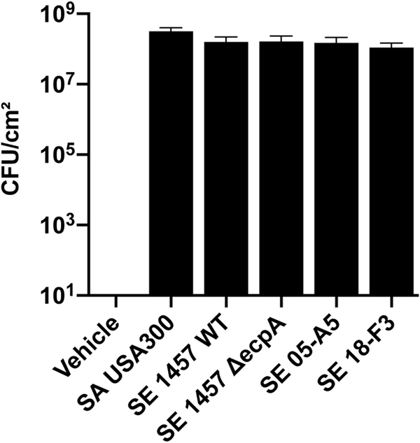
Methods: The protease activity of S epidermidis isolates was compared with that of other staphylococcal species. The capacity of S epidermidis to degrade the barrier and induce inflammation was examined by using human keratinocyte tissue culture and mouse models. Skin swabs from atopic and healthy adult subjects were analyzed for the presence of S epidermidis genomic DNA and mRNA.

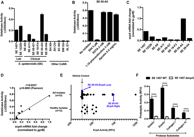
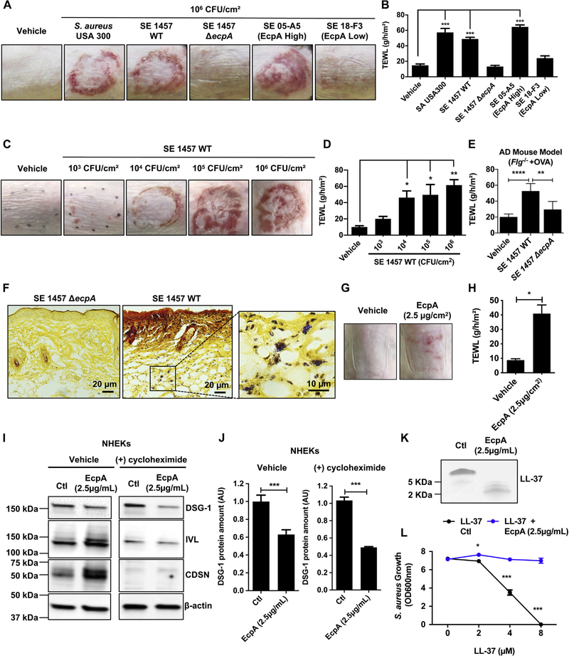
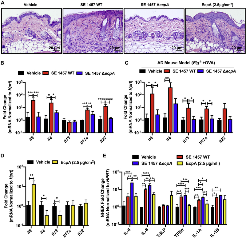
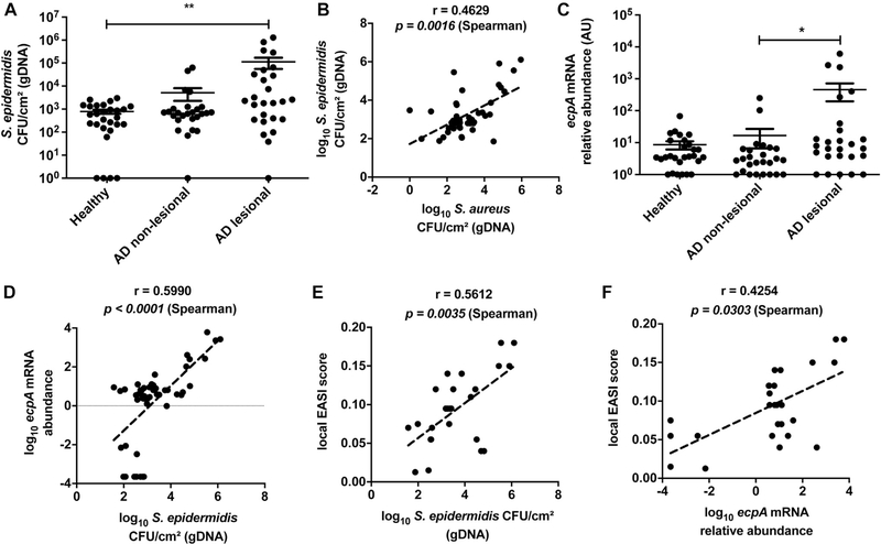
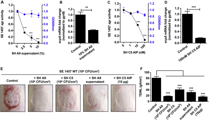
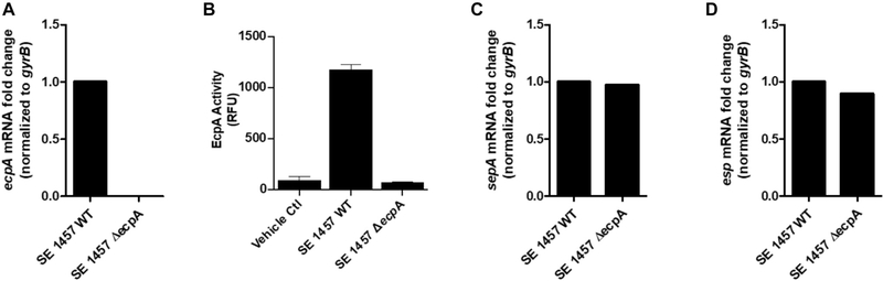
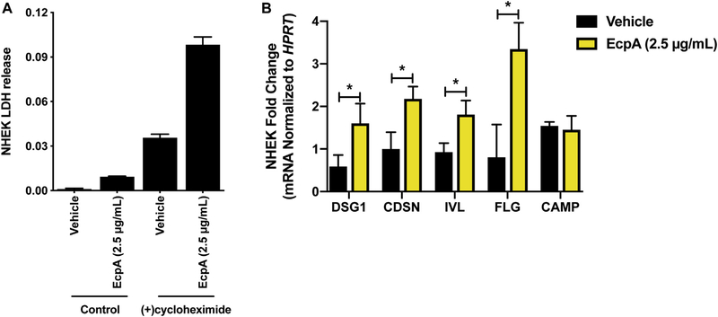
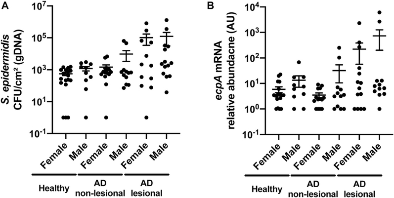
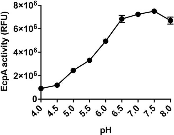
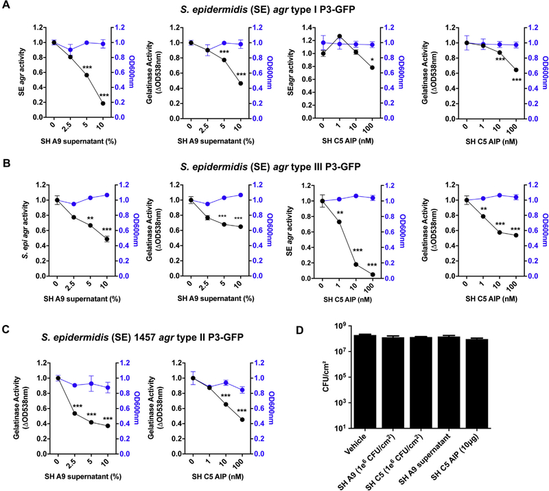
Results: S epidermidis strains were observed to produce strong cysteine protease activity when grown at high density. The enzyme responsible for this activity was identified as EcpA, a cysteine protease under quorum sensing control. EcpA was shown to degrade desmoglein-1 and LL-37 in vitro, disrupt the physical barrier, and induce skin inflammation in mice. The abundance of S epidermidis and expression of ecpA mRNA were increased on the skin of some patients with AD, and this correlated with disease severity. Another commensal skin bacterial species, Staphylococcus hominis, can inhibit EcpA production by S epidermidis.

Conclusion: S epidermidis has commonly been regarded as a beneficial skin microbe, whereas S aureus has been considered deleterious. This study suggests that the overabundance of S epidermidis found on some atopic patients can act similarly to S aureus and damage the skin by expression of a cysteine protease.

Keywords: Atopic dermatitis; Staphylococcus epidermidis; cytokine; dysbiosis; epidermal barrier; inflammation; microbiome; protease; skin.


Conflict of interest statement

Disclosure of potential conflict of interest: R. L. Gallo is a cofounder, scientific advisor, and consultant of MatriSys Biosciences and has equity in the company; in addition, he receives income from and has equity in Sente. The rest of the authors declare that they have no relevant conflicts of interest.

Figures

FIG 1.
FIG 1.
The cysteine protease EcpA secreted by S epidermidis (SE) presents a unique proteolytic activity. A, Protease activity against gelatin measured in the supernatant of CoNS strains cultured for 24 hours: SE, S hominis (SH), S capitis (SC), S warneri (SW), and S lugdunensis (SL) (n = 3). B, Protease activity against gelatin measured in the supernatant of SE 05-A5 cultured for 24 hours in the presence of various protease inhibitors: E64 (a cysteine protease inhibitor), EDTA, 1,10-phenanthroline (a metalloprotease inhibitor), and aprotinin (a serine protease inhibitor) (n = 3). C and D, ecpA mRNA levels across SE strains and Pearson correlation to gelatinase activity. E, Assessment of EcpA specific activity (FRET substrate) in supernatant of SE isolates from both healthy (n = 12) and atopic (n = 33) individuals after the isolates had been cultured for 24 hours (n = 3). F, Protease activity against gelatin, elastin, collagen I, and collagen IV substrates measured in the supernatant of SE 1457 WT or ΔecpA strains cultured for 24 hours. All data are representative of at least 2 independent experiments, and the results are means ± SEMs. F, Student t tests were used to determine statistical significance: *P < .05; **P < .01; ***P < .001; **** P < .0001. RFU, Relative fluorescence unit.
FIG 2.
FIG 2.
S epidermidis (SE) cysteine protease EcpA disrupts the skin barrier and degrades DSG-1 and LL-37. A and B, Representative pictures of the murine back skin treated with 106 CFUs/cm2 SA or SE isolates for 48 hours and TEWL measurements (n = 5). C and D, Representative pictures of the back skin after colonization with 103to 106 CFUs/cm2 SE 1457 WT for 48 hours and TEWL measurements (n = 4). E, TEWL measurements of AD mouse model back skin (Balb/c Flg−/− + ovalbumin [OVA]) colonized for 24 hours with live 106 CFUs/cm2 SE 1457 WT or SE 1457 ΔecpA (n = 5 or 6). F, Gram-positive bacteria staining (in purple) of skin sections from mice treated with either 106 CFUs/cm2 of SE 1457 WT or SE 1457 ΔecpA (n = 5). G and H, Assessment of C57BL/6 murine back skin and TEWL measurements after treatment with 2.5 μg/cm2 of EcpA or vehicle for 24 hours (n = 3). I and J, Differentiated NHEKs were treated for 20 hours with 2.5 μg/mL of EcpA ± 20 μg/mL of cycloheximide (protein synthesis inhibitor). Immunoblotting for DSG-1, involucrin (IVL), corneodesmosin (CDSN), and β-actin. Quantification of DSG-1 was normalized on β-actin (n = 3) (see Table E4). K and L, Coomassie blue staining of in vitro proteolysis of LL-37 by EcpA and S aureus (strain 113) growth inhibition assay (n = 3). All data are representative of at least 2 independent experiments, and results are means ± SEMs. One-way ANOVAs (B, D, and E), Student t tests (H and J), and 2-way ANOVA (L) were used to determine statistical significance: *P < .05; **P < .01; ***P < .001, ****P < .0001. Ctl, Control.
FIG 3.
FIG 3.
EcpA induces skin inflammation in combination with other S epidermidis toxins. A, Representative hematoxylin and eosin staining of mouse skin sections across treatments. B and C, Analysis of the mRNA level of various cytokines after treatment of murine back skin with 106 CFUs/cm2 of S epidermidis (SE) 1457 WT or SE 1457 ΔecpA for 48 hours in the C57BL/6 mouse model (B) or AD mouse model (BALB/c Fig−/− 1 ovalbumin [OVA]) (C), respectively (n = 4–6). mRNA levels normalized by using the housekeeping gene Hprt. D, Analysis of the mRNA level of various cytokines after treatment of C57BL/6 murine back skin with vehicle or 2.5 μg/cm2 of EcpA for 24 hours. E, Differentiated NHEK treated for 8 hours with 2.5 μg/mL of EcpA or with 5% bacterial supernatant followed by analysis of the mRNA level of various cytokines normalized to the housekeeping gene HPRT (n = 3). All data are representative of at least 2 independent experiments, and results are means ± SEMs. One-way ANOVAs (B, C, and E) and Student t tests (D) were used to determine statistical significance: *P < .05; **P < .01; ***P < .001; **** P < .0001.
FIG 4.
FIG 4.
S epidermidis colonization and ecpA expression are increased on some subjects with AD. A, Measurement of gDNA absolute abundance of S epidermidis CFUs/cm2 from swabs of healthy control and AD nonlesional and lesional skin normalized to skin area. B, Spearman correlation between the gDNA absolute abundance of S epidermidis and S aureus from skin swabs. C, Relative abundance of S epidermidis ecpA mRNA isolated from swabs of healthy control and AD nonlesional and lesional skin normalized to skin area. D, Spearman correlation between S epidermidis EcpA mRNA relative abundance and S epidermidis CFUs/cm2 from swabs of AD skin. E, Spearman correlation between the local Eczema Area and Severity Index (EASI) score and S epidermidis CFUs/cm2 from swabs of AD lesional skin. F, Spearman correlation between the local EASI score and ecpA mRNA relative abundance from swabs of AD lesional skin. Each dot represents a single swab, and the bars represent means ± SEMs. A and C, A nonparametric unpaired Kruskal-Wallis analysis was used to determine statistical significance. *P < .05; **P < .01; ***P < .001; ****P < .0001.
FIG 5.
FIG 5.
EcpA production is downregulated by CoNS AIPs. A and C, Modulation of the S epidermidis (SE) 1457 agr type II P3-GFP reporter strain activity by S hominis (SH) A9 supernatant (A) or synthetic SH C5 AIP (C) after 24 hours of culturing (n = 3). OD values at 600 nm used to assess bacterial growth at the hour 24 time point. B and D, ecpA mRNA levels after 12 hours of culturing of SE 1457 agr reporter strain with SH A9 supernatant (10%) (B) or SH C5 AIP (100 nM) (n = 3) (D). E and F, C57BL/6 murine back skin was topically treated with 106 CFUs/cm2 of SE 1457 for 48 hours either alone (control) or with 106 CFUs/cm2 of S hominis (SH) A9,106 CFUs/cm2 of SH C5, SH A9 supernatant (5 μL), or SH C5 synthetic AIP (10 μg). E, Representative pictures of the treated back skin and measurement of TEWL (F) (n = 4). All data are representative of at least 2 independent experiments, and the results are means ± SEMs. One-way ANOVAs (A, C, and F) and Student t tests (B and D) were used to determine statistical significance: *P < .05; **P < .01; ***P < .001; ****P < .0001.
FIG E1.
FIG E1.
Characterization of S epidermidis (SE) 1457 ΔecpA. A, Analysis of the mRNA levels of ecpA in both the SE 1457 WT or ΔecpA strain after 12 hours of culturing. B, EcpA activity with use of a specific FRET substrate measured in the sterile-filtered supernatant of SE 1457 WT or ΔecpA after 24 hours of culturing (n = 3). C and D, Analysis of mRNA levels for 2 other SE-secreted protease genes, sepA and esp, in both SE 1457 WT or ΔecpA after 12 hours of culturing. Data are representative of at least 2 independent experiments, and the results are represented as means ± SEMs. RFU, Relative fluorescence unit.
FIG E2.
FIG E2.
Characterization of S epidermidis (SE) protease activity. A-C, Measurement of protease activity against the substrates elastin (A), collagen I (B), and collagen IV (C) with CoNS sterile-filtered supernatants that had been cultured for 24 hours (n = 3). D, Protease activity against the substrates elastin, collagen I, and collagen IV measured in the sterile-filtered supernatant of SE 05-A5 (after 24 hours of culturing) in the presence of various protease inhibitors: E64 (a cysteine protease inhibitor), EDTA, and 1,10-phenanthroline (a metalloprotease inhibitor), and aprotinin (a serine protease inhibitor) (n = 3). Data are representative of at least 2 independent experiments, and results are represented as means ± SEMs. SH, S hominis; SC, Staphylococcus capitis; SW, Staphylococcus warneri; SL, Staphylococcus lugdunensis.
FIG E3.
FIG E3.
Quantification of live staphylococci on 8- to 10-week-old female C57BL/6 mice skin after topical treatment with S aureus (SA) or S epidermidis (SE) live bacteria for 48 hours.
FIG E4.
FIG E4.
Effect of EcpA on cultured keratinocytes. A, Evaluation of the cytotoxicity of EcpA on differentiated NHEKs by measuring lactate dehydrogenase (LDH) release in the culture medium after 20 hours of treatment alone or in the presence of 20 μg/mL of cycloheximide. B, Differentiated NHEKs were treated for 8 hours with 2.5 μg/ mL of EcpA followed by analysis of the mRNA level of several epidermal differentiation markers and CAMP by qPCR (n = 3). Data are representative of at least 2 independent experiments, and results are represented as means ± SEMs. B, Student t tests were used to determine statistical significance: *P < .05; **P < .01; ***P < .001; ****P < .0001.
FIG E5.
FIG E5.
Sex differences in S epidermidis colonization and EcpA expression on AD skin. A and B, Measurement of gDNA absolute abundance of S epidermidis CFUs/cm2 or ecpA mRNA levels from swabs of healthy control and AD nonlesional and lesional skin normalized to skin area. Data are represented as means ± SEMs. A and B, Nonparametric unpaired Kruskal-Wallis analysis and Spearman correlation were used to determine statistical significance.
FIG E6.
FIG E6.
Colonization of AD skin by S aureus and correlation to disease severity. A, Measurement of gDNA absolute abundance of S aureus CFUs/cm2 from swabs of healthy control and AD nonlesional and lesional skin normalized to skin area. B, Spearman correlation between the local Eczema Area and Severity Index (EASI) score and S aureus CFUs/cm2 from swabs of AD lesional skin. Each dot represents a single swab and the bars represent means ± SEMs. A, Nonparametric unpaired Kruskal-Wallis analysis was used to determine statistical significance: *P < .05; **P < .01; ***P < .001; ****P < .0001.
FIG E7.
FIG E7.
pH-dependent activity of S epidermidis EcpA. EcpA (100 ng) was incubated with EcpA FRET substrate (250 nM) in sweat-like buffer (40 mM NaCl, 10 mM KCl, 1 mM sodium dihydrogen phosphate, 5 mM l-cysteine) at different pH ranges (4–8) for 1 hour at 37°C (n = 3). Data are representative of at least 2 independent experiments, and results are represented as means ± SEMs.
FIG E8.
FIG E8.
Modulation of S epidermidis (SE) agr activity by some S hominis (SH) strains. A, Modulation of the agr activity and gelatinase activity of SH 12228 agr type I P3-GFP reporter strain by either SH A9 supernatant or SH C5 synthetic AIP (n = 3). B, Modulation of the agr activity and gelatinase activity of SE 8247 agr type III P3-GFP reporter strain by either SH A9 supernatant or SH C5 synthetic AIP (n = 3). c, Modulation of the gelatinase activity of SE 1457 agr type II P3-GFP reporter strain by either SH A9 supernatant or SH C5 synthetic AIP (n = 3). A-C, OD values recorded at 600 nm to approximate bacterial growth overtime. D, Quantification of live staphylococci on 8-to 10-week-oldfemaleC57BL/6mice skin after topical treatment with SE 1457 live bacteria for 48 hours either alone (vehicle) or along with SH A9, C5 live bacteria, SH A9 supernatant (5 μL), or SH C5AIP (n = 4). Data are representative of at least 2 independent experiments, and results are represented as means ± SEMs. A-C, One-way ANOVAs were used to determine statistical significance: *P < .05; **P < .01; *** P <.001; ****P <.0001.

References

    1. Bieber T Atopic dermatitis. N Engl J Med 2008;358:1483–94. - PubMed
    1. Kapoor R, Menon C, Hoffstad O, Bilker W, Leclerc P, Margolis DJ. The prevalence of atopic triad in children with physician-confirmed atopic dermatitis. J Am Acad Dermatol 2008;58:68–73. - PubMed
    1. Carroll CL, Balkrishnan R, Feldman SR, Fleischer AB Jr, Manuel JC. The burden of atopic dermatitis: impact on the patient, family, and society. Pediatr Dermatol 2005;22:192–9. - PubMed
    1. Nutten S Atopic dermatitis: global epidemiology and risk factors. Ann Nutr Metab 2015;66(Suppl 1):8–16. - PubMed
    1. Benn CS, Melbye M, Wohlfahrt J, Bjorksten B, Aaby P. Cohort study of sibling effect, infectious diseases, and risk of atopic dermatitis during first 18 months of life. BMJ 2004;328:1223. - PMC - PubMed

Publication types

MeSH terms