

doi: 10.1016/j.neuroimage.2020.117127.
Epub 2020 Jul 4.
Neuroharmony: A new tool for harmonizing volumetric MRI data from unseen scanners
Affiliations
- PMID: 32634595
- PMCID: PMC7573655
- DOI: 10.1016/j.neuroimage.2020.117127
Item in Clipboard
Neuroharmony: A new tool for harmonizing volumetric MRI data from unseen scanners
Neuroimage.
.
Abstract
- •
We present Neuroharmony, a harmonization tool for images from unseen scanners.
- •
We developed Neuroharmony using a total of 15,026 sMRI images.
- •
The tool was able to reduce scanner-related bias from unseen scans.
- •
Neuroharmony represents a significant step towards imaging-based clinical tools.
- •
Neuroharmony is available at
https://github.com/garciadias/Neuroharmony .
Figures
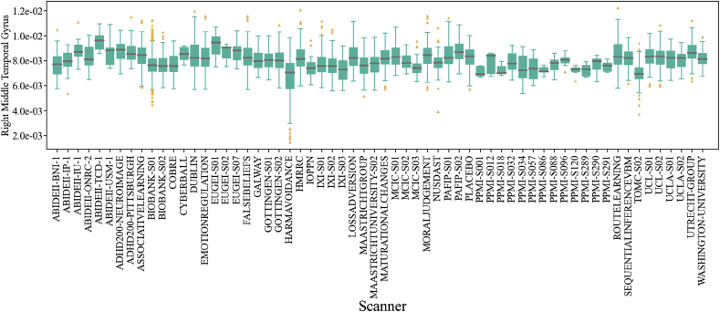
Box plot of the right middle temporal gyrus relative volumes for all scanners included in our study. A grey horizontal line marks the median value in each dataset, the solid green boxes present the inter-quantile ratio in each dataset. The vertical green lines cover 90% of the measurements in each dataset. The yellow triangles represent data points outside the 5–95% interval.
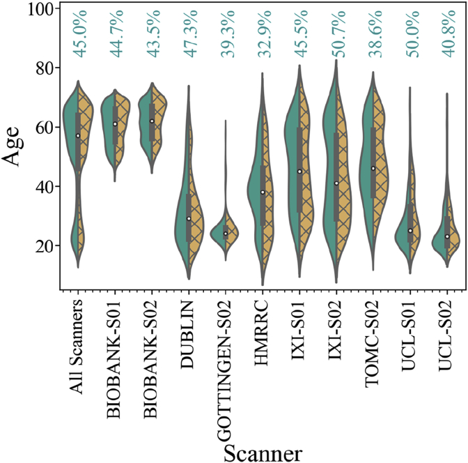
Violin plot showing age distribution for male (in green, left distribution) and female (in yellow, right distribution) subjects for all datasets along with the individual distribution of the 10 largest scanner samples.
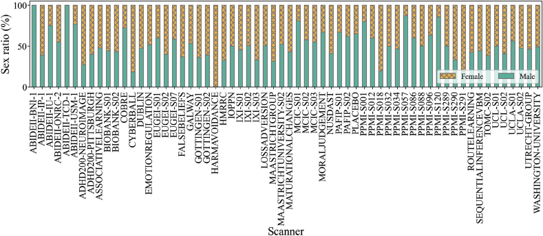
The ratio of subjects of each sex in the data from all included scanners. The plot shows bars corresponding to 100% of the subjects measured with each scanner. The portion of male subjects is colored in green and the portion of female subjects is colored in yellow and X-hatched.
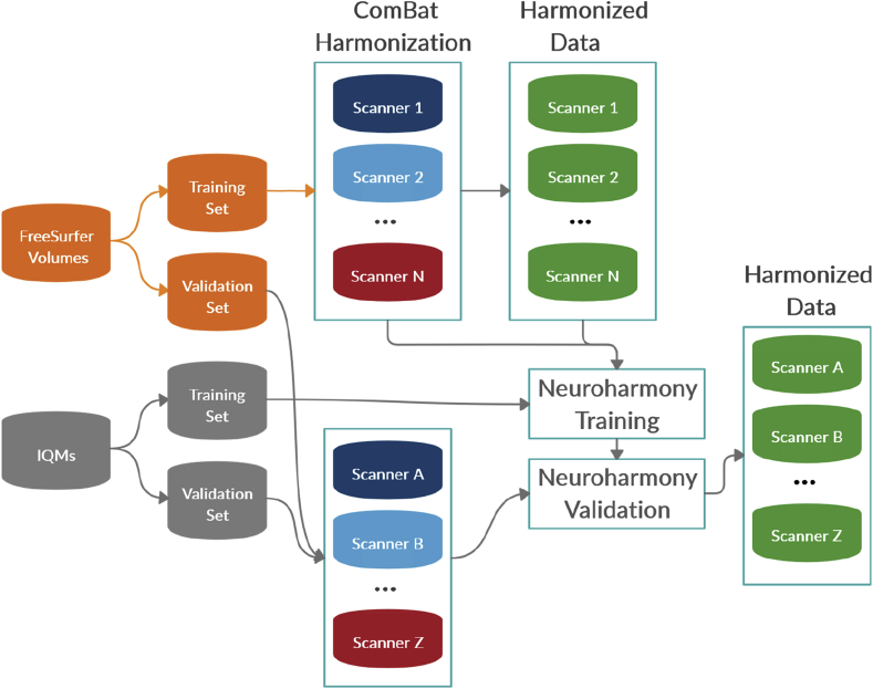
Diagram showing the data splits to train Neuroharmony.
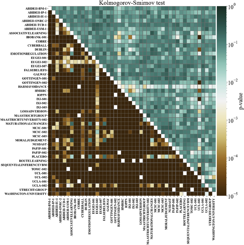
The minimum p-values for the K–S test among all ROIs, before and after ComBat harmonization. The cells under the main diagonal of the matrix represent the K–S test p-values before harmonization, while the values in the top of the main diagonal represent the p-values after the ComBat harmonization. Each cell corresponds to a pair of scanners. Cells are colored as shown on the color bar.
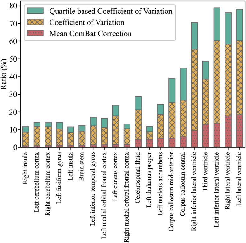
The median ROI volume divided by the median ComBat correction. From left to right, the first 10 bars show the ROIs with the smallest correction ratios while the next 10 show the ROIs with the largest correction ratio (red dotted bars). The X-hatched yellow bars show coefficient of variation and the green bars show the quartile-based coefficient of variation.
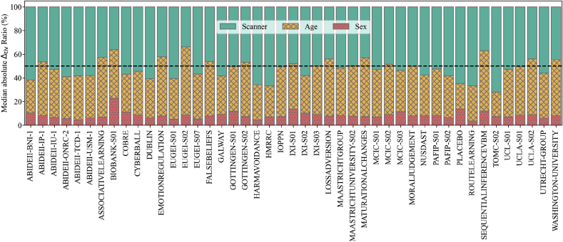
Relative contributions of each confound to the final ComBat correction. Each scanner is represented as a vertical bar divided in portions equivalent to the contributions that either scanner (green), age (yellow, X-hatched) or sex (red, filled with dots) made to the correction. The black dashed line marks the 50% level.
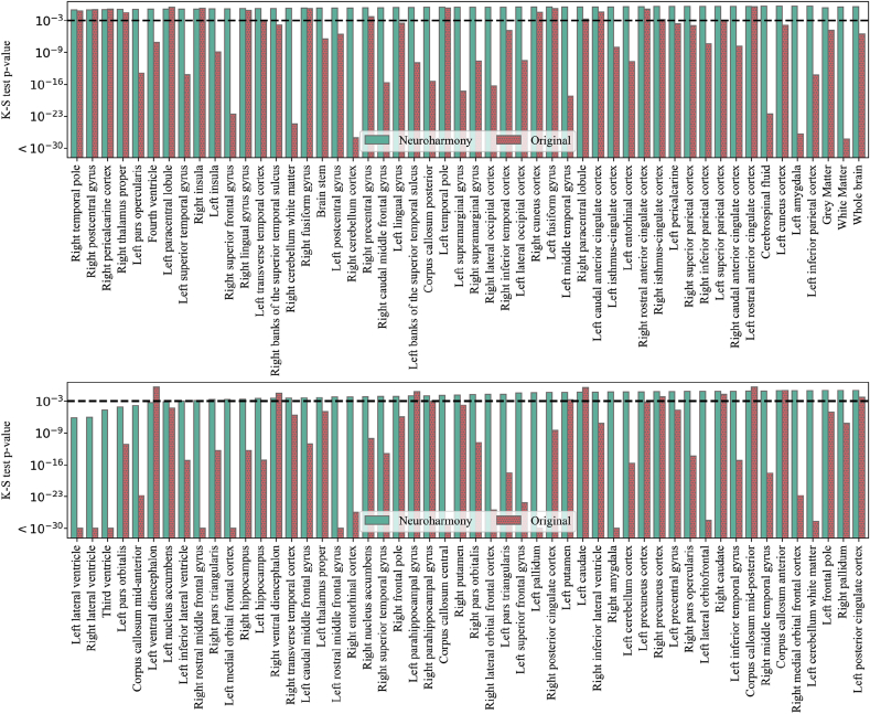
The p-values for the K–S test for the comparison between the validation set harmonized with Neuroharmony and the training sample harmonized with ComBat. From left to right, each bar in the pairs of bars represents the p-value of the K–S test for the data corrected by Neuroharmony (green) and the data without any correction (red, filled with dots). A horizontal black dashed line marks the 0.001 threshold.
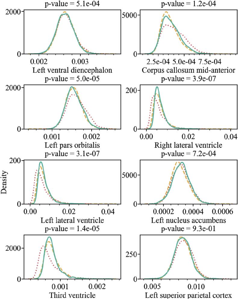
The kernel density plot for the relative volume of the regions as labelled in the x-axis of each plot. The title of each plot includes the p-value of the K–S test comparing the training set harmonized with ComBat (yellow dashed lines) and the validation set harmonized with Neuroharmony (green solid lines). The validation set without harmonization is shown as a red dotted line.
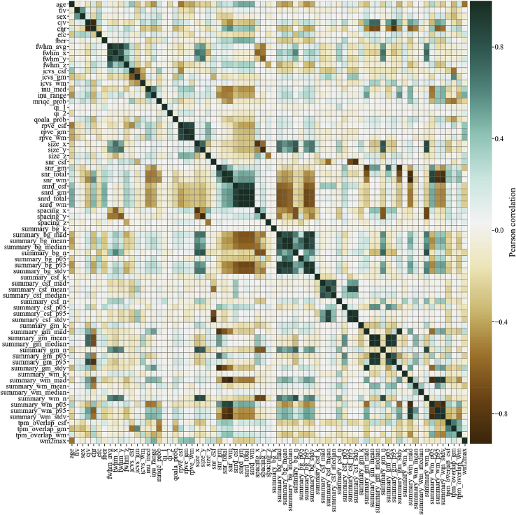
The Pearson correlation matrix for all IQMs, sex and age.
References
-
- Benetti S., Pettersson-Yeo W., Hutton C., Catani M., Williams S.C.R., Allen P. Elucidating neuroanatomical alterations in the at risk mental state and first episode psychosis: a combined voxel-based morphometry and voxel-based cortical thickness study. Schizophr. Res. 2013;150(2–3):505–511. doi: 10.1016/j.schres.2013.08.030. - DOI - PMC - PubMed
-
- Buitinck L., Louppe G., Blondel M., Pedregosa F., Mueller A., Grisel O. API design for machine learning software: experiences from the scikit-learn project. 2013. http://arxiv.org/abs/1309.0238 Retrieved from.
Publication types
MeSH terms
Grants and funding
LinkOut - more resources
Full Text Sources
Other Literature Sources
Medical
