

Robotic management of painful Zinner syndrome, case report and review of literature
- PMID: 32634620
- PMCID: PMC7338681
- DOI: 10.1016/j.ijscr.2020.06.078
Robotic management of painful Zinner syndrome, case report and review of literature
Abstract
Background: Zinner Syndrome is a congenital pathology due to an embryologic anomaly occurring between the 4th and 13th gestational week. This embryologic defect leads to unilateral renal agenesis, ipsilateral seminal vesicle cyst and ejaculatory duct obstruction. Most of the time patients are asymptomatic and do not need any treatment but for symptomatic cases, only surgical removal of the cyst and seminal vesicle are 100% effective.
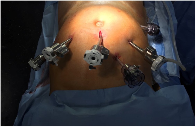
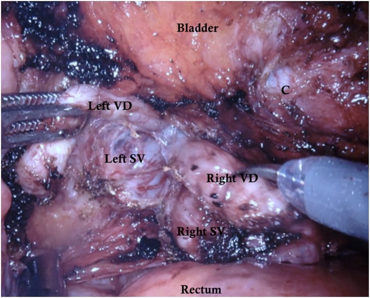
Case: The case presented here is that of a healthy 33-year old man with symptomatic right seminal vesicle cyst and ipsilateral renal agenesis. First a conservative approach was attempted but each time the symptoms ended up reappearing. We decided to use robot-assisted laparoscopy to completely resect the cyst and the right seminal vesicle. There was no postoperative complication and the patient's symptoms improved immediately. After a 6 months follow-up the patient remains completely asymptomatic.
Conclusion: Complete excision of the seminal vesicle cyst is the only 100% effective treatment option for symptomatic patients with Zinner syndrome. Minimally invasive approaches like conventional laparoscopy or robotic assisted laparoscopy are safe and effective and should currently be considered as the surgical gold standard.
Keywords: Robotic management; Seminal vesicle cyst; Zinner syndrome.
Copyright © 2020 The Authors. Published by Elsevier Ltd.. All rights reserved.
Figures


References
-
- van den Ouden D., Blom J.H.M., Bangma C., de Spiegeleer A.H.V.C. Diagnosis and management of seminal vesicle cysts associated with ipsilateral renal agenesis: a pooled analysis of 52 cases. Eur. Urol. 1998;33:433–440. - PubMed
-
- Pereira, Sousa, Azinhais, Conceição, Borges, Leão, Brandão, Temido, Retroz, Sobral Zinner’s syndrome: an up‐to‐date review of the literature based on a clinical case. Andrologia. 2009;41(5):322–330. - PubMed
-
- Agha R.A., Borrelli M.R., Farwana R., Koshy K., Fowler A., Orgill D.P., For the SCARE Group The SCARE 2018 statement: updating consensus surgical CAse REport (SCARE) guidelines. Int. J. Surg. 2018;60:132–136. - PubMed
Publication types
LinkOut - more resources
Full Text Sources
