

Ensemble Deep Learning for Cervix Image Selection toward Improving Reliability in Automated Cervical Precancer Screening
- PMID: 32635269
- PMCID: PMC7400120
- DOI: 10.3390/diagnostics10070451
Ensemble Deep Learning for Cervix Image Selection toward Improving Reliability in Automated Cervical Precancer Screening
Abstract
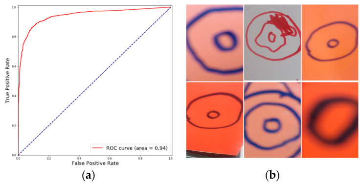
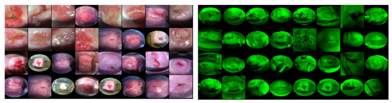
Automated Visual Examination (AVE) is a deep learning algorithm that aims to improve the effectiveness of cervical precancer screening, particularly in low- and medium-resource regions. It was trained on data from a large longitudinal study conducted by the National Cancer Institute (NCI) and has been shown to accurately identify cervices with early stages of cervical neoplasia for clinical evaluation and treatment. The algorithm processes images of the uterine cervix taken with a digital camera and alerts the user if the woman is a candidate for further evaluation. This requires that the algorithm be presented with images of the cervix, which is the object of interest, of acceptable quality, i.e., in sharp focus, with good illumination, without shadows or other occlusions, and showing the entire squamo-columnar transformation zone. Our prior work has addressed some of these constraints to help discard images that do not meet these criteria. In this work, we present a novel algorithm that determines that the image contains the cervix to a sufficient extent. Non-cervix or other inadequate images could lead to suboptimal or wrong results. Manual removal of such images is labor intensive and time-consuming, particularly in working with large retrospective collections acquired with inadequate quality control. In this work, we present a novel ensemble deep learning method to identify cervix images and non-cervix images in a smartphone-acquired cervical image dataset. The ensemble method combined the assessment of three deep learning architectures, RetinaNet, Deep SVDD, and a customized CNN (Convolutional Neural Network), each using a different strategy to arrive at its decision, i.e., object detection, one-class classification, and binary classification. We examined the performance of each individual architecture and an ensemble of all three architectures. An average accuracy and F-1 score of 91.6% and 0.890, respectively, were achieved on a separate test dataset consisting of more than 30,000 smartphone-captured images.
Keywords: cervical cancer; cervix/non-cervix; deep learning; ensemble; one-class classification.
Conflict of interest statement
The authors declare no conflict of interest.
Figures


References
-
- Human Papillomavirus (HPV) and Cervical Cancer. [(accessed on 2 October 2019)]; Available online: https://www.who.int/en/news-room/fact-sheets/detail/human-papillomavirus....
-
- Egede J., Ajah L., Ibekwe P., Agwu U., Nwizu E., Iyare F. Comparison of the accuracy of papanicolaou test cytology, Visual Inspection with Acetic acid, and Visual Inspection with Lugol Iodine in screening for cervical neoplasia in southeast Nigeria. J. Glob. Oncol. 2018;4:1–9. doi: 10.1200/JGO.17.00127. - DOI - PMC - PubMed
-
- Sritipsukho P., Thaweekul Y. Accuracy of visual inspection with acetic acid (VIA) for cervical cancer screening: A systematic review. J. Med. Assoc. Thai. 2010;93:254–261. - PubMed
-
- Jeronimo J., Massad L.S., Castle P.E., Wacholder S., Schiffman M., National Institutes of Health (NIH)-American Society for Colposcopy and Cervical Pathology (ASCCP) Research Group Interobserver agreement in the evaluation of digitized cervical images. Obstet. Gynecol. 2007;110:833–840. doi: 10.1097/01.AOG.0000281665.63550.8f. - DOI - PubMed
LinkOut - more resources
Full Text Sources
