Review
. 2020 Jul 4;12(7):1793.
doi: 10.3390/cancers12071793.

Autophagy Roles in Genome Maintenance

Affiliations
Review

Autophagy Roles in Genome Maintenance

Susanna Ambrosio et al. Cancers (Basel). .

Abstract

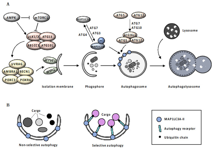
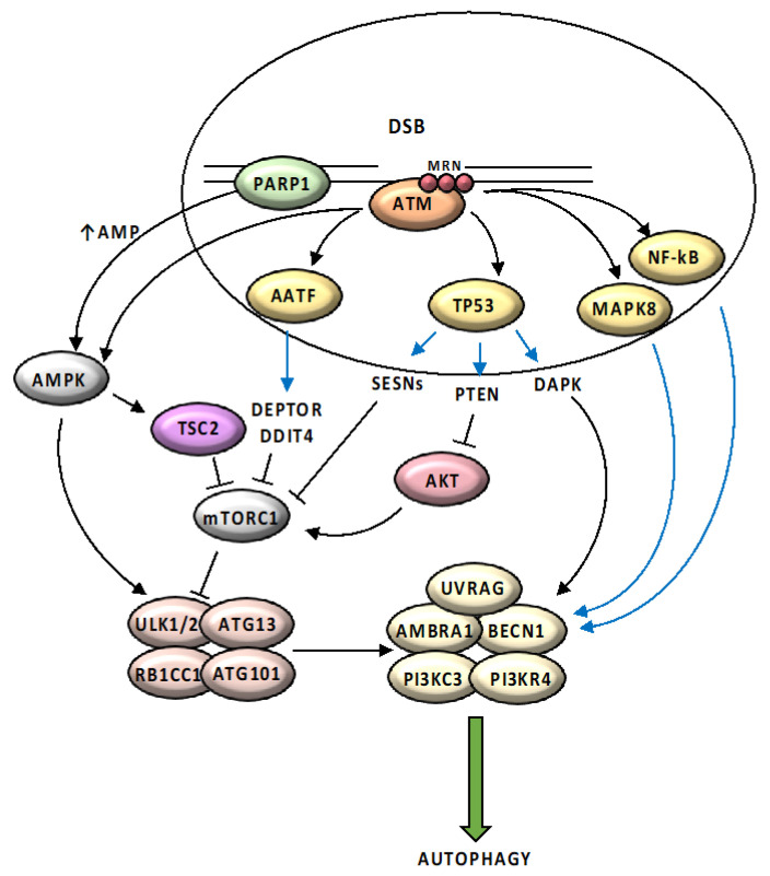
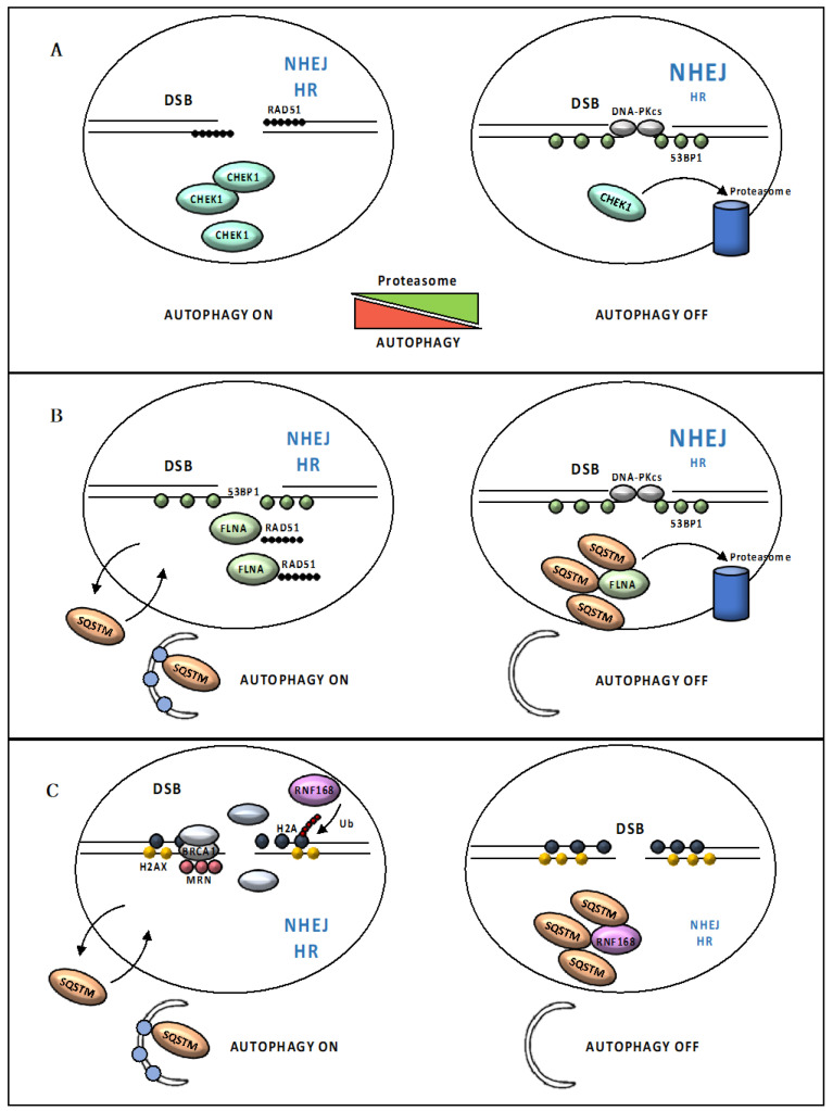
In recent years, a considerable correlation has emerged between autophagy and genome integrity. A range of mechanisms appear to be involved where autophagy participates in preventing genomic instability, as well as in DNA damage response and cell fate decision. These initial findings have attracted particular attention in the context of malignancy; however, the crosstalk between autophagy and DNA damage response is just beginning to be explored and key questions remain that need to be addressed, to move this area of research forward and illuminate the overall consequence of targeting this process in human therapies. Here we present current knowledge on the complex crosstalk between autophagy and genome integrity and discuss its implications for cancer cell survival and response to therapy.

Keywords: DNA damage repair; DNA damage response; autophagy; cancer therapy.


Conflict of interest statement

No potential conflicts of interest were disclosed.

Figures

Figure 1
Figure 1
Autophagic Machinery. Box 1: Mechanism of autophagy. Autophagy initiates with the elongation of a precursor structure, called phagophore, surrounding cargo, to generate a double-membrane vesicle, called autophagosome [8,9]. Autophagosomes biogenesis is orchestrated by a subset of autophagy-related (ATG) proteins [10]. Upon autophagic stimuli, activation of AMPK (adenosine monophosphate (AMP)-activated protein kinase) and inhibition of mTORC1 (mammalian target of rapamycin complex 1) signaling pathway [11,12] leads to the activation of the ULK1/ULK2 (UNC51-like autophagy activating kinase 1/2) complex, which in turn activates the nucleation complex, including ATG13, RB1CC1 (RB1-inducible coiled-coil 1) and ATG101, which orchestrates the recruitment of further ATG proteins on the phagophore, promoting membrane elongation [13]. Nucleation complex phosphorylates AMBRA1 (autophagy and beclin 1 regulator 1) and BECN1 (beclin1), disrupting the inhibitory association with BCL2 (BCL2 apoptosis regulator) and allowing PI3KC3 (phosphatidylinositol 3-kinase catalytic subunit type 3) complex assembly [14]. The PI3KC3 complex, including the PI3KC3, PIK3R4 (PI3KC3 regulatory subunit 4), UVRAG (UV radiation resistance associated), AMBRA1 and BECN1, controls the membrane nucleation stage and initial phagophore formation [15]. PI3P (phosphatidylinositol 3-phosphate) that is generated by PI3K activity in the newly formed membranes, serves as a landing pad, known as omegasomes, for effector proteins such as ZFYVE1 (zinc finger FYVE-type containing 1) and WIPI2 (WD repeat domain phosphoinositide-interacting 2), to promote the formation of isolation membrane [16]. Elongation of the isolation membrane requires the involvement on two ubiquitin-like conjugation systems. In the first system, the protease ATG4 cleaves the precursor form of MAP1LC3A (microtubule associated protein 1 light chain 3 alpha), allowing ATG7 (E1 ubiquitin-activating enzyme) and ATG3 (E2 ubiquitin-conjugating enzyme) to catalyze the MAP1LC3A-I conjugation with phosphatidylethanolamine, to form MAP1LC3A-II [17,18]. At the same time, ATG12 is covalently conjugated to the ATG5 protein through the action of ATG7 and ATG10 (E2 ubiquitin-like enzyme) proteins [17,18]. Then, recruitment of the ATG16L1 protein (E3 ubiquitin-protein ligase) stabilizes the ATG12-ATG5 complex. ATG12-5-16L1 oligomers facilitates MAP1LC3A-II localization to the phagophore membrane, where it drives autophagosomal maturation. Once autophagosome maturation is finished, ATG4 catalyzes the reverse modification reaction of MAP1LC3A-II to MAP1LC3A-I [18]. Following completion and closure, the autophagosome ultimately undergoes fusion with a lysosome (Figure 1A). The fusion process is regulated by lysosomal membrane and cytoskeletal proteins. Finally, the fusion results in the exposure of autophagosomal cargo to the lysosomal acid hydrolases required for degradation and recycling [18]. Autophagy can be classified as non-selective or cargo-specific [19]. Selectivity of autophagy is ensured by two groups of autophagic cargo receptors, the ubiquitin-binding type, exemplified by SQSTM1 (sequestosome-1) [20] and CALCOCO2 (calcium binding and coiled-coil domain 2) [21], and the trans-membrane type, exemplified by BNIP3 (BCL2 interacting protein 3) and BNIP3L (BCL2 interacting protein 3 like) [22,23]. Although not essential for autophagosomal biogenesis itself, receptor proteins specifically bind the cargo material and the autophagosomal membrane, acting as a bridge that promotes cargo sequestration by the nascent autophagosome [24] (Figure 1B).
Figure 2
Figure 2
Mechanisms of DSB repair. (a) Following the recruitment and activation of ATM (ATM serine/protein kinase), the nuclease MRN (MRE11 (MRE11 homolog, double strand break repair nuclease)/RAD50 (RAD50 double strand break repair protein)/NBN (nibrin), along with its cofactor RBBP8 (RB binding protein 8) and other accessory proteins, such as BRCA1(BRCA1 DNA repair associated), initiates the 5′-3′ resection of the DNA ends, resulting in the creation of 3′-OH ssDNA tails on both ends of the break. Next, long range resection is achieved by EXO1 (exonuclease 1), DNA2 (DNA replication helicase/nuclease 2) and BLM (BLM RecQ like helicase). (b) The ssDNA-ends are coated and stabilized by RPA (replication protein A). (c) RPA is replaced from ssDNA ends by RAD51 (RAD51 recombinase) in a BRCA2 (BRCA2 DNA repair associated)-dependent process and formation of the RAD51 multimeric filament on ssDNA occurs. (d) RAD51-ssDNA filament can then strand invade a homologous dsDNA, leading to displacement on one strand of the intact DNA duplex and formation of a D-loop. (e) Following strand exchange, the resulting Holliday junction is resolved. (f) NHEJ (Non-homologous end joining) pathway initiates with the ATM-mediated phosphorylation of 53BP1, required for the recruitment of the anti-resection proteins RIF1 (replication timing regulatory factor 1) to the DSBs (double strand breaks) and inhibition of DNA resection (g). 53BP1 (TP53 (tumor protein p53)-binding protein 1) promotes the binding of XRCC5/XRCC6 (X-ray repair cross-complementing protein 6 and 5) heterodimer to the two ends of the DSB. (h) DNA-PKcs (DNA-dependent protein kinase catalytic subunit) acts as a scaffold protein that tethers the broken ends together. (i) The LIG4 (DNA ligase 4)/XRCC4 (X-ray repair cross complementing 4)/NHEJ1 (non-homologous end joining factor 1) complex ligates the DNA ends. Black arrows (↑) and perpendicular lines (⊥) indicate activation and repression, respectively.
Figure 3
Figure 3
Schematic representation of main pathways regulating the DDR-mediated autophagic response. ATM is activated in response to DNA damage by the MRN complex and initiates a pathway that results in activation of AMPK and its target TSC2 (TSC complex subunit 2), which removes the inhibitory effect of MTORC1 on autophagy, promoting ULK1-dependent autophagosome formation. ATM directly phosphorylates and stabilizes TP53, which activates the expression of several regulators of the autophagic pathway including SESNs (Sestrin family genes), DAPK (death-associated protein kinase 1) and PTEN (phosphatase and tensin homolog). ATM contributes to the activation of AATF (apoptosis antagonizing transcription factor) and leads to increased transcription of two mTOR inhibitors, DDIT4 (DNA damage inducible transcript 4) and DEPTOR (DEP domain containing MTOR interacting protein). DNA damage response activates MAPK8 (mitogen-activated protein kinase 8) and NFκB (nuclear factor kappa-light-chain-enhancer of activated B cells) pathways, that induce expression of several autophagy related genes, including BECN1. PARP1 activation in response to DNA damage causes reduction in NAD+ (nicotinamide adenine dinucleotide) and ATP (adenosine triphosphate) pool depletion; elevated AMP (adenosine monophosphate) levels are sensed by AMPK, leading to its activation and induction of autophagy. Black arrows (↑) and perpendicular lines (⊥) indicate activation and repression, respectively. Blue arrows indicate transcriptional regulation.
Figure 4
Figure 4
Loss of autophagy impairs DSB repair. (A) Autophagy indirectly regulates the CHEK1 (serine/threonine checkpoint kinase 1) levels through inhibition of its proteasome-mediated degradation. Loss of autophagy leads to deficiency in CHEK1 as a result of uncontrolled CHEK1 degradation, compromising RAD51 foci formation and causing HR deficiency. (B) Nuclear SQSTM1 targets FLNA (filamin A) and RAD51 for degradation via the proteasome within the nucleus, resulting in reduced levels of nuclear RAD51 and facilitating NHEJ at the expense of HR. (C) Increased SQSTM1 levels also suppress the E3 ligase activity of RNF168. RNF168-induced histone polyubiquitination is hampered and results in reduced recruitment of DDR proteins following DNA damage.
Figure 5
Figure 5
Schematic representation of main pathways linking oxidative stress to autophagy. Mitochondria, the main source of cellular ROS; in order to limit ROS damage, autophagy clears damaged and dysfunctional mitochondria by mitophagy. ROS-mediated ATG4 oxidation causes MAP1LC3A-II accumulation, promoting its association with the autophagosome membrane. Accumulation of ROS activates autophagy by inhibiting mTORC1 activity via the ATM/STK11/AMPK axis. ROS promote HMGB1 (high mobility group box 1) translocation from the nucleus to the cytosol, where it induces autophagy disrupting the inhibitory interaction of BCL2 with BECN1. In response to oxidative stress, JNK1 (c-Jun-N-terminal kinase 1) becomes activated and in turn induces autophagy by phosphorylating BCL2, thereby disrupting the BCL2/BECN1 interaction. SQSTM1 directly interacts and sequesters KEAP1 into autophagosomes, disrupting the KEAP1-mediated NFE2L2 (nuclear factor, erythroid 2 like 2) ubiquitination and leading to NFE2L2 release into the nucleus and thereby activation of specific transcription program. During ROS-induces Hypoxia, HIF-1 (Hypoxia-inducible factor 1) activate the expression of autophagy key proteins, including mitophagy receptors BNIP3 and BNIP3L, to mitigate hypoxic condition and mitochondrial ROS generation. Black arrows (↑) and perpendicular lines (⊥) indicate activation and repression, respectively. Blue arrows indicate transcriptional regulation.
Figure 6
Figure 6
Autophagy acts as a barrier to malignant transformation. Telomeres shorten as a result of cellular replication, leading to replicative senescence. Autophagy promotes senescence through degradation of the nuclear lamina; loss of nuclear lamina leads to extrusion of fragments of chromatin and portions of nucleus, generating CCFs (cytoplasmic chromatin fragments) which are exposed to DNA-sensing pathway, promoting SASP (senescence-associated secretory phenotype). When senescence is bypassed, due to dysfunctional cell cycle check-points, cells continue to divide and could undergo breakage/fusion/bridge cycle, that involves repeated formation of chromosomal fusions and subsequent breaks initiated by the loss of telomere protection. Telomere dysfunction generates CCFs that activate the DNA-sensing CGAS–STING (cyclic GMP-AMP synthase-stimulator of interferon response cGAMP interactor 1) pathway, required for a form of cell death known as replicative crisis, preventing malignant transformation.

References

    1. Ravanan P., Srikumar I.F., Talwar P. Autophagy: The spotlight for cellular stress responses. Life Sci. 2017;188:53–67. doi: 10.1016/j.lfs.2017.08.029. - DOI - PubMed
    1. Dikic I., Elazar Z. Mechanism and medical implications of mammalian autophagy. Nat. Rev. Mol. Cell Biol. 2018;19:349–364. doi: 10.1038/s41580-018-0003-4. - DOI - PubMed
    1. Yang Y., Klionsky D.J. Autophagy and disease: Unanswered questions. Cell Death Differ. 2020;27:858–871. doi: 10.1038/s41418-019-0480-9. - DOI - PMC - PubMed
    1. Yang Z., Klionsky D.J. Mammalian autophagy: Core molecular machinery and signaling regulation. Curr. Opin. Cell Biol. 2010;22:124–131. doi: 10.1016/j.ceb.2009.11.014. - DOI - PMC - PubMed
    1. Bento C.F., Renna M., Ghislat G., Puri C., Ashkenazi A., Vicinanza M., Menzies F.M., Rubinsztein D.C. Mammalian Autophagy: How Does It Work? Annu. Rev. Biochem. 2016;85:685–713. doi: 10.1146/annurev-biochem-060815-014556. - DOI - PubMed

LinkOut - more resources