. 2020 Jul 4;12(7):1795.
doi: 10.3390/cancers12071795.

Rapid Depletion of Subcutaneous Adipose Tissue during Sorafenib Treatment Predicts Poor Survival in Patients with Hepatocellular Carcinoma

Affiliations

Rapid Depletion of Subcutaneous Adipose Tissue during Sorafenib Treatment Predicts Poor Survival in Patients with Hepatocellular Carcinoma

Kenji Imai et al. Cancers (Basel). .

Abstract

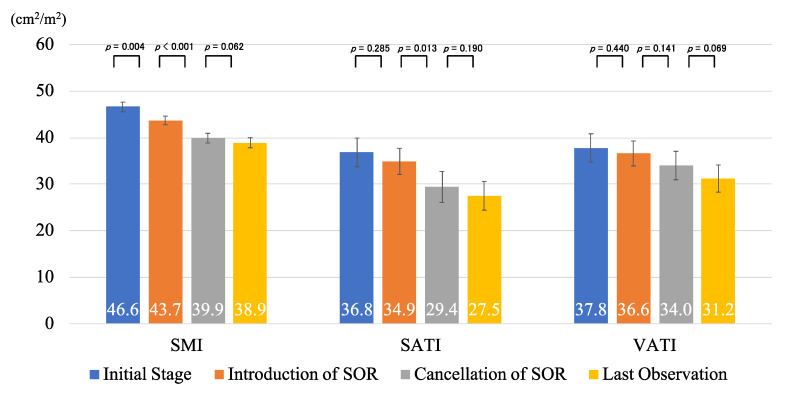
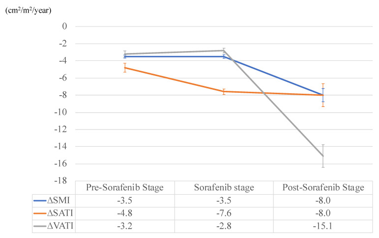
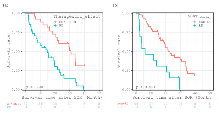
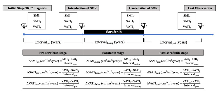
The aim of this study was to assess the annualized changes in body composition, including skeletal muscle, subcutaneous adipose tissue (SAT), and visceral adipose tissue (VAT) before, during, and after sorafenib treatment in patients with hepatocellular carcinoma (HCC). This retrospective study evaluated 61 HCC patients treated with sorafenib. Annualized changes (Δ; cm2/m2/year) in skeletal muscle index (SMI), SAT index (SATI), and VAT index (VATI), which were defined as the cross-sectional areas (cm2) of those areas on computed tomography normalized by the square of one's height (m2), before (pre), during (during), and after (post) sorafenib treatment, were calculated. Patients within the 20th percentile cutoffs for these indices were classified into the rapid depletion group and the effects of these values on survival were analyzed using the Kaplan-Meier analysis and Cox proportional-hazards model. Annualized depletion rates of SMI (ΔSMIpre: -3.5, ΔSMIduring: -3.5, ΔSMIpost: -8.0) and VATI (ΔVATIpre: -3.2, ΔVATIduring: -2.8, ΔVATIpost: -15.1) accelerated after the cancellation of sorafenib, whereas that of SATI (ΔSATIpre: -4.8, ΔSATIduring; -7.6, ΔSATIpost; -8.0) had already accelerated during sorafenib treatment. Patients with rapid depletion of ΔSATIduring experienced significantly worse survival rates (p < 0.001), and it was an independent predictor of survival (p = 0.009), together with therapeutic effect (p < 0.001). Rapid depletion of SAT during sorafenib treatment can be used to predict survival in patients with HCC.

Keywords: body composition; hepatocellular carcinoma; prognostic factor; skeletal muscle; sorafenib; subcutaneous fat mass.


Conflict of interest statement

The authors declare no conflict of interest.

Figures

Figure 1
Figure 1
The values of skeletal muscle index (SMI), subcutaneous adipose tissue index (SATI), and visceral adipose tissue index (VATI) at the initial stage, introduction of sorafenib (SOR), cancellation of SOR, and last observation time. Error bars represent standard errors.
Figure 2
Figure 2
Annualized changes in skeletal muscle index (SMI), subcutaneous adipose tissue index (SATI), and visceral adipose tissue index (VATI) at the pre-sorafenib, duration of sorafenib treatment, and post-sorafenib stages. Error bars represent standard errors.
Figure 3
Figure 3
Kaplan-Meier curves for overall survival time after introducing sorafenib (SOR) divided into therapeutic effect (CR/PR/SD vs. PD) (a), and the rapid depletion (RD) or non-rapid depletion (non-RD) groups in ΔSATIduring (b).
Figure 4
Figure 4
Outline and formula for ΔSMIpre, ΔSATIpre, ΔVATIpre, ΔSMIduring, ΔSATIduring, ΔVATIduring, ΔSMIpost, ΔSATIpost, and ΔVATIpost (cm2/m2/year). Skeletal muscle index (SMI) was defined as the cross-sectional area of the muscle (cm2) at the L3 level of the computed tomography image, normalized by the square of the height (m2). Subcutaneous adipose tissue index (SATI) and visceral adipose tissue index (VATI) were defined as the cross-sectional areas of the subcutaneous and visceral fat (cm2), respectively, at the umbilical point, normalized by the square of the height (m2).

References

    1. El-Serag H.B. Hepatocellular carcinoma. N. Engl. J. Med. 2011;365:1118–1127. doi: 10.1056/NEJMra1001683. - DOI - PubMed
    1. Njei B., Rotman Y., Ditah I., Lim J.K. Emerging trends in hepatocellular carcinoma incidence and mortality. Hepatology. 2015;61:191–199. doi: 10.1002/hep.27388. - DOI - PMC - PubMed
    1. Singal A.G., El-Serag H.B. Hepatocellular Carcinoma From Epidemiology to Prevention: Translating Knowledge into Practice. Clin. Gastroenterol. Hepatol. 2015;13:2140–2151. doi: 10.1016/j.cgh.2015.08.014. - DOI - PMC - PubMed
    1. Galle P.R., Forner A., Llovet J.M., Mazzaferro V., Piscaglia F., Raoul J.L., Schirmacher P., Vilgrain V. EASL Clinical Practice Guidelines: Management of hepatocellular carcinoma. J. Hepatol. 2018;69:182–236. doi: 10.1016/j.jhep.2018.03.019. - DOI - PubMed
    1. Bruix J., Sherman M. American Association for the Study of Liver Diseases Management of hepatocellular carcinoma: An update. Hepatology. 2011;53:1020–1022. doi: 10.1002/hep.24199. - DOI - PMC - PubMed