Review
. 2020 May;22(2):185-202.
doi: 10.5853/jos.2020.00227. Epub 2020 May 31.

Unfavorable Vascular Anatomy during Endovascular Treatment of Stroke: Challenges and Bailout Strategies

Affiliations
Review

Unfavorable Vascular Anatomy during Endovascular Treatment of Stroke: Challenges and Bailout Strategies

Francisco José Arruda Mont Alverne et al. J Stroke. 2020 May.

Abstract

The benefit of mechanical thrombectomy (MT) in acute ischemic stroke (AIS) due to large vessel intracranial occlusions is directly related to the technical success of the procedures in achieving fast and complete reperfusion. While a precise definition of refractoriness is lacking in the literature, it may be considered when there is reperfusion failure, long procedural times, or high number of passes with the MT devices. Detailed knowledge about the causes for refractory MT in AIS is limited; however, it is most likely a multifaceted problem including factors related to the vascular anatomy and the underlying nature of the occlusive lesion amongst other factors. We aim to review the impact of several key unfavorable anatomical factors that may be encountered during endovascular AIS treatment and discuss potential bail-out strategies to these challenging situations.

Keywords: Brain ischemia; Reperfusion; Stroke; Thrombectomy.


Figures

Figure 1.
Figure 1.
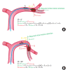
Some situations impose more challenges for supra-aortic access during mechanical thrombectomy and should be promptly identified: (A) a common origin for the innominate and left common carotid arteries (CCAs), (B) the left CCA originates from the innominate artery. a., artery.
Figure 2.
Figure 2.
Aortic arch configurations. Type 2 and 3 aortic arch may be obstacles for rapid supra-aortic access. The vertical distance from the origin of the innominate artery to the top of the arch determines the arch type. This distance is <1 diameter of the left common carotid artery (CCA) in a type 1 arch (A), between 1 and 2 diameters of the left CCA in a type 2 arch (B), and >2 diameters of the left CCA in a type 3 arch (C).
Figure 3.
Figure 3.
Strategies for supra-aortic access using coaxial technique with a 125-cm 5F Vitek diagnostic catheter (VDC; blue) over a 9F balloon-guiding catheter (BGC; green) to avoid an exchanging guiding maneuver. (A) VDC engaged in left common carotid artery then a hydrophilic 0.035" or 0.038" guidewire is advanced in the internal carotid artery (ICA). (B) BGC can be advanced in the ICA over the guidewire while the VDC is still at the origin of the proximal supraaortic vessel. (C) In case of severe proximal tortuosity first VDC is engaged, then the guidewire is progressed distally, after that (D) VDC is navigated over the guidewire into the ICA and finally the BGC is advanced over the VDC/guidewire (E). In case of more proximal angulation with a short proximal segment of CCA for engagement, BGC is initially advanced in the proximal CCA over the VDC, then it is maximally inflated to stabilize (F). Once the support is ensured, a guidewire and VDC are sequentially advanced more distally (G). Finally, the balloon is deflated, and the BGC is advanced over the VDC (H).
Figure 4.
Figure 4.
Changing the curvature of diagnostic catheter in coaxial technique is an option to achieve supra-aortic access. (A) Brachiocephalic trunk injection with a Vitek diagnostic catheter in coaxial system with a Neuron™ MAX 088 shuttle (Penumbra Inc.) depicting an extreme tortuosity that was an obstacle for progression of the guiding catheter. (B) A Neuron™ MAX 088 shuttle was placed at the origin of supra-aortic vessel (long black arrow) and three 0.014” exchange wires were consecutively advanced (long white arrow). (C) Once a stable support was obtained, a 6F diagnostic catheter with a Berenstein curve (black arrowhead) was advanced into the right common carotid artery allowing the progression of the Neuron™ MAX 088 shuttle.
Figure 5.
Figure 5.
Use of multiple guidewires in very tortuous extracranial vessel access. (A) Anteroposterior roadmap view of left common carotid artery depicting a recurrent origin at the aortic arch and a proximal angle turn after its origin (long black arrow), precluding advance of 0.035” guidewire. (B) Three 0.014” guidewires were navigated one after the other (long white arrow), (C) allowing enough support for progression of the guiding catheter.
Figure 6.
Figure 6.
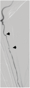
Right radial angiogram depicting a hypoplasia of right radial artery with associated tortuosity (black arrowheads), preventing the use of this route for mechanical thrombectomy.
Figure 7.
Figure 7.
Direct carotid puncture is valuable option is very difficult aortic arch. (A) Anteroposterior acquisition of a recurrent left common carotid artery in type III aortic arch, preventing advance of a coaxial system guiding catheter over a hydrophilic guide wire. (B) Anteroposterior roadmap view for direct puncture guidance with a 18 G needle (long black arrow) with concomitant use of an ultrasound. (C) A 6F sheath was placed in the left common carotid artery and an ACE 68 aspiration catheter (Penumbra Inc.) was advanced the internal carotid artery (long white arrow). (D) Selective anteroposterior carotid angiogram at the beginning of thrombectomy disclosing proximal M1 occlusion (short black arrow). Thrombectomy was performed using a combined technique (ACE 68 aspiration catheter; 4×30 mm Trevo XP Stentriever, Stryker) allowing a thrombolysis in cerebral infarction (TICI) grade 3 reperfusion after one pass as shown in anteroposterior (short white arrow) (E) and lateral view (F). After procedure, we retrieved the sheath and closed the punctured site by manual compression.
Figure 8.
Figure 8.
Tortuous vascular anatomy impairs the optimal positioning of the guiding catheter and subsequent distal progression of catheters. We should identify variations of carotid artery that may be a barrier to distal cervical and intracranial access like coiling and kinking. Modified of Nagata et al. [49], with permission from Elsevier. ICA, internal carotid artery.
Figure 9.
Figure 9.
A 65-year-old man with left middle cerebral artery (MCA). (A) Left anteroposterior internal carotid angiogram through an 8F balloon-guiding catheter (BGC) shows a M1 occlusion (short white arrow) and a looping in the mid-cervical segment of internal carotid artery (long black arrow) that precluded the advance of the BGC. (B) An aspiration catheter (long white arrow) was used but it could not be advanced distally due to instability and low position of the BGC. A Solitaire stent-retriever (Medtronic) 4×40 mm was then deployed over the clot (black arrowhead). (C) Lateral view of internal carotid artery (ICA) discloses a long dissection in the internal carotid artery (black arrowheads). (D) Oblique view of ICA shows a carotid occlusion at the level of dissection. (E) ICA was remodeled with two flexible stents but control angiogram of ICA M1 segment was occluded and was refractory to new attempts for revascularization (modified thrombolysis in cerebral infarction [mTICI] of 0).
Figure 10.
Figure 10.
Decomposition of the original vector of force applied in the stent-retriever (SR) during mechanical thrombectomy. (A) Schematic representation of a M1 occlusion in a straight vessel. Vector decomposition during retrieval. The angle Θ (yellow) between the plan of the vector of retrieval force (FR, black) in SR and direction of required motion to displace the SR (plan A, green) is 0°. The plan B (red) is perpendicular to plan A, it creates a detrimental force to pullback (FR detrimental, red). The effective pullback force (FR effective, green) will be towards the direction of the main axis of the occluded artery, in this situation equal to FR. Schematic representation of a M1 occlusion in a curved vessel. (B) Decomposition of vectors during retrieval, where the angle Θ (yellow) between FR in SR and required direction of motion (plan A, green) is 90°. The FR effective in this case is the force required to displace the SR in the “diving” distal M1 segment that is towards the vertical plan A. The FR effective will be equal to zero the SR would not be displaced and the FR detrimental is maximum and equal to FR.
Figure 11.
Figure 11.
Tapering phenomenon during stent retrieval. (A) Stent-retriever deployed in a curved middle cerebral artery with clot within the struts of stent, note in yellow the cell size at rest. (B) Once a retrieval force (FR) is applied to the stent, there is decomposition of forces in the curve of the artery, (C) an effective resultant force (FR effective; green arrow) favoring stent retraction and a deleterious force perpendicular to this movement (FR detrimental; red arrow) that (D) diminishes full expansion of stent, reduces stent cell size (yellow) and forces the clot outside the stent.
Figure 12.
Figure 12.
Degree of tortuosity of the cavernous segment of internal carotid artery (cICA). When anterior and posterior genu of cICA configurations are opened it is a type I, that can be further divides in type Ia (posterior genu angle [P] is >90°) and type Ib (P=90°). Type II has a more acute angle of anterior genu (A) in comparison to type I. Type III has a superior deflection of posterior genu. Type IV is the most tortuous with a Simmons catheter shape. The higher the tortuosity (represented by the ratio H/[A+P]), the more complex may be the procedure. H, height between the trough of anterior genu and the peak of posterior genu. Modified from Lin et al. [62] with permission from BMJ.
Figure 13.
Figure 13.
A 77-year-old patient with 1-hour right middle cerebral artery (MCA). (A) Right anteroposterior internal carotid angiogram depicting a distal occlusion in a curved (or diving) M1 segment (long black arrow). (B) Anteroposterior internal carotid artery (ICA) angiographic acquisition with a Solitaire stent-retriever (Medtronic) 4×40 mm in place delineating the curved MCA (short black arrowhead). (C) Anteroposterior angiogram of ICA after two passes with Solitaire stent-retriever 4×40 mm showing a reperfusion failure (modified thrombolysis in cerebral infarction [mTICI] of 0) (long white arrow). (D) Solitaire stent-retriever 4×40 mm was deployed in the inferior branch of a MCA bifurcation (short black arrow). (E) A Rebar 18 Microcatheter (Medtronic) 0.021” was navigated over a wire 0.014” in the antero-superior branch (short white arrow) then (F) a second Solitaire stent-retriever 4×40 mm was deployed in Y-configuration (black arrowheads). (G) Unsubstracted anteroposterior and (H) lateral view showing Solitaire stent-retriever 4×40 mm deployed in the inferior and superior branch of a MCA (white arrowheads). After one pass of Y-configuration stent-retrievers full reperfusion (mTICI of 3) was obtained as shown in (I) anteroposterior (J) and lateral acquisition of ICA.

References

    1. Albers GW, Marks MP, Kemp S, Christensen S, Tsai JP, OrtegaGutierrez S, et al. Thrombectomy for stroke at 6 to 16 hours with selection by perfusion imaging. N Engl J Med. 2018;378:708–718. - PMC - PubMed
    1. Berkhemer OA, Fransen PS, Beumer D, van den Berg LA, Lingsma HF, Yoo AJ, et al. A randomized trial of intraarterial treatment for acute ischemic stroke. N Engl J Med. 2015;372:11–20. - PubMed
    1. Bracard S, Ducrocq X, Mas JL, Soudant M, Oppenheim C, Moulin T, et al. Mechanical thrombectomy after intravenous alteplase versus alteplase alone after stroke (THRACE): a randomised controlled trial. Lancet Neurol. 2016;15:1138–1147. - PubMed
    1. Campbell BC, Mitchell PJ, Kleinig TJ, Dewey HM, Churilov L, Yassi N, et al. Endovascular therapy for ischemic stroke with perfusion-imaging selection. N Engl J Med. 2015;372:1009–1018. - PubMed
    1. Goyal M, Demchuk AM, Menon BK, Eesa M, Rempel JL, Thornton J, et al. Randomized assessment of rapid endovascular treatment of ischemic stroke. N Engl J Med. 2015;372:1019–1030. - PubMed

LinkOut - more resources