. 2020 Jul;19(14):1740-1753.
doi: 10.1080/15384101.2020.1777804. Epub 2020 Jun 17.

miR-96-5p regulated TGF-β/SMAD signaling pathway and suppressed endometrial cell viability and migration via targeting TGFBR1

Affiliations

miR-96-5p regulated TGF-β/SMAD signaling pathway and suppressed endometrial cell viability and migration via targeting TGFBR1

Silei Chen et al. Cell Cycle. 2020 Jul.

Retraction in

Abstract

We previously performed high throughput RNA-seq in paired eutopic and ectopic endometrial specimen of endometriosis patients, and validated the results by qRT-PCR in endometriosis endometrial tissues. MiR-96-5p was significantly downregulated in ectopic endometrial tissues compared to eutopic tissues. In order to identify the role of miR-96-5p in endometriosis and endometrial cells, and investigate the underlying mechanisms, the Ishikawa and End1/E6E7 cell lines were transfected with miR-96-5p mimics, miR-96-5p inhibitors or TGFBR1 siRNA. The expression of TGF-β/SMAD signaling pathway components and epithelial-mesenchymal transition (EMT) markers were examined by qRT-PCR and western blot, and cell viability and migration were determined by CCK-8, transwell and wound healing assays, respectively. We discovered miR-96-5p to be significantly downregulated while TGFBR1 was distinctly up-regulated in endometriosis. Overexpression of miR-96-5p inhibited endometrial cells viability and migration, while inhibition of miR-96-5p had opposite effect. Furthermore, we confirmed TGFBR1 was a direct target of miR-96-5p. Overexpression of miR-96-5p could block the TGF-β/SMAD signaling pathway via targeting TGFBR1 and reverse the TGF-β1 induced EMT in endometrial cell lines. In conclusion, we demonstrated that miR-96-5p interacted with TGF-β/SMAD signaling pathway and blocked the TGF-β1 induced EMT in endometrial cells via directly targeting TGFBR1.

Keywords: EMT; TGF-β/SMAD signaling pathway; TGFBR1; endometriosis; miR-96-5p.


Conflict of interest statement

All authors declare no conflicts of interest.

Figures
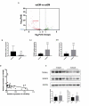
Figure 1.
Figure 1.
Down-regulation of miR-96-5p and up-regulation of TGFBR1 in ectopic endometrial tissue. (a) Volcano plots demonstrate differentially expressed microRNAs between ectopic and eutopic endometrium of endometriosis patients. Green points indicated upregulated while red points indicated downregulated microRNAs. (b) RT-PCR showed miR-96-5p was downregulated in ectopic endometrial tissue (***, P< 0.001). (c) RT-PCR showed mRNA expression of TGFBR1 and SMAD3 was upregulated in ectopic endometrial tissue (***, P< 0.001). (d) A significant correlation between the levels of miR-96-5p and TGFBR1 in ectopic endometrium tissues was demonstrated using Pearson’s correlation coefficient analysis (r = −0.3315, P = 0.0482). (e) Western blot showed protein expression of TGFBR1 and SMAD3 was upregulated in ectopic endometrial tissue (*, P < 0.05).
Figure 2.
Figure 2.
Overexpression of miR-96-5p inhibited endometrial cell viability and migration. (a) The ectopic expression of miR-96-5p in Ishikawa and End1/E6E7 cell lines transfected with mimics was confirmed by RT-PCR (**, P< 0.01). (b)CCK-8 assay showed miR-96-5p overexpression inhibited endometrial cells viability (*, P< 0.05). (c) Transwell assay showed miR-96-5p overexpression inhibited endometrial cell migration. (d) Wound assay showed overexpression of miR-96-5p inhibited endometrial cell migration (*, P< 0.05; **, P< 0.01).
Figure 3.
Figure 3.
Inhibition of miR-96-5p promoted endometrial cell viability and migration. (a) The downregulation of miR-96-5p in Ishikawa and End1/E6E7 cell lines was confirmed by RT-PCR (***, P < 0.001). (b) CCK-8 assay showed miR-96-5p downregulation promoted endometrial cells viability (*, P < 0.05). (c) Transwell assay showed miR-96-5p downregulation promoted endometrial cell migration. (d) Wound assay showed inhibition of miR-96-5p promoted endometrial cell migration (**, P < 0.01; ***, P < 0.001).
Figure 4.
Figure 4.
Silencing TGFBR1 inhibited endometrial cell migration and cell viability. (a)The downregulation of TGFBR1 in endometrial cell lines by siRNA was confirmed by RT-PCR (***, P < 0.001). (b) CCK-8 assay showed TGFBR1 downregulation by siRNA inhibited endometrial cells viability (*, P < 0.05). (c) Transwell assay showed TGFBR1 downregulation inhibited endometrial cell migration. (d) Wound assay showed silencing TGFBR1 inhibited endometrial cell migration (*, P < 0.05; **, P < 0.01).
Figure 5.
Figure 5.
TGFBR1 is a direct target of miR-96-5p. (a) The predicted miR-96-5p binding sites of TGFBR1 3ʹUTR. (b) Dual-luciferase assay confirmed that miR-96-5p directly targets TGFBR1 (*, P < 0.05). (c), (d) PCR and western blot showed that the expression of TGFBR1 was diminished by overexpression of miR-96-5p (*, P < 0.05).
Figure 6.
Figure 6.
TGF-β1 activated TGF-β/SMAD signaling pathway and induced EMT in endometrial cells. (a) PCR showed that after TGF-β1 treatment, RNA expression of TGF-β/SMAD signaling pathway components, TGFBR1, TGFBR2 and SMAD3 was increased in Ishikawa and End1/E6E7 cell lines. (b) Western blot showed that after TGF-β1 treatment, protein expression of TGF-β/SMAD signaling pathway components, TGFBR1, TGFBR2 and SMAD3 was increased in Ishikawa and End1/E6E7 cell lines. Expression of epithelial marker E-cadherin was decreased, and expression of mesenchymal markers N-cadherin and Vimentin was increased following TGF-β1 treatment in Ishikawa and End1/E6E7 cell lines.
Figure 7.
Figure 7.
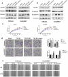
miR-96-5p reversed the TGF-β1 activated TGF-β/SMAD signaling pathway and TGF-β1 induced EMT. (a) Western blot showed miR-96-5p overexpression decreased the expression of TGFBR1 and SMAD3 but not TGFBR2 in endometrial cell lines treated with TGF-β1. (b) Western blot showed miR-96-5p overexpression increased the expression of E-cadherin and decreased the expression of N-cadherin and Vimentin in endometrial cell lines treated with TGF-β1. (c) CCK-8 showed TGF-β1 increased cell viability, miR-96-5p overexpression reduced the promotive effect of TGF-β1 (*, P < 0.05; **, P < 0.01). (d) Transwell showed miR-96-5p overexpression attenuates the cell migration ability induced by TGF-β1 (**, P < 0.01). (e) Wound assay showed miR-96-5p overexpression inhibited the TGF-β1 induced cell migration ability (**, P < 0.01).

References

    1. Giudice LC, Kao LC.. Endometriosis [J]. Lancet. 2004;364(9447):1789–1799. - PubMed
    1. Giudice LC. Clinical practice. Endometriosis [J]. N Engl J Med. 2010;362(2010):2389–2398. - PMC - PubMed
    1. Shafrir AL, Farland LV, Shah DK, et al. Risk for and consequences of endometriosis: A critical epidemiologic review [J]. Best Pract Res Clin Obstetrics Gynaecol. 2018. August;51(51):1–15. - PubMed
    1. Tomassetti C, D’Hooghe T. Endometriosis and infertility: insights into the causal link and management strategies [J]. Best Pract Res Clin Obstetrics Gynaecol. 2018. August;51(51):25–33. - PubMed
    1. Matalliotakis M, Matalliotaki C, Goulielmos GN, et al. Association between ovarian cancer and advanced endometriosis [J]. Oncol Lett. 2018;15(5):7689–7692. - PMC - PubMed

Publication types

MeSH terms

Substances

LinkOut - more resources