. 2020 Jul 21;117(29):16928-16937.
doi: 10.1073/pnas.1820132117. Epub 2020 Jul 7.

A plant DNA virus replicates in the salivary glands of its insect vector via recruitment of host DNA synthesis machinery

Affiliations

A plant DNA virus replicates in the salivary glands of its insect vector via recruitment of host DNA synthesis machinery

Ya-Zhou He et al. Proc Natl Acad Sci U S A. .

Abstract

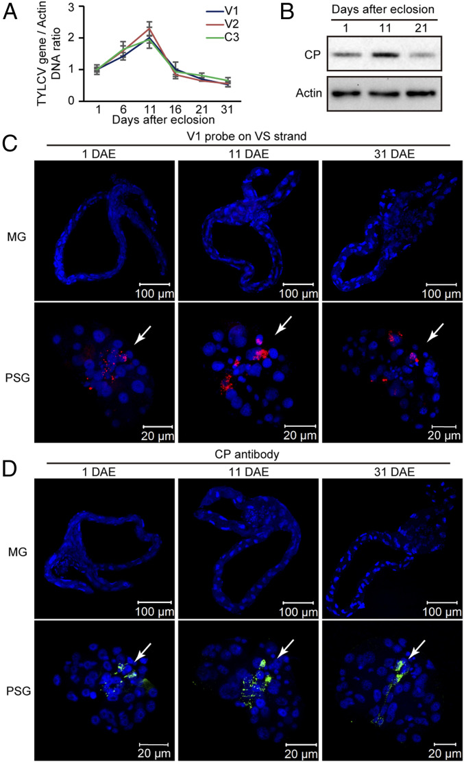
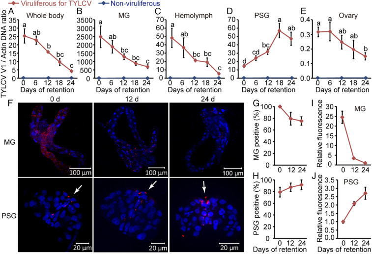
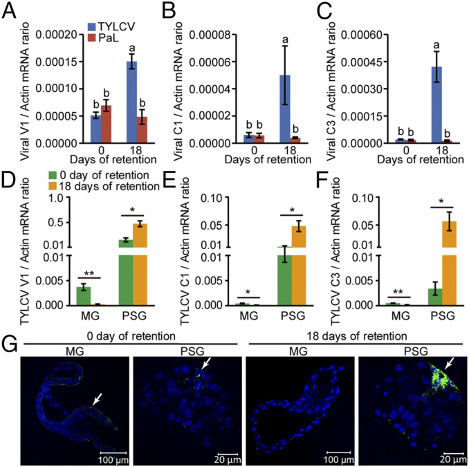
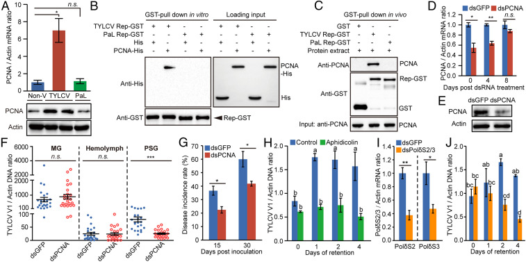
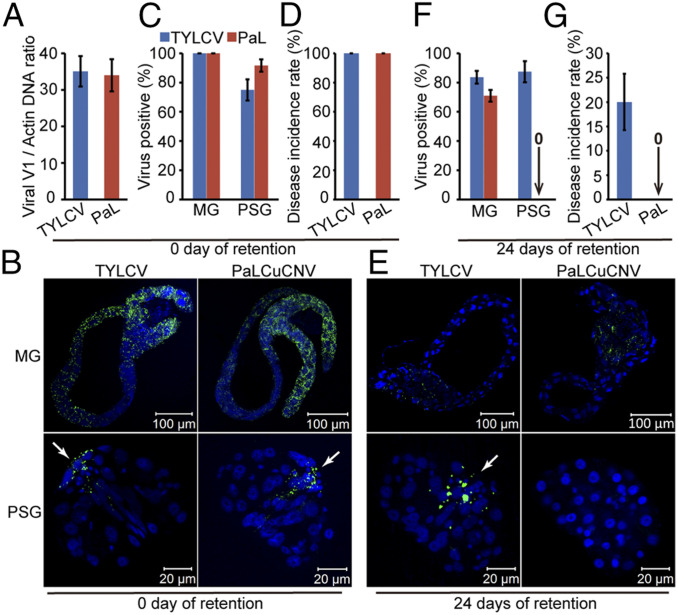
Whereas most of the arthropod-borne animal viruses replicate in their vectors, this is less common for plant viruses. So far, only some plant RNA viruses have been demonstrated to replicate in insect vectors and plant hosts. How plant viruses evolved to replicate in the animal kingdom remains largely unknown. Geminiviruses comprise a large family of plant-infecting, single-stranded DNA viruses that cause serious crop losses worldwide. Here, we report evidence and insight into the replication of the geminivirus tomato yellow leaf curl virus (TYLCV) in the whitefly (Bemisia tabaci) vector and that replication is mainly in the salivary glands. We found that TYLCV induces DNA synthesis machinery, proliferating cell nuclear antigen (PCNA) and DNA polymerase δ (Polδ), to establish a replication-competent environment in whiteflies. TYLCV replication-associated protein (Rep) interacts with whitefly PCNA, which recruits DNA Polδ for virus replication. In contrast, another geminivirus, papaya leaf curl China virus (PaLCuCNV), does not replicate in the whitefly vector. PaLCuCNV does not induce DNA-synthesis machinery, and the Rep does not interact with whitefly PCNA. Our findings reveal important mechanisms by which a plant DNA virus replicates across the kingdom barrier in an insect and may help to explain the global spread of this devastating pathogen.

Keywords: DNA synthesis machinery; insect vector; plant DNA virus; replication; salivary glands.


Conflict of interest statement

The authors declare no competing interest.

Figures

Fig. 1.
Fig. 1.
Dynamics of TYLCV in adult offspring of viruliferous MEAM1 whiteflies. (A) Relative concentration of TYLCV DNA in whole bodies of F1 adults at different developmental stages obtained by amplifying portions of the V1, V2, and C3 genes using qPCR. Mean ± SEM of three independent experiments is shown. (B) Accumulation of TYLCV CP in F1 adults at different developmental stages. (C and D) Localization of TYLCV VS DNA (C) and CP (D) in MGs and PSGs of F1 adults at different developmental stages. For TYLCV VS DNA localization, MGs and PSGs were hybridized with a Cy3-labeled VS strand-specific probe (V1 probe; red). TYLCV CP was detected by use of a mouse anti-CP monoclonal antibody and goat anti-mouse IgG labeled with Dylight 488 (green) secondary antibody. Cell nucleus was stained with DAPI (blue). The white arrow indicates of the virus signal in the PSG. For each time point, 20 samples were analyzed, and a similar trend was observed.
Fig. 2.
Fig. 2.
Dynamics of TYLCV in whole body and various tissues of MEAM1 whiteflies during long-term retention. (AE) Relative concentration of TYLCV DNA in the whole body (A), MG (B), hemolymph (C), PSG (D), and ovary (E) of MEAM1 whiteflies. Total DNA was extracted from the whole body, MG, hemolymph, PSG, and ovary for assay by qPCR. Mean ± SEM of three independent experiments is shown. P < 0.05 (one-way ANOVA, least significant difference [LSD] test). (F) Localization of TYLCV VS DNA in MGs and PSGs of whiteflies after different times of retention. MGs and PSGs were hybridized with a Cy3-labeled VS strand-specific probe (V1 probe; red). Cell nucleus was stained with DAPI (blue). The white arrow indicates TYLCV VS DNA signal in the PSG. (G and H) The proportion of TYLCV-positive MGs (G) and PSGs (H) at each time point. MGs, n = 24; PSGs, n = 24. (I and J) Relative fluorescence density of TYLCV VS DNA signal in MGs (I) and PSGs (J). For MGs and PSGs, the fluorescence density was set at one at days 24 and 0, respectively. (GJ) Mean ± SEM of three independent experiments is shown.
Fig. 3.
Fig. 3.
Expression of viral genes in MEAM1 whiteflies during long-term retention. (AC) Relative expression levels of V1 (A), C1 (B), and C3 (C) of TYLCV and PaLCuCNV (PaL) in whitefly whole bodies after 0 and 18 d of retention as detected by qRT-PCR. Mean ± SEM of three independent experiments is shown. P < 0.05 (one-way ANOVA, LSD test). (DF) Relative expression levels of TYLCV V1 (D), C1 (E), and C3 (F) genes in MGs and PSGs of whiteflies after 0 and 18 d of retention as detected by qRT-PCR. Mean ± SEM of three independent experiments is shown. *P < 0.05; **P < 0.01 (independent-sample t test). (G) Localization of TYLCV Rep in MGs and PSGs of whiteflies after 0 and 18 d of retention. Rep was detected by use of a rabbit anti-Rep polyclonal antibody and goat anti-rabbit IgG labeled with Dylight 488 (green) secondary antibody. Cell nucleus was stained with DAPI (blue). The white arrow indicates the immune-reactive signal of TYLCV Rep. For each time point, 20 samples were analyzed, and a similar trend was observed.
Fig. 4.
Fig. 4.
The function of whitefly PCNA and DNA Polδ in TYLCV replication. (A) PCNA expression was induced by TYLCV infection, but not by PaLCuCNV (PaL). Non-V, nonviruliferous. (B and C) Both recombinant PCNA (B) and whitefly endogenous PCNA (C) interacted with GST-fused TYLCV Rep, but not with GST-fused PaL Rep. (D) PCNA mRNA levels in whiteflies at several time points after dsRNA treatment. (E) PCNA protein levels in whiteflies at 0 d after dsRNA treatment. (F) dsPCNA treatment decreased TYLCV load in PSGs, whereas it did not affect the loads in the MGs and hemolymph. One dot represents one MG, PSG, or the hemolymph of one female whitefly. Mean ± SEM is shown. n.s., not significant. ***P < 0.001 (nonparametric Mann–Whitney U test). The results were reproduced at least two times. (G) The disease incidence rate of the tomato plants with TYLCV fed upon by dsPCNA-/dsGFP-treated whiteflies. (H) The effect of aphidicolin treatment on TYLCV replication in whiteflies. (I) Relative mRNA levels of DNA Polδ subunit 2 (PolδS2) and subunit 3 (PolδS3) after feeding with the mixture of dsPolδS2 and dsPolδS3. (J) The effect of dsPolδS2/3 treatment on TYLCV replication in whiteflies. (A, D, G, and I) Mean ± SEM of three independent experiments is shown. n.s., not significant. *P < 0.05; **P < 0.01 (independent-sample t test). (H and J) Mean ± SEM of three independent experiments is shown. P < 0.05 (one-way ANOVA, LSD test).
Fig. 5.
Fig. 5.
Replication of TYLCV in whitefly PSGs contributes to viral infectivity persistence after long-term retention. (A) Relative abundance of TYLCV and PaLCuCNV (PaL) in whitefly whole bodies immediately after a 48-h AAP on TYLCV- or PaL-infected tomato plants. (B) Localization of TYLCV or PaL in MGs and PSGs of whiteflies immediately after the 48-h AAP. (C) The proportion of TYLCV- or PaL-positive MGs and PSGs immediately after the 48-h AAP. MGs, n = 24; PSG, n = 24. (D) The disease incidence rate of the tomato plants with TYLCV or PaL fed upon by whiteflies that immediately after the 48-h AAP. (E) Localization of TYLCV or PaLCuCNV in MGs and PSGs of whiteflies after 24 d of retention. (F) The proportion of TYLCV- or PaL-positive MGs and PSGs after 24 d of retention. MGs, n = 24; PSGs, n = 24. (G) The disease incidence rate of the tomato plants with TYLCV or PaL fed upon by whiteflies after 24 d of retention. (B and E) TYLCV and PaLCuCNV CP was detected by use of a mouse anti-CP monoclonal antibody and goat anti-mouse IgG labeled with Dylight 488 (green) secondary antibody. Cell nucleus was stained with DAPI (blue). The white arrow indicates the viral CP signal in the PSG. For each time point, 24 samples were analyzed, and a similar trend was observed. (A, C, D, F, and G) Mean ± SEM of three independent experiments is shown.

References

    1. Gray S. M., Banerjee N., Mechanisms of arthropod transmission of plant and animal viruses. Microbiol. Mol. Biol. Rev. 63, 128–148 (1999). - PMC - PubMed
    1. Eigenbrode S. D., Bosque-Pérez N. A., Davis T. S., Insect-borne plant pathogens and their vectors: Ecology, evolution, and complex interactions. Annu. Rev. Entomol. 63, 169–191 (2018). - PubMed
    1. Hogenhout S. A., Ammar D., Whitfield A. E., Redinbaugh M. G., Insect vector interactions with persistently transmitted viruses. Annu. Rev. Phytopathol. 46, 327–359 (2008). - PubMed
    1. Ng J. C. K., Falk B. W., Virus-vector interactions mediating nonpersistent and semipersistent transmission of plant viruses. Annu. Rev. Phytopathol. 44, 183–212 (2006). - PubMed
    1. Lequime S., Paul R. E., Lambrechts L., Determinants of arbovirus vertical transmission in mosquitoes. PLoS Pathog. 12, e1005548 (2016). - PMC - PubMed

Publication types

LinkOut - more resources