. 2020 Jul 7;13(639):eaax6313.
doi: 10.1126/scisignal.aax6313.

A redox-active switch in fructosamine-3-kinases expands the regulatory repertoire of the protein kinase superfamily

Affiliations

A redox-active switch in fructosamine-3-kinases expands the regulatory repertoire of the protein kinase superfamily

Safal Shrestha et al. Sci Signal. .

Abstract

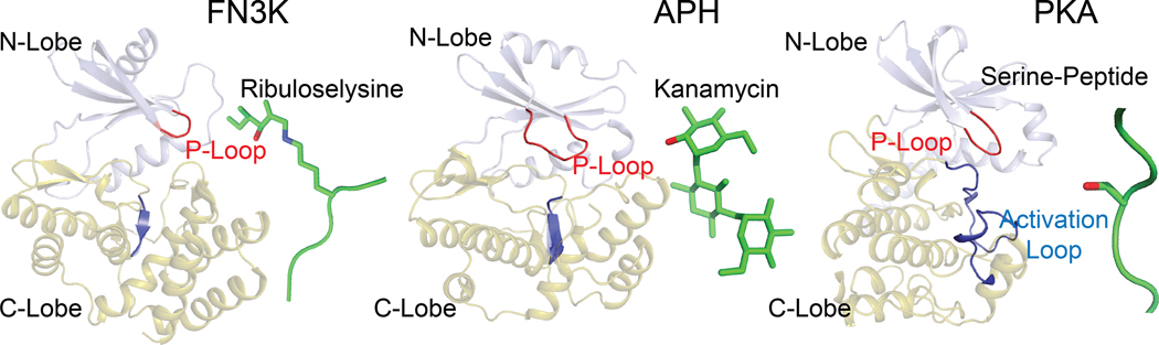
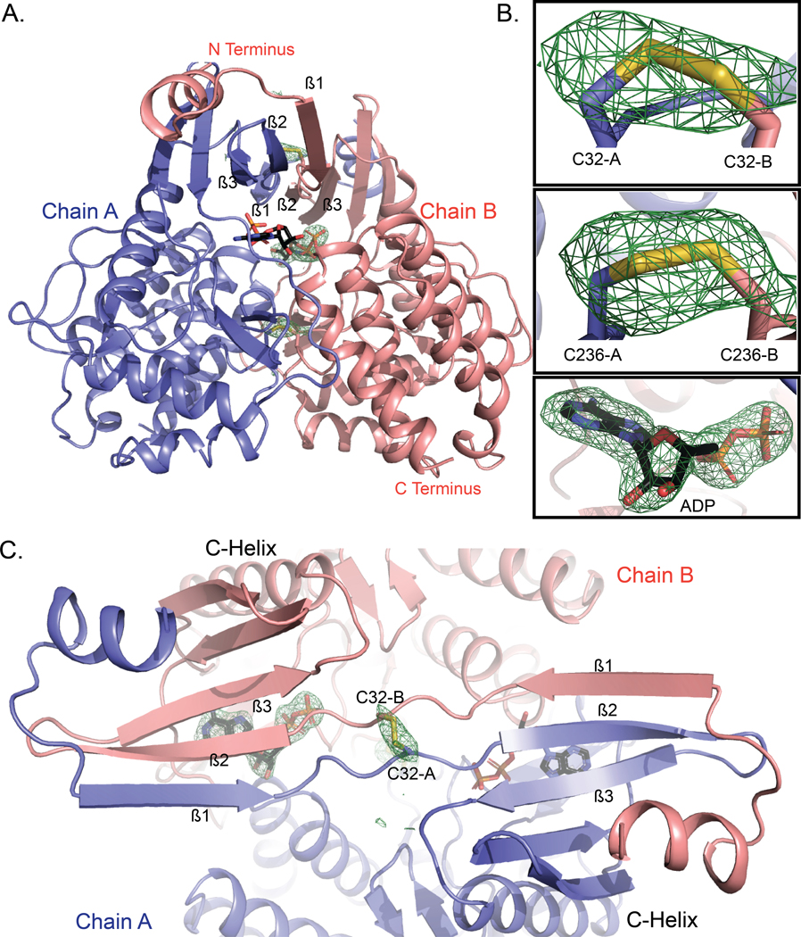
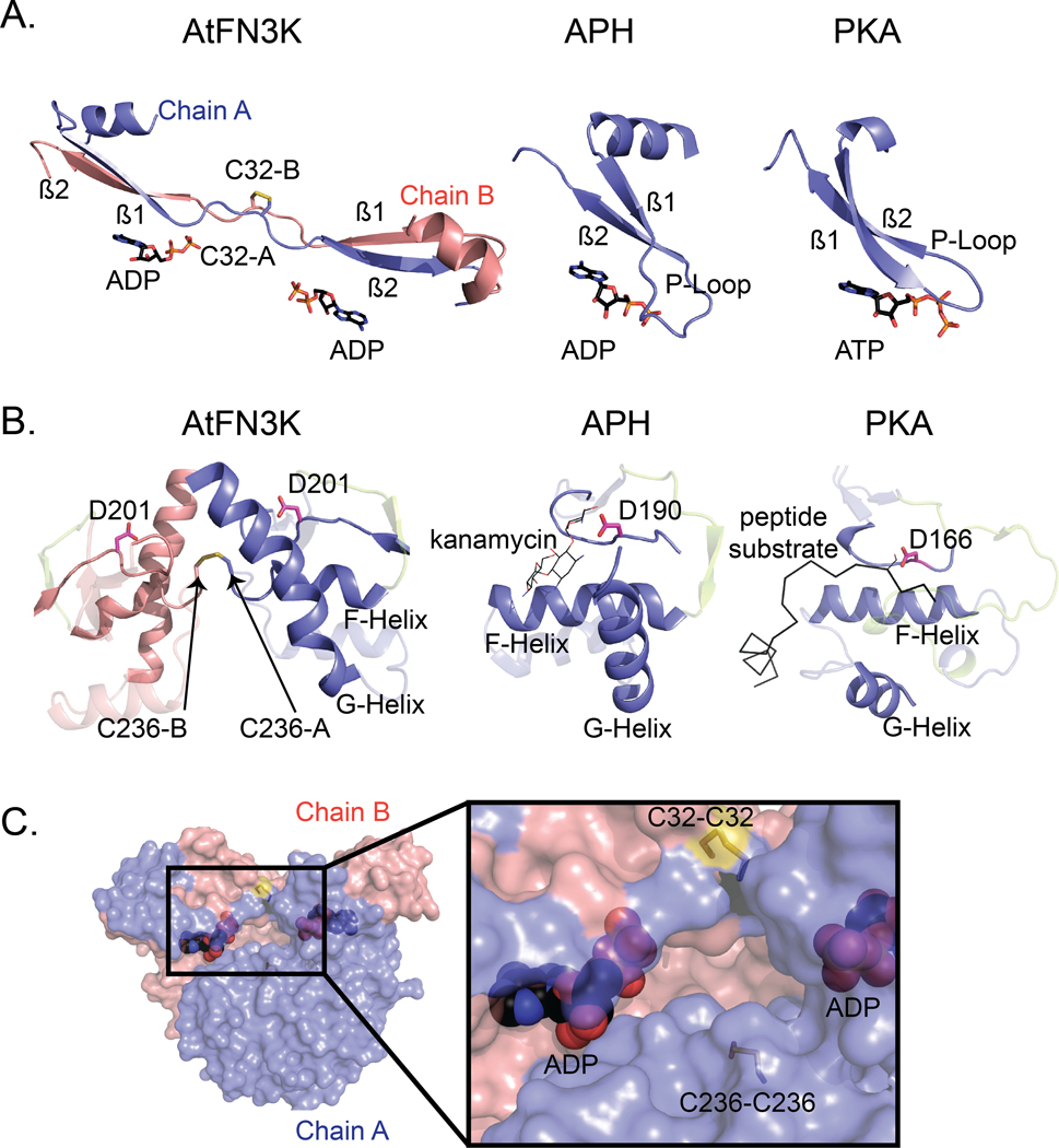
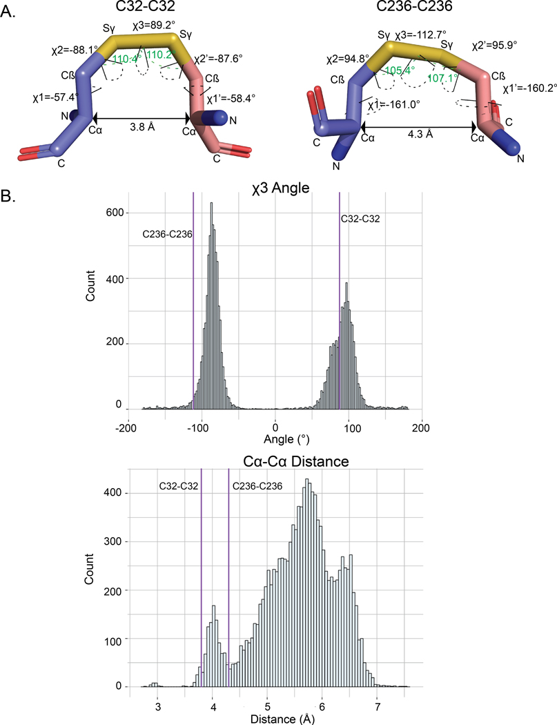
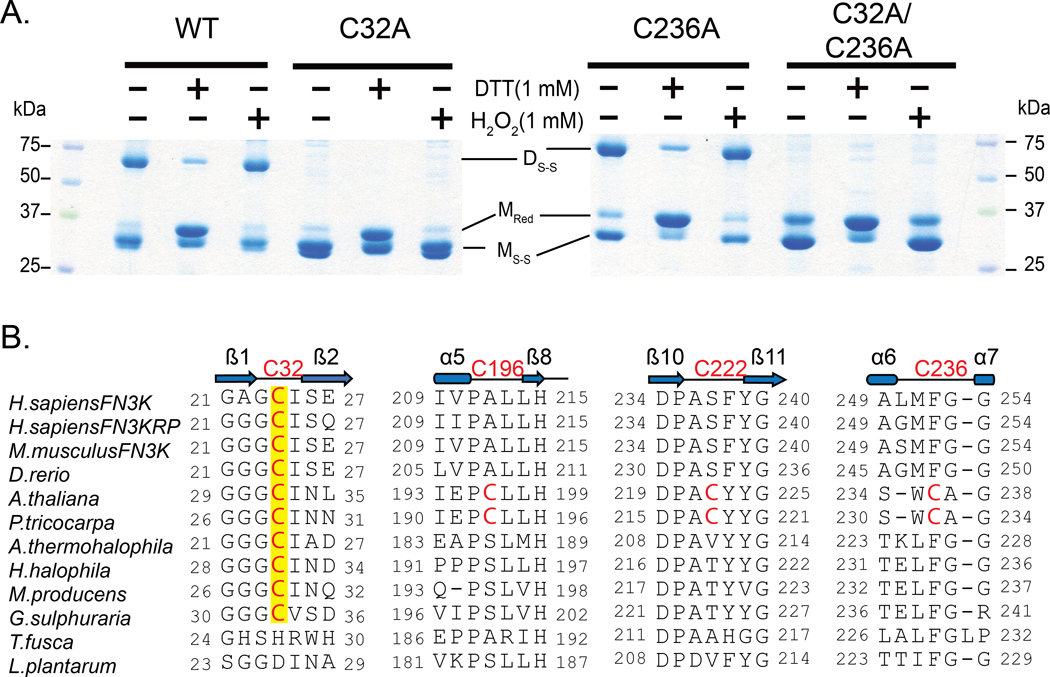
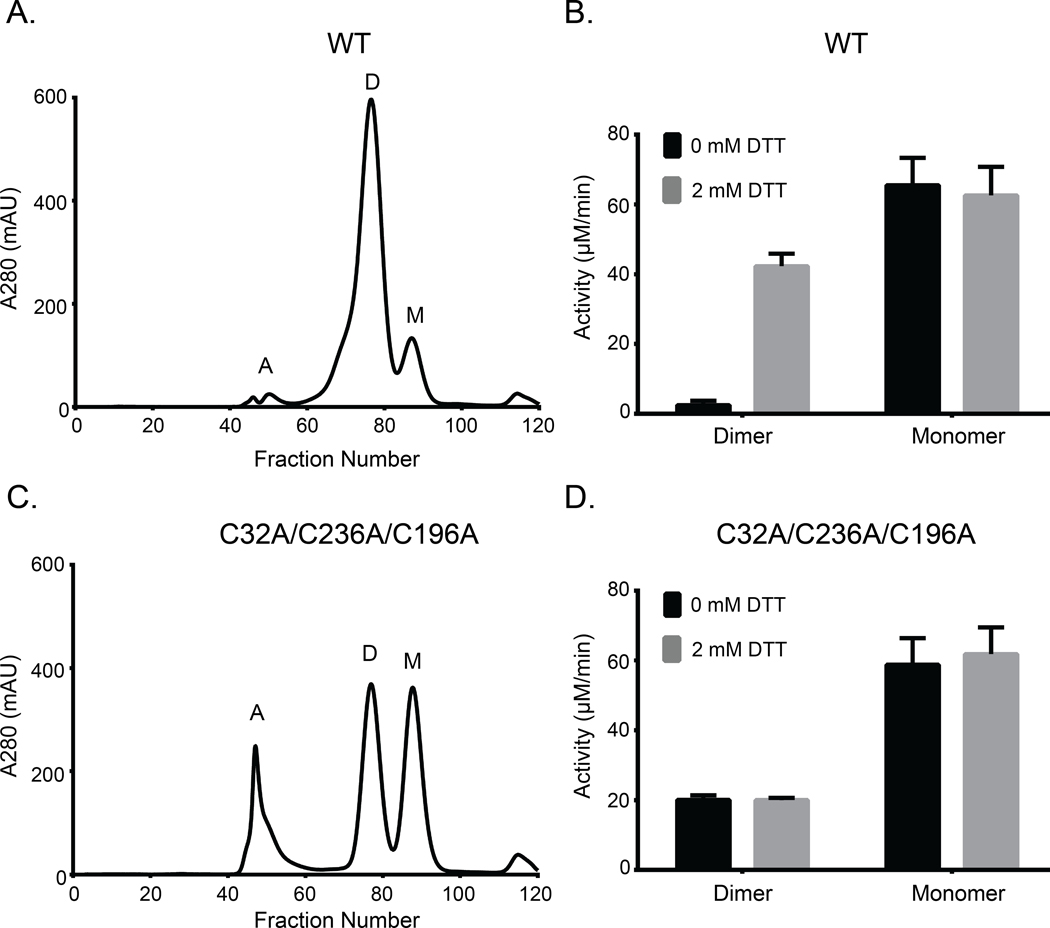
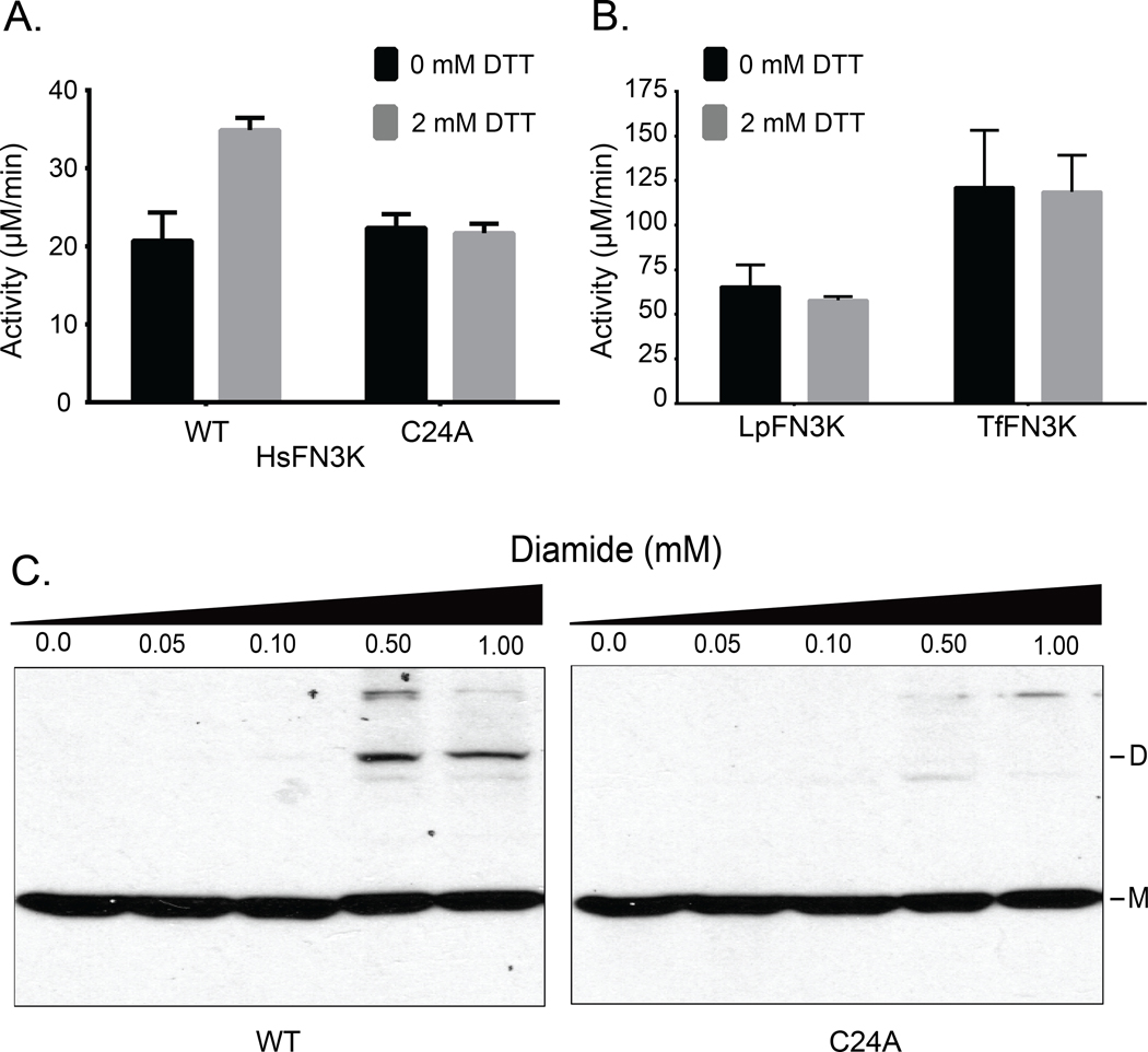
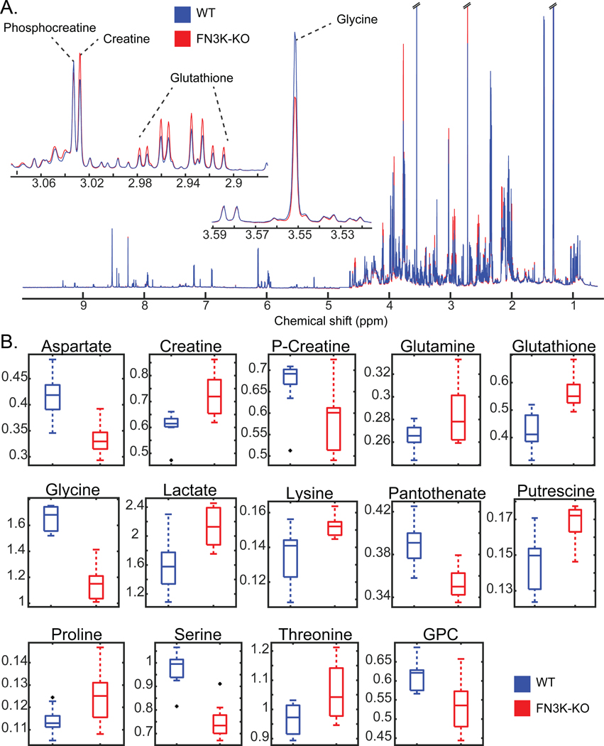
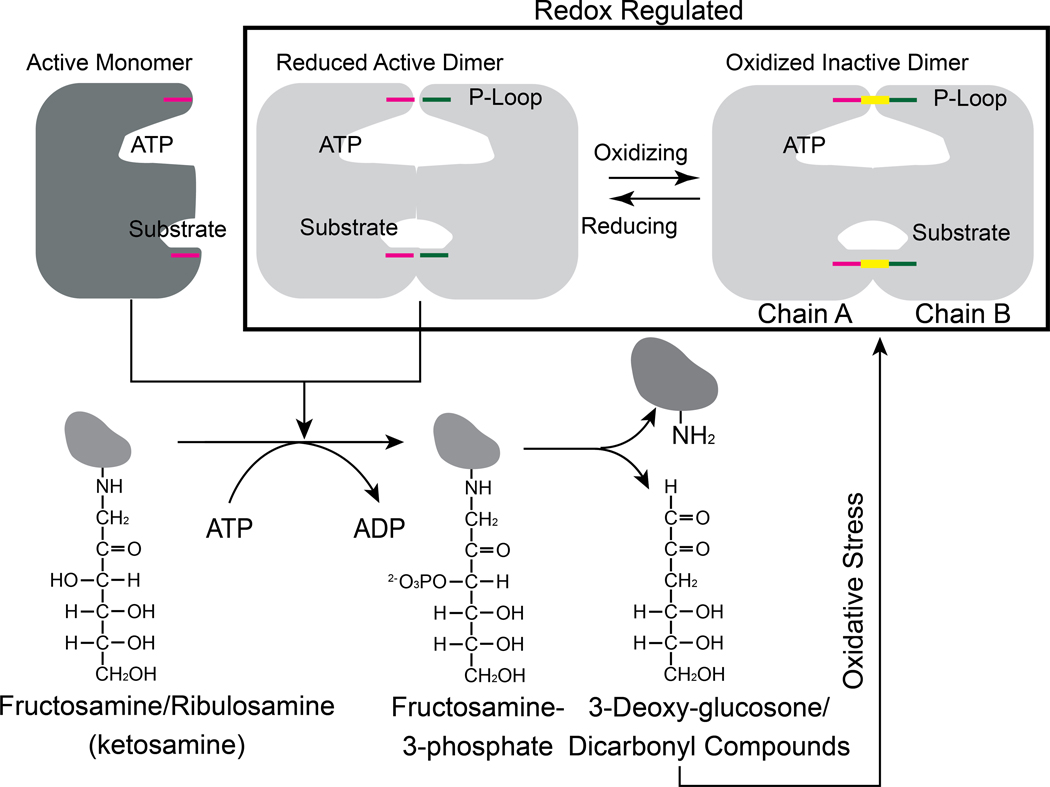
Aberrant regulation of metabolic kinases by altered redox homeostasis substantially contributes to aging and various diseases, such as diabetes. We found that the catalytic activity of a conserved family of fructosamine-3-kinases (FN3Ks), which are evolutionarily related to eukaryotic protein kinases, is regulated by redox-sensitive cysteine residues in the kinase domain. The crystal structure of the FN3K homolog from Arabidopsis thaliana revealed that it forms an unexpected strand-exchange dimer in which the ATP-binding P-loop and adjoining β strands are swapped between two chains in the dimer. This dimeric configuration is characterized by strained interchain disulfide bonds that stabilize the P-loop in an extended conformation. Mutational analysis and solution studies confirmed that the strained disulfides function as redox "switches" to reversibly regulate the activity and dimerization of FN3K. Human FN3K, which contains an equivalent P-loop Cys, was also redox sensitive, whereas ancestral bacterial FN3K homologs, which lack a P-loop Cys, were not. Furthermore, CRISPR-mediated knockout of FN3K in human liver cancer cells altered the abundance of redox metabolites, including an increase in glutathione. We propose that redox regulation evolved in FN3K homologs in response to changing cellular redox conditions. Our findings provide insights into the origin and evolution of redox regulation in the protein kinase superfamily and may open new avenues for targeting human FN3K in diabetic complications.


Figures

Fig. 1.
Fig. 1.. FN3K adopts a protein kinase fold.
Comparison of the overall fold of FN3K from T. fusca (TfFN3K; PDB ID: 3F7W), aminoglycoside phosphotransferase (APH; PDB ID: 1L8T) (85) and protein kinase A (PKA; PDB ID: 1ATP) (86). The structures are shown as a cartoon where the N-lobe is colored in light blue and the C-lobe in olive green. The substrates are shown as either sticks (ribuloselysine, kanamycin) or cartoons (serine-peptide), and colored green. The oxygen atom on the hydroxyl group where the phosphate group is transferred is colored in red. The P-loop and activation loop are colored in red and blue, respectively.
Fig. 2.
Fig. 2.. WT AtFN3K is a beta-strand exchange disulfide-mediated dimer.
(A) Cartoon representation of the crystal structure of A. thaliana FN3K (AtFN3K) homodimer. The two disulfide bridges between two Cys32 and two Cys236 as well as the ADP molecules are shown as sticks. (B) Simulated annealing omit difference maps (Fo-Fc) calculated at 2.4 Å resolution and contoured at 4.5 rmsd. Maps were calculated after substituting both cysteines with alanine (top two panels) and removing the ADP molecule (bottom panel). (C) Top view of AtFN3K showing the beta-strand exchange.
Fig. 3.
Fig. 3.. Comparison of the ATP- and substrate-binding regions in AtFN3K with APH and PKA.
(A) Comparison of the P-loop of AtFN3K with that of APH and PKA. PDBs 1L8T and 1ATP were used for APH and PKA respectively. Carbon atoms of ADP and ATP molecules are colored in black, and the oxygen atoms are colored in red. Chain A and chain B of AtFN3K are colored in slate and salmon, respectively. (B) Comparison of the substrate binding lobe of AtFN3K with APH and PKA. Catalytic aspartate is shown as sticks with carbon atoms colored in magenta. The activation loop is colored in limon. The APH substrate kanamycin is shown as lines with carbon atoms colored in magenta. The PKA peptide substrate is shown as ribbon and colored in black. The serine residue in the peptide is modelled and shown as lines. PDBs used as in (A). (C) Surface representation of AtFN3K. Chains A and B and ADP-associated carbons are colored as described in (A). The two disulfide bridges, C32-C32 and C236-C236 are indicated with sticks with sulfur atoms colored in yellow.
Fig. 4.
Fig. 4.. Geometric analysis of the disulfides in AtFN3K.
(A) Distances and dihedral values labeled for the disulfide bridges between Cys32-Cys32 and Cys236-Cys236 between chain A and chain B. The angle (Cβ-Sγ-Sγ) is shown with a green dashed line. The dihedrals are shown with dashed line colored in black. PyMOL version 2.3 (87) was used to calculate the distances and the dihedral angles. (B) Distribution of the Cα-Cα distance and χ3 angle (Cβ-Sγ- Sγ- Cβ) for PDB structures with resolution less than 1.5 Å. The data was retrieved from (65). Values for the Cys32-Cys32 and Cys236-Cys236 disulfides in AtFN3K is represented by the purple vertical lines.
Fig. 5.
Fig. 5.. P-loop cysteine of AtFN3K (Cys32) is critical for the formation of disulfide-linked dimer species.
(A) Non-reducing SDS-PAGE of WT and Cys-to-Ala mutant AtFN3K. 15 μg of protein was incubated with 1 mM DTT or 1mM H2O2 for 20 min and then subjected to SDS-PAGE under non-reducing conditions. DS-S: Disulfide linked dimer; MRed: Monomer Reduced; MS-S: Monomer with intramolecular disulfide. Blots are representative of 3 experiments. (B) Multiple sequence alignment of FN3K orthologs. Two additional cysteines (Cys196 and Cys222) specific to plant FN3Ks are shown. The alignment was generated using MUSCLE (68).
Fig. 6.
Fig. 6.. Both WT and triple-cysteine-mutant (C32A/C236A/C196A) AtFN3K exist as two distinct species in solution, and the WT dimer is redox sensitive.
(A) Size exclusion chromatography (SEC) of AtFN3K WT protein. Each fraction was 1 ml in volume. A: aggregates, D: dimer, M: monomer. (B) PK/LDH assay using 1 μg of protein to assess the activity of WT protein in the presence or absence of 2 mM DTT. Ribulose-N-α-Ac-lysine was used as the substrate. Data are means ± standard error of six independent experiments. (C and D) As in (A) and (B), respectively, for the triple-cysteine-mutant protein.
Fig. 7.
Fig. 7.. P-loop cysteine contributes to redox-sensitivity in HsFN3K.
(A and B) PK/LDH assays performed with 10.0 μg of HsFN3K (A) or 1.0 μg each of TfFN3K and L. plantarum FN3K (LpFN3K) (B). Proteins were incubated with buffer (0 mM DTT) or 2 mM DTT, and ribulose-N-α-Ac-lysine was used as the substrate. Data are mean ± standard error of three independent experiments. (C) Effect of different diamide concentrations on transfected Flag-tagged WT and C24A HsFN3K in HEK293 cells. Total cell lysates were immunoblotted for Flag. DS-S: disulfide-linked dimer, M: monomer. Blot is representative of 3 experiments.
Fig. 8.
Fig. 8.. Redox-sensitive metabolites are altered in HsFN3K knockout cells.
(A)1H NMR spectra of WT and FN3K-knockout (FN3K-KO) HepG2 cells. Traces are the average for each group (WT N=10, FN3K-KO N=9). Insets highlight examples of regions containing annotated metabolites observed to be significantly different between cell lines. (B) Box and whisker plots of significant (FDR p-value < 0.05) metabolites annotated with highest confidence. Black points indicate outliers. Two-tailed T-test was performed, and the p-values were corrected for false discovery rate using the Benjamini-Hochberg method.
Fig. 9.
Fig. 9.. Proposed redox feedback regulation of plant and mammalian FN3Ks.
Cartoon showing the possible relationship between redox regulation of FN3K activity and its physiological function. The disulfide is colored in yellow.

References

    1. Maillard LC, The action of amino acids on sugar; the formation of melanoidin by a methodic route. Comptes Ren- dus Hebdomadaires Des Seances De L Academie Des Sci- ences 154, 66–68 (1912).
    1. Teodorowicz M, van Neerven J, Savelkoul H, Food Processing: The Influence of the Maillard Reaction on Immunogenicity and Allergenicity of Food Proteins. Nutrients 9, (2017); published online EpubAug 04 (10.3390/nu9080835). - DOI - PMC - PubMed
    1. Hodge JE, The Amadori rearrangement. Adv Carbohydr Chem 10, 169–205 (1955). - PubMed
    1. Higgins H. F. B. a. P. J., Reaction of Monosaccharides with Proteins: Possible Evolutionary Significance. Science 213, 222–224 (1981). - PubMed
    1. Baynes JW, Watkins NG, Fisher CI, Hull CJ, Patrick JS, Ahmed MU, Dunn JA, Thorpe SR, The Amadori product on protein: structure and reactions. Prog Clin Biol Res 304, 43–67 (1989). - PubMed

Publication types

MeSH terms

Substances

LinkOut - more resources