. 2020 Jul 7;11(1):3384.
doi: 10.1038/s41467-020-17153-0.

A nanotrap improves survival in severe sepsis by attenuating hyperinflammation

Affiliations

A nanotrap improves survival in severe sepsis by attenuating hyperinflammation

Changying Shi et al. Nat Commun. .

Abstract

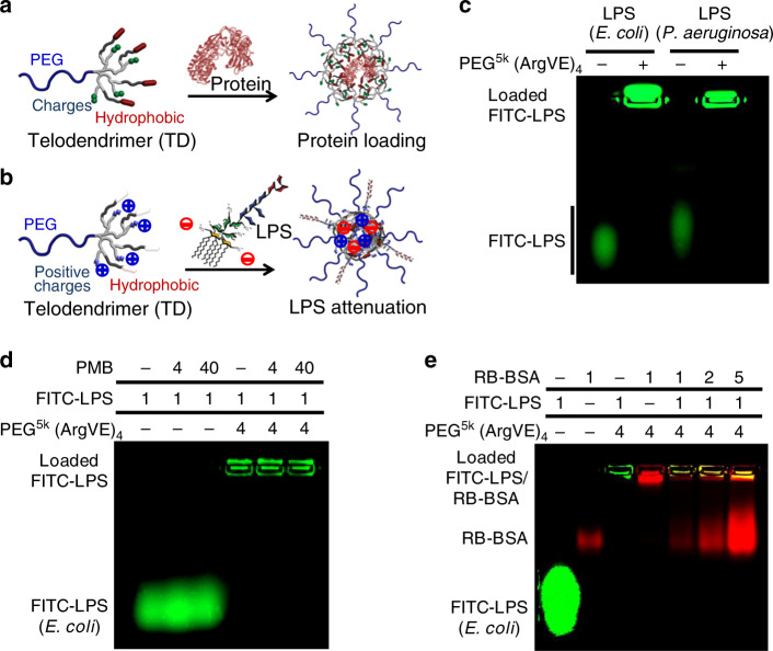
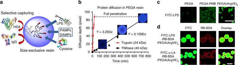
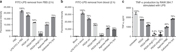
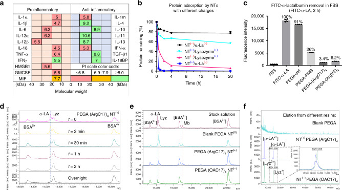
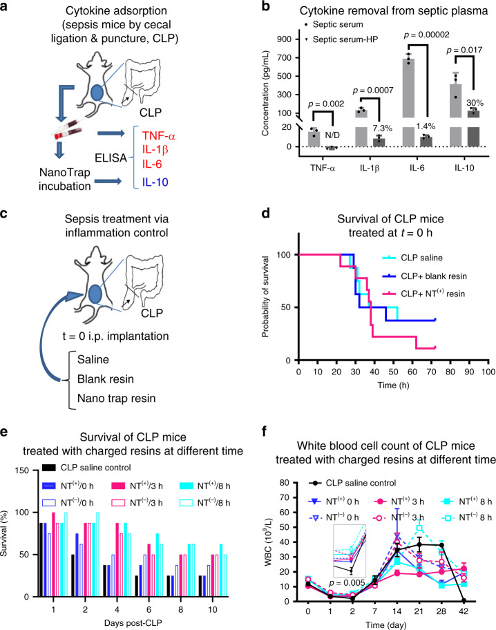
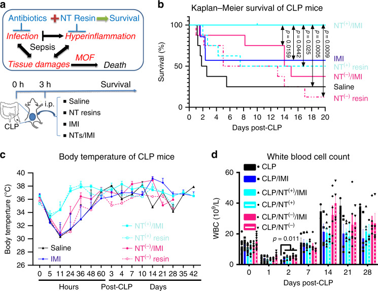
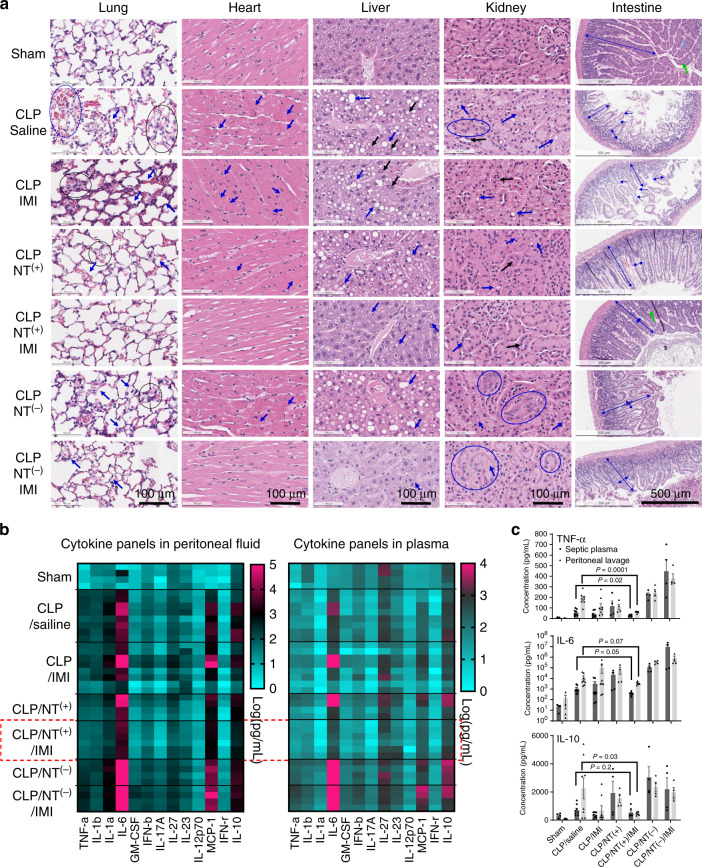
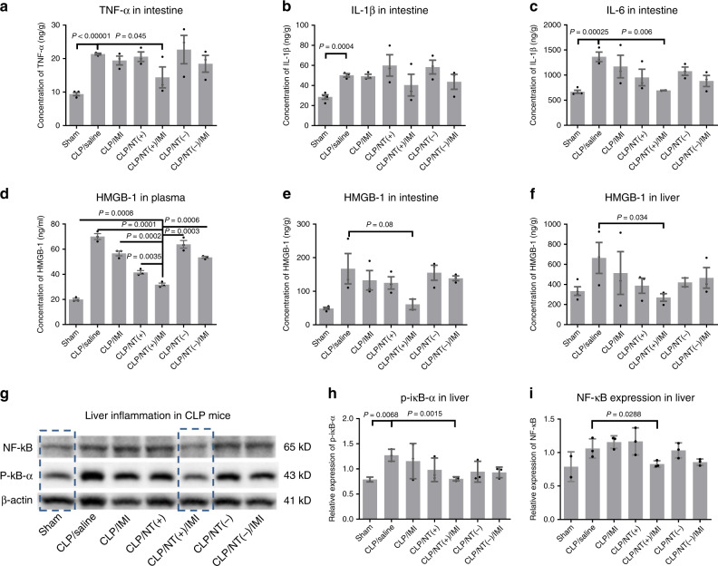
Targeting single mediators has failed to reduce the mortality of sepsis. We developed a telodendrimer (TD) nanotrap (NT) to capture various biomolecules via multivalent, hybrid and synergistic interactions. Here, we report that the immobilization of TD-NTs in size-exclusive hydrogel resins simultaneously adsorbs septic molecules, e.g. lipopolysaccharides (LPS), cytokines and damage- or pathogen-associated molecular patterns (DAMPs/PAMPs) from blood with high efficiency (92-99%). Distinct surface charges displayed on the majority of pro-inflammatory cytokines (negative) and anti-inflammatory cytokines (positive) allow for the selective capture via TD NTs with different charge moieties. The efficacy of NT therapies in murine sepsis is both time-dependent and charge-dependent. The combination of the optimized NT therapy with a moderate antibiotic treatment results in a 100% survival in severe septic mice by controlling both infection and hyperinflammation, whereas survival are only 50-60% with the individual therapies. Cytokine analysis, inflammatory gene activation and tissue histopathology strongly support the survival benefits of treatments.


Conflict of interest statement

C.S., L.W., and J.L. are inventors on a related patent submitted by State University of New York Upstate Medical University (International Application No. PCT/US2018/0497, published March 14, 2019). The remaining authors declare no competing interests.

Figures

Fig. 1
Fig. 1. Protein and LPS binding in telodendrimer nanoparticles.
Schematic illustration of a protein and b LPS captured by telodendrimer nanoparticles via the combination of charge and hydrophobic interactions. ce Agarose gel electrophoresis profiles reveal the complex formation of the FITC-labled LPS with telodendrimer PEG5k(ArgVE)4, as indicated by the lost mobility in migration (repeated independently a minimum of twice). c LPS originated from both E. coli and P. aeruginosa can be captured efficiently by the telodendrimer PEG5k(ArgVE)4. d PMB form less-stable complex with LPS in electrophoresis, which was also unable to dissociate LPS– PEG5k(ArgVE)4 nanocomplex with 40-fold excess in mass ratio. e The stability of LPS–PEG5k(ArgVE)4 nanocomplex was also observed to be stable in the presence of serum protein (RB-BSA) at different mass ratios.
Fig. 2
Fig. 2. TD NT resins for selective adsorption based on size effects.
a Schematic representation of selective LPS and cytokine removal by nanotrap-immobilized size-exclusive resin. b Kinetic diffusion of proteases Trypsin (24 kDa) and TNKase (45 kDa) in PEGA resins conjugated with the corresponding substrates: beads become fluorescent upon substrate cleavage by enzymes releasing the quencher (nitrotyrosine) from the fluorescent dye (Abz) (see Supplementary Fig. 9). c Fluorescent microscopy images showing the adsorption of FITC-LPS by blank PEGA, PEGA-PMB, and PEGA-(ArgVE)4 resins (scale bar: 500 µm). d Confocal images of PEGA-(ArgVE)4 resins incubated with the mixture of FITC-LPS or FITC-α-LA with RB-BSA (1:100 mass ratio), indicating the effective penetration of smaller LPS and α-LA and restriction of larger BSA binding on the surface of resin (scale bar: 100 µm). c, d Repeated independently a minimum of twice.
Fig. 3
Fig. 3. LPS attenuation by TD NT resins.
The removal of FITC-LPS (12.5 µg/mL) by nanotrap PEGA hydrogel resins in comparison with commercial LPS-removal resins (a) in FBS and in (b) whole blood after 2-h incubation, respectively. c The stock LPS solution was pretreated with or without different LPS-adsorption resins before adding to RAW 264.7 cells at an untreated LPS concentration of 500 ng per mL. TNF-α production in the culture medium after overnight incubation was analyzed by ELISA assay (n = 4, mean ± SEM). Source data are available in the Source Data file.
Fig. 4
Fig. 4. Selective protein adsorption in TD NT resins based on charge interactions.
a Summary of the molecular weights and isoelectric points (PIs) of key proinflammatory and anti-inflammatory cytokines in human sepsis. b The kinetic adsorption profiles of FITC-labeled α-LA(−) and lysozyme(+) by nanotrap (NT) resins with positive (arginine) or negative (oxalic acid (OA)) charges, respectively. NT(+): PEGA-(ArgC17)4; NT(−): PEGA-(OAC17)4 (n = 2, mean ± SD). c The adsorption efficiency of negatively charged FITC-labeled α-lactalbumin (α-LA, 14.2 kDa, PI: 4.5) by various positively charged resins in FBS after 2-h incubation (n = 4, mean ± SEM). d MALDI-TOF MS analysis of the protein mixture solution of α-LA (14.2 kDa, PI: 4.5, 0.5 mg per mL), lysozyme (Lyz, 14.4 kDa, PI: 10.7, 0.5 mg/mL), and BSA (66 kDa, PI: 4.2, 5 mg/mL) before and after incubation with NT(+) PEGA-(ArgC17)4 resin for different time at a bead/solution ratio of 1:10 v/v. Selective α-LA adsorption was observed. e MALDI-TOF MS analysis of the protein mixture solution of α-LA(−) (0.1 mg per mL), lysozyme(+) (Lyz, 0.1 mg per mL), myoglobin(0) (Mb, 0.1 mg per mL, PI: 7.1, 16.7 kDa), and BSA(−) (1 mg per mL, PI: 4.8–5.4, 66.4 kDa) before and after incubation with blank-acetylated PEGA, positive NT(+) PEGA-(ArgC17)4, and negative NT(−) PEGA-(OAC17)4 resins, respectively, at bead/solution ratio of 1:4 v/v. Charge-specific protein adsorption was observed. f MALDI-TOF MS analysis of proteins eluted from nanotrap resins after protein adsorption with 8 M urea: weak signals were observed from blank resin eluent, and strong signal and charge selectivity were observed for charged TD resins. Source data are available in the Source Data file.
Fig. 5
Fig. 5. Immune modulation via TD NT resins.
a Schematic illustration of sepsis mouse model induced by cecal ligation and punctuation (CLP) procedure, and septic blood was collected 24 h post CLP for ex vivo bead incubation. b Key cytokines TNF-α, IL-1β, IL-6, and IL-10 in plasma were quantified via ELISA assays before and after 2-h incubation (n = 3, duplicated measurements, mean ± SEM. Statistical significance was measured by paired one-sided Student’s test). c Experimental design of the spontaneous intraperitoneal treatments of CLP mice (n = 8) with saline, blank PEGA resin, or PEGA-(ArgC17)4 NT(+) resin in situ right after CLP procedure. d The survival of the animal was monitored for 3 days. Surprisingly, higher mortality was observed for spontaneous NT(+) treatment, which may be due to the spread of the infection with the disabled innate immune response by effective resin attenuation. e The survival of CLP mice (n = 8) treated with intraperitoneal implantation of NT(−) PEGA-(OAC17)4, or NT(+) PEGA-(ArgC17)4 at 0 h, 3 h, or 8 h after CLP without antibiotics. f The white blood cells were monitored for the survived CLP mice in the above treatments over 42 days (statistical significance was measured by unpaired one-sided Student’s test). Source data are available in the Source Data file.
Fig. 6
Fig. 6. Sepsis treatment via TD NT resins.
a Schematic illustration of the mechanism of sepsis associated with multiple organ failure (MOF) and death driven by both infection and hyperinflammation and our therapeutic strategy by combining antibiotics and immune modulation via NT resin scavenging to control both infection and hyperinflammation through i.p. implantation on day 3 post-CLP in mouse sepsis models. b The survival of CLP mice (n = 8, except for IMI group, n = 7) treated with NT(−) PEGA-(OAC17)4 or NT(+) PEGA-(ArgC17)4 3 h post CLP with or without antibiotics imipenem/cilastatin (IMI, 50/50 mg per kg body weight) treatments. As a result, the combination of IMI and NT(+) provides a 100% survival by treating both infection and hyperinflammation. After 1 week post CLP, animals in NT(−) and NT(−)/antibiotic groups were continuously sacrificed upon severe necrotic abscess observed (statistical significance was analyzed by Log-rank (Mantel–Cox) test, and a significant level of 0.05 was used for comparison). c The reduced hypothermia and fast body-temperature recovery were observed in the groups treated with NT(+) w/wo IMI. d The white blood cells (WBC) were monitored for the survived CLP mice in the above treatments over 4 weeks (n = 8), and reduce over time as shown in b. Significant low WBC was observed in CLP control group on day 2, and stable white blood cell counts were observed in NT(+)/IMI treatment groups with all mice survived (mean ± SEM, statistical significance was measured by unpaired one-sided Student’s test, and a significant level of 0.05 was used for comparison). Source data are available in the Source Data file.
Fig. 7
Fig. 7. Reduced tissue damage and attenuated hyperinflammation in severe sepsis.
a Tissue histology (n = 2) stained with hematoxylin and eosin for major organs of septic mice with the most severe hypothermia at 24 h post CLP in different groups. Histopathological findings: Lung: alveolar congestion (black circle), hemorrhage (blue circle), alveolar thickening, and hyaline membrane formation (arrow); Heart: intracellular edema and contraction bands (arrow), indicating early cell death; Liver: predominantly macrovascular steatosis (blue arrow) and nuclear inclusion (black arrow); Kidney: proximal tubular edema and vacuolization (blue arrow), luminal epithelial desquamation (black arrow), and epithelial nuclear enlargement (circle); Intestine: villous shortening, villous edema, villous necrosis (blue arrow), and loss of goblet cells (green arrow). b The heatmap of multiple inflammatory cytokine profiles, and c quantitative analysis of key cytokines (IL-6, TNF-α, IL-1β, and MCP-1) in both plasma and peritoneal lavage fluids in mice at 24 h post CLP in different treatment groups in comparison with sham mice (n = 4, mean ± SEM. Statistical significance was measured by unpaired one-sided Student’s test). Source data are available in the Source Data file.
Fig. 8
Fig. 8. Reduced tissue damage and organ inflammation in sepsis survivor.
ac Critical cytokine expression levels in the intestine. df HMGB-1 expression levels in the plasma, liver, and intestine as a DAMP indicator for tissue damage. gi The expression levels of NF-κB and its activation via phosphorylation of IκB-α in the liver as an indicator of remote organ inflammation (n = 3, mean ± SEM. Statistical significance was measured by unpaired one-sided Student’s test). Source data are available in the Source Data file.

References

    1. Zuev SM, Kingsmore SF, Gessler DDG. Sepsis progression and outcome: a dynamical model. Theor. Biol. Med. Model. 2006;3:8–8. - PMC - PubMed
    1. Iskander KN, et al. Sepsis: multiple abnormalities, heterogeneous responses, and evolving understanding. Physiol. Rev. 2013;93:1247–1288. - PMC - PubMed
    1. Gotts JE, Matthay MA. Sepsis: pathophysiology and clinical management. BMJ. 2016;353:i1585. - PubMed
    1. Harrison C. Sepsis: calming the cytokine storm. Nat. Rev. Drug Discov. 2010;9:360–361. - PubMed
    1. Wang H, Ma S. The cytokine storm and factors determining the sequence and severity of organ dysfunction in multiple organ dysfunction syndrome. Am. J. Emerg. Med. 2008;26:711–715. - PubMed

Publication types