. 2020 Nov;13(6):958-968.
doi: 10.1038/s41385-020-0318-2. Epub 2020 Jul 7.

IL-17A both initiates, via IFNγ suppression, and limits the pulmonary type-2 immune response to nematode infection

Affiliations

IL-17A both initiates, via IFNγ suppression, and limits the pulmonary type-2 immune response to nematode infection

Jesuthas Ajendra et al. Mucosal Immunol. 2020 Nov.

Abstract

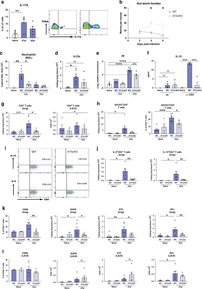
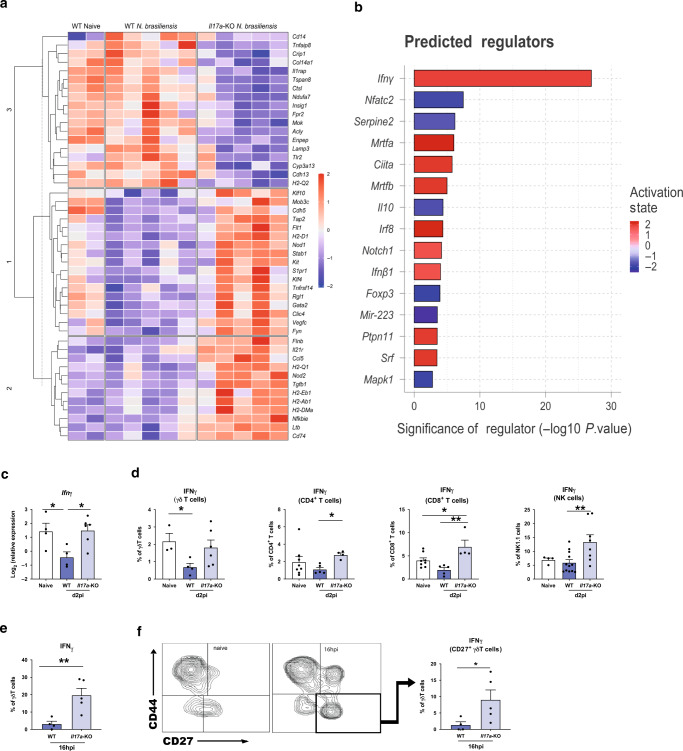
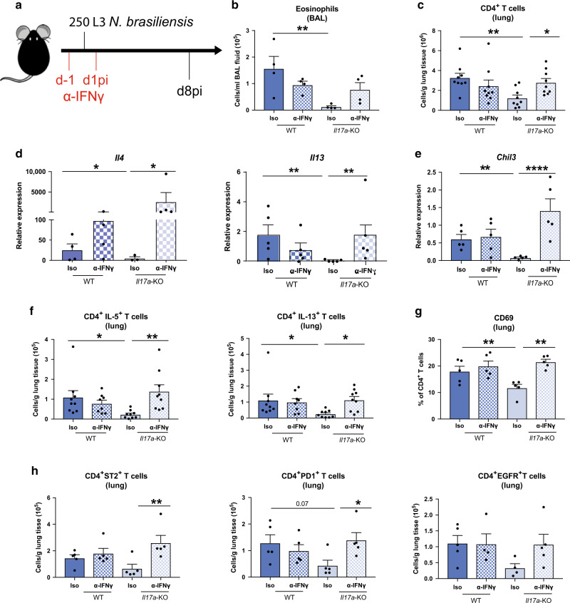
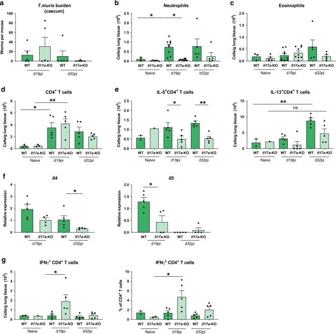
Nippostrongylus brasiliensis is a well-defined model of type-2 immunity but the early lung-migrating phase is dominated by innate IL-17A production. In this study, we confirm previous observations that Il17a-KO mice infected with N. brasiliensis exhibit an impaired type-2 immune response. Transcriptional profiling of the lung on day 2 of N. brasiliensis infection revealed an increased Ifng signature in Il17a-KO mice confirmed by enhanced IFNγ protein production in lung lymphocyte populations. Depletion of early IFNγ rescued type-2 immune responses in the Il17a-KO mice demonstrating that IL-17A-mediated suppression of IFNγ promotes type-2 immunity. Notably, later in infection, once the type-2 response was established, IL-17A limited the magnitude of the type-2 response. IL-17A regulation of type-2 immunity was lung-specific and infection with Trichuris muris revealed that IL-17A promotes a type-2 immune response in the lung even when infection is restricted to the intestine. Together our data reveal IL-17A as a major regulator of pulmonary type-2 immunity such that IL-17A supports early development of a protective type-2 response by suppression of IFNγ but subsequently limits excessive type-2 responses. A failure of this feedback loop may contribute to conditions such as severe asthma, characterised by combined elevation of IL-17 and type-2 cytokines.


Conflict of interest statement

The authors declare no competing interests.

Figures

Fig. 1
Fig. 1. Mice deficient in IL-17A mount a diminished type-2 response at the site of infection.
C57BL/6 (WT) and Il17a-KO mice were infected with 250 N. brasiliensis L3s and cell frequencies and cytokines were measured at different time points post infection compared with WT naïve mice. Frequencies of IL-17A-producing γδ T cells on d1pi and d2pi and representative flow-plot at d1pi (a). Worm burden in small intestine assessed in WT and Il17a-KO mice on days 2, 4 and 6 post N. brasiliensis infection (b). Absolute numbers of neutrophils (Ly6G+CD11b+) (c) and lung ILC2s (Lineage KLRG+CD127+CD90.2+ST2+) as measured via flow cytometry (d). Relative mRNA expression of cytokine Il4 in whole lung as quantified by qRT-PCR (log2 expression relative to actb (β-actin)) (e). Secreted IL-13 levels from unstimulated (us) or 72 h α-CD3 treated single-suspension lung cells (f). Absolute numbers of live CD4+ T cells in lung tissue and lung draining lymph nodes (LdLN) (g). Frequency and absolute numbers of GATA3+ CD4+ T cells in the lung (h). Representative flow-plots showing the frequency of IL-5 and IL-13 production by CD4+ T cells d7pi in lung from WT and Il17a-KO infected mice (i). Absolute numbers of IL-5+ and IL-13+ CD4+ T cells in the lung (j). Expression of CD69 on CD4+T cells and absolute numbers of EGFR+CD4+T cells, ST2+CD4+T cells and PD-1+CD4+T cells in lung (k) and LdLNs (l). Data are representative (mean ± s.e.m.) of at least 3 individual experiments (a, cl) or pooled data from three experiments (b). Data were tested for normality using Shapiro-Wilk test and analysed using one-way ANOVA followed by Sidak’s multiple comparisons test for selected groups. NS – not significant. Data in (e) were log2 transformed to achieve normal distribution and statistical tests were performed on transformed data *P < 0.05, **P < 0.01, ***P < 0.001, ****P < 0.0001.
Fig. 2
Fig. 2. Presence of IL-17A leads to a downregulation of early IFNγ during N. brasiliensis infection.
Whole lung RNA from C57BL/6 (WT) and Il17a-KO mice on d2pi with N. brasiliensis compared with WT naïve mice, were analysed by Nanostring. Unsupervised, hierarchically clustered heat map showing significant differentially expressed genes between infected WT, Il17a-KO mice and uninfected (naïve) WT (a). Top differentially regulated genes from (a) between infected WT and Il17a-KO mice were run in Ingenuity pathway analyzer, with top predicted regulators shown in (b). Relative expression of Ifng in whole lung of naïve WT and d2 N. brasiliensis infected WT and Il17a-KO mice (log2 expression relative to actb (β-actin)) (c). Frequencies of IFNγ+ γδ T cells, CD4+ T cells, CD8+ T cells and NK cells in WT and Il17a-KO mice d2pi compared with WT naïve mice as assessed by flow cytometry (d). Frequency of IFNγ+ γδ T cells 16 h post N. brasiliensis infection in WT and Il17a-KO mice (e). Representative flow plots showing CD44 and CD27 γδ T cell subsets in naïve mice and mice 16 h post N. brasiliensis infection as well as frequency of IFNγ+ CD27+ γδ T cells 16 h post N. brasiliensis infection in WT and Il17a-KO mice (f). Data (cf) are expressed as mean ± s.e.m. and are representative of at least 2 individual experiments with at least three mice per infected group. Data were tested for normality using Shapiro-Wilk test and analysed using one-way ANOVA followed by Sidak’s multiple comparisons test for selected groups or student’s t test. *P < 0.05, **P < 0.01.
Fig. 3
Fig. 3. IFNγ neutralization in Il17a-KO mice rescues the impaired type-2 immune response.
C57BL/6 (WT) and Il17a-KO mice and were treated with α-IFNγ or isotype control on days −1 and 1pi with 250 L3 larvae of N. brasiliensis (a). Absolute numbers of eosinophils per mL of BAL (b) or CD4+ T cells per gram lung tissue (c) as measured via flow cytometry on d8pi. Relative mRNA expression of type-2 cytokines Il4 and Il13 (d) and type-2 marker Chil3 (e) from whole lung. Absolute numbers of IL-5+ and IL-13+ CD4+ T cells (f). Frequency of CD69+ CD4+ T cells (g) and numbers of EGFR+, ST2+ and PD1+ CD4+ T cells per gram lung tissue (h). Data (b, d, eg) are representative (mean ± s.e.m.) of two individual experiments with at least three mice per group (per experiment) or pooled data from two experiments (c, f). Data were tested for normality using Shapiro-Wilk test and analysed using one-way ANOVA followed by Sidak’s multiple comparisons test for selected groups. Data in (d, e) were log2 transformed to achieve normal distribution and statistical tests were performed on transformed data. *P < 0.05, **P < 0.01, ***P < 0.001.
Fig. 4
Fig. 4. Late stage IL-17A suppresses type-2 immune responses during N.brasiliensis infection.
C57BL/6 WT mice were treated with α-IL-17A or isotype control on days 3, 4, and 5pi with 250 L3 N. brasiliensis and responses measured at d7pi compared with uninfected (naïve) mice (a). Cells per gram lung tissue and frequency of ILC2s in live lung cells (b). Absolute number of IL-5+ and IL-13+ ILC2s per gram lung tissue (n = 5 for naïve, n = 11–12 for d7 N. brasiliensis infected groups) (c). Absolute numbers of CD4+ T cells (d) and IL-5+ and IL-13+ CD4+ T cells per gram of lung tissue (n = 3 for naïve and n = 6 for d7 N. brasiliensis infected groups) (e). Data pooled from two independent experiments (b, c) or are representative (mean ± s.e.m.) of 3 individual experiments with at least 3 mice per group (per experiment) (d, e). Data were tested for normality using Shapiro-Wilk test and analysed by a one-way ANOVA followed by Sidak’s multiple comparisons test for selected groups. *P < 0.05, **P < 0.01, ***P < 0.001.
Fig. 5
Fig. 5. Lack of IL-17A impairs concurrent type-2 immune responses in the lung following infection with Trichuris muris.
C57BL/6 WT and Il17a-KO mice were infected with a high dose of T. muris and immune parameters investigated at d19 and d32pi compared with uninfected (naïve) C57BL/6 WT and Il17a-KO mice. Worms counts in the caecum (a). Absolute numbers of neutrophils (b), eosinophils (c) and CD4+ T cells (d) per gram of lung at d19pi and d32pi compared with naïve mice. Absolute numbers of IL-5+ and IL-13+ CD4 + T cells per gram of lung tissue (e). Relative mRNA expression of cytokines Il4 and Il5 from whole lung (log2 expression relative to actb (β-actin)) of infected mice (f). Absolute numbers and frequency of IFNγ+ CD4+ T cells per gram of lung tissue (g). Data are expressed as mean ± s.e.m. and are representative of 3 individual experiments with at least four mice per infected group and one mouse per control group. Data were tested for normality using Shapiro-Wilk test and analysed with one-way ANOVA followed by Sidak’s multiple comparisons test for selected groups. Data in (f) were log2 transformed to achieve normal distribution and statistical tests were performed on transformed data *P < 0.05, **P < 0.01.

Comment in

  • IL-17A moonlighting in lung type 2 immunity.
    Ruscher R, Giacomin PR. Ruscher R, et al. Mucosal Immunol. 2020 Nov;13(6):849-851. doi: 10.1038/s41385-020-0329-z. Epub 2020 Jul 29. Mucosal Immunol. 2020. PMID: 32728131 Free PMC article. No abstract available.

References

    1. Papotto PH, Ribot JC, Silva-Santos B. IL-17 + γδ T cells as kick-starters of inflammation. Nat. Immunol. 2017;18:604–611. - PubMed
    1. Iwakura Y, Ishigame H, Saijo S, Nakae S. Functional specialization of interleukin-17 family members. Immunity. 2011;34:149–162. - PubMed
    1. Allen JE, Maizels RM. Diversity and dialogue in immunity to helminths. Nat. Rev. Immunol. 2011;11:375–388. - PubMed
    1. Mbow M, et al. T-helper 17 cells are associated with pathology in human schistosomiasis. J. Infect. Dis. 2013;207:186–195. - PMC - PubMed
    1. Rutitzky LI, Stadecker MJ. Exacerbated egg-induced immunopathology in murine Schistosoma mansoni infection is primarily mediated by IL-17 and restrained by IFN-γ. Eur. J. Immunol. 2011;41:2677–2687. - PMC - PubMed

Publication types