. 2020 Jul 7;10(1):11163.
doi: 10.1038/s41598-020-68074-3.

Tumor microenvironment characterization in head and neck cancer identifies prognostic and immunotherapeutically relevant gene signatures

Affiliations

Tumor microenvironment characterization in head and neck cancer identifies prognostic and immunotherapeutically relevant gene signatures

Mengqi Huo et al. Sci Rep. .

Abstract

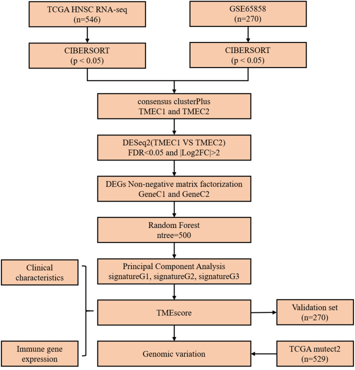
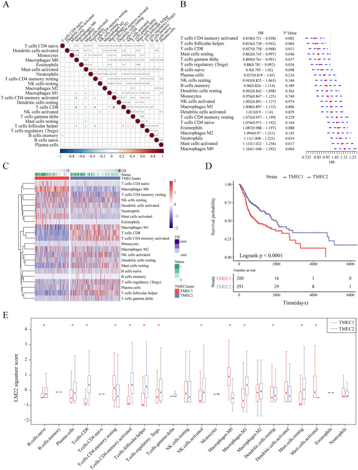
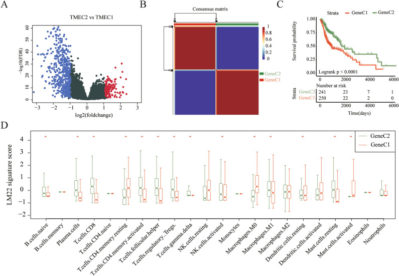
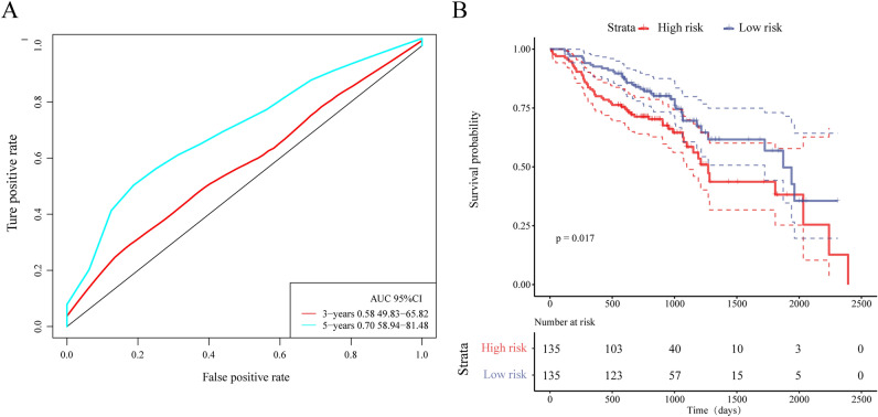
The tumor microenvironment (TME) is of great clinical significance for predicting the therapeutic effect of tumors. Nonetheless, there was no systematic analysis of cellular interactions in the TME of head and neck cancer (HNSC). This study used gene expression data from 816 patients with HNSC to analyze the scores of 22 immune cells. On this basis, we have established a novel TMEscore-based prognostic risk model. The relationship between TMEscore and clinical and genomic characteristics was analyzed. The sample was divided into risk-H and risk-L groups based on the prognosis risk model of TMEscore, with significant differences in overall survival between the two groups (log rank p < 0.001). In terms of clinical features, the TMEscore is closely related to the T staging, Grade, and HPV. As for genomic characteristics, the genomic features of the Risk-H samples are a low expression of immune-related genes and high-frequency mutations of TP53 and CEP152. This model was validated in an external test set, in which the prognosis for Risk-H group and Risk-L group was also significantly different (log rank p = 0.017). A quantitative method of TME infiltration pattern is established, which may be a potential predictor of HNSC prognosis.


Conflict of interest statement

The authors declare no competing interests.

Figures

Figure 1
Figure 1
Flow chart of the experiment.
Figure 2
Figure 2
(A) The correlation of 22 immune cells of TME, in which the dot size and color indicate correlation, blue indicates negative correlation, red indicates positive correlation, white area in the figure indicates insignificant, and the number in the upper right corner of the figure represents correlation coefficient; (B) Forest map of 22 immune cells of TME; (C) The heat map of 22 immune cell scores. Red indicates high scores, blue indicates low scores; (D) KM survival curves of two types of TMEC; (E) Distribution box plot of 22 immune cell scores in two types of TMEC, * indicates significant difference.
Figure 3
Figure 3
(A) Volcano map of DEGs between TMEC1 and TMEC2. (B) Consistency matrix heatmap of NMF algorithm; (C) KM survival curve of GeneC1 and GeneC2; (D) Box plot of 22 immune cell scores in GeneC1 and GeneC2.
Figure 4
Figure 4
(A) GO enrichment analysis of 160 genes; (B) KEGG enrichment analysis of 160 genes; (C) k-means clustering results of 160 genes; (D) heat map of 160 gene expression levels; (E) Comparison of TMEscore between GeneC1 and GeneC2; (F) TMEscore distribution of GeneC1 and GeneC2; (G) KM survival curves for the Risk-H and Risk-L.
Figure 5
Figure 5
(A) heatmap of immune activation genes expression of TCGA samples; (B) heatmap of the immune checkpoint gene expression of TCGA samples; (C) heatmap of TGF pathway genes expression of TCGA samples.
Figure 6
Figure 6
Relationship between TMEscore and genomic mutations. The horizontal axis represents the sample, the vertical axis represents the gene, the black rectangle represents the mutation, and the gray represents the unmutated.
Figure 7
Figure 7
(A) ROC curve and AUC values in the test set; (B) KM survival curves for the two groups of samples in the test set.

References

    1. Leemans CR, Snijders PJ, Brakenhoff RH. The molecular landscape of head and neck cancer. Nat. Rev. Cancer. 2018;18(5):269. doi: 10.1038/nrc.2018.11. - DOI - PubMed
    1. Du E, Mazul AL, Farquhar D, Brennan P, Anantharaman D, Abedi-Ardekani B, Weissler MC, Hayes DN, Olshan AF, Zevallos JP. Long-term survival in head and neck cancer: impact of site, stage, smoking, and human papillomavirus status. Laryngoscope. 2019;129(11):2506–2513. doi: 10.1002/lary.27807. - DOI - PMC - PubMed
    1. Elkashty, O. A. et al. Cancer stem cells enrichment with surface markers CD271 and CD44 in human head and neck squamous cell carcinomas. Carcinogenesis. 11(19), pii: bgz182 (2019). - PMC - PubMed
    1. Vargo JA, et al. A multi-institutional comparison of SBRT and IMRT for definitive reirradiation of recurrent or second primary head and neck cancer. Int. J. Radiat. Oncol. Biol. Phys. 2018;100(3):595–605. doi: 10.1016/j.ijrobp.2017.04.017. - DOI - PMC - PubMed
    1. Noronha, V. et al. Once-a-week versus once-every-3-weeks cisplatin chemoradiation for locally advanced head and neck cancer: a phase III randomized noninferiority trial. J. Clin. Oncol. 2017749457 (2017). - PubMed

Publication types