. 2020 Jun 29:15:4639-4657.
doi: 10.2147/IJN.S242359. eCollection 2020.

Magnetically Directed Enzyme/Prodrug Prostate Cancer Therapy Based on β-Glucosidase/Amygdalin

Affiliations

Magnetically Directed Enzyme/Prodrug Prostate Cancer Therapy Based on β-Glucosidase/Amygdalin

Jie Zhou et al. Int J Nanomedicine. .

Abstract

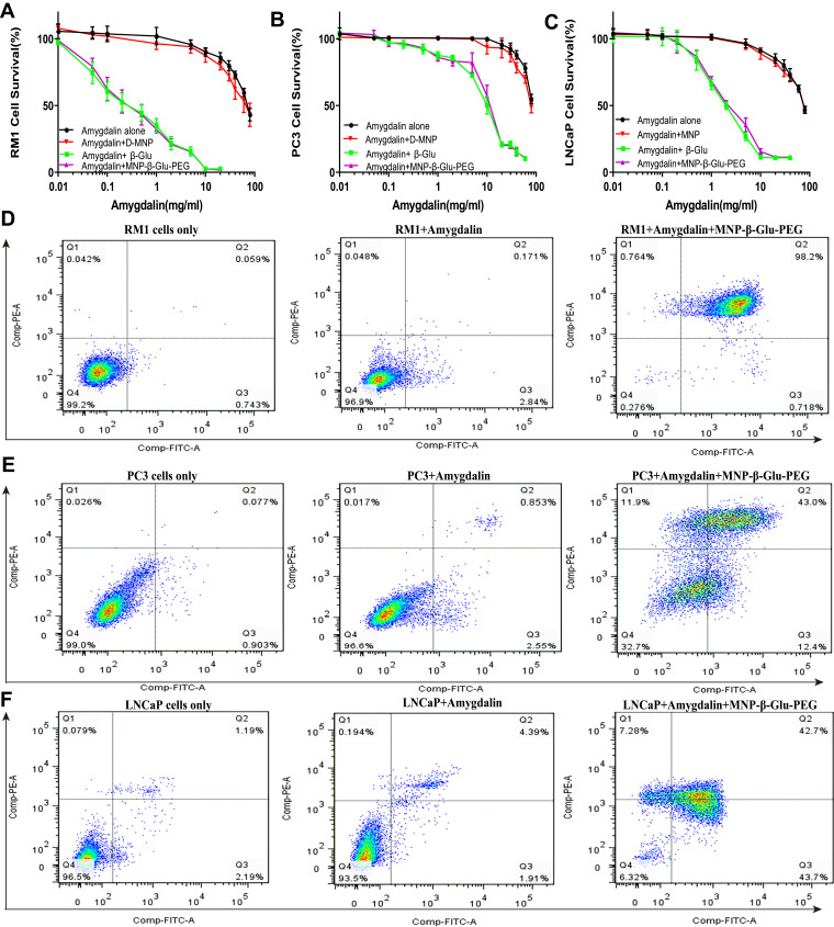
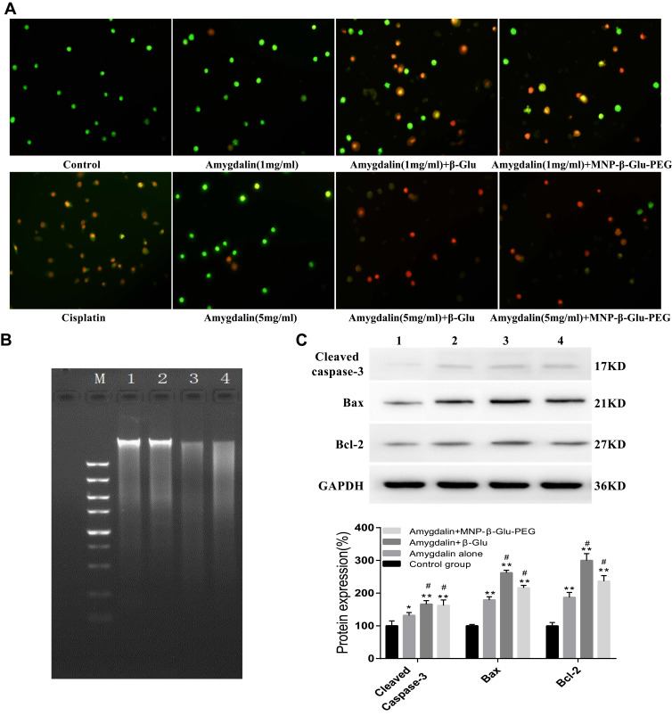
Background: β-Glucosidase (β-Glu) can activate amygdalin to kill prostate cancer cells, but the poor specificity of this killing effect may cause severe general toxicity in vivo, limiting the practical clinical application of this approach.

Materials and methods: In this study, starch-coated magnetic nanoparticles (MNPs) were successively conjugated with β-Glu and polyethylene glycol (PEG) by chemical coupling methods. Cell experiments were used to confirm the effects of immobilized β-Glu on amygdalin-mediated prostate cancer cell death in vitro. Subcutaneous xenograft models were used to carry out the targeting experiment and magnetically directed enzyme/prodrug therapy (MDEPT) experiment in vivo.

Results: Immobilized β-Glu activated amygdalin-mediated prostate cancer cell death. Tumor-targeting studies showed that PEG modification increased the accumulation of β-Glu-loaded nanoparticles in targeted tumor tissue subjected to an external magnetic field and decreased the accumulation of the nanoparticles in the liver and spleen. Based on an enzyme activity of up to 134.89 ± 14.18mU/g tissue in the targeted tumor tissue, PEG-β-Glu-MNP/amygdalin combination therapy achieved targeted activation of amygdalin and tumor growth inhibition in C57BL/6 mice bearing RM1 xenografts. Safety evaluations showed that this strategy had some impact on liver and heart function but did not cause obvious organ damage.

Conclusion: All findings indicate that this magnetically directed enzyme/prodrug therapy strategy has the potential to become a promising new approach for targeted therapy of prostate cancer.

Keywords: amygdalin; magnetic nanoparticles; magnetically directed enzyme/prodrug therapy; prostate cancer; β-glucosidase.


Conflict of interest statement

The authors report no conflicts of interest in the work

Figures

Figure 1
Figure 1
Preparation process and characterization analyses of MNP-β-Glu-PEG. (A) Schematic diagram of the preparation process. (B) Representative TEM images of D-MNP, MNP-β-Glu and MNP-β-Glu-PEG. Various particles showed no significant morphological differences. (C) Infrared spectrum analysis. A characteristic β-Glu peak located at 1540 cm−1 is visible in the MNP-β-Glu spectrum. Characteristic β-Glu and PEG peaks located at 1540 cm−1 and 1100 cm−1 are visible in the MNP-β-Glu-PEG spectrum. (D) The magnetization properties of D-MNP, MNP-β-Glu and MNP-β-Glu-PEG. Neither hysteresis nor remnant magnetization was observed for all particles. (E) Magnetophoretic mobility curves for D-MNP, MNP-β-Glu and MNP-β-Glu-PEG (n=3). Relative Fe concentrations for the nanoparticle suspensions indicate the ratio of noncaptured nanoparticles in the magnetic field.
Figure 2
Figure 2
Proliferation inhibition and apoptosis analyses of prostate cancer cells. (AC) The growth inhibition effects of amygdalin, amygdalin/MNP, amygdalin/β-Glu and amygdalin/MNP-β-Glu-PEG on RM1 cells, PC3 cells and LNCaP cells. Data show the mean±standard deviation of measurements conducted in quadruplicate. (DF) Representative annexin V-FITC/PI flow cytometry analysis of RM1, PC3 and LNCaP cells after amygdalin or amygdalin/MNP-β-Glu-PEG treatment. Cells were defined as viable (PI, annexin V, lower left quadrant), early apoptotic (PI, annexin V+, lower right quadrant), late-stage apoptotic (PI+, annexin V+, upper right quadrant) or necrotic (PI+, annexin V, upper left quadrant).
Figure 3
Figure 3
Mechanism of prostate cancer cell death resulting from amygdalin combined with β-Glu or MNP-β-Glu-PEG treatment. (A) AO/EB staining analyzing the effects of amygdalin combined with β-Glu or MNP-β-Glu-PEG on prostate cancer. Green indicates live cells; cells with orange pyknotic nuclei in the cisplatin group are apoptotic cells; yellow swollen cells in the combined drug administration groups (β-Glu/amygdalin or MNP-β-Glu-PEG/amygdalin) are necrotic cells; and red indicates cells in the late phases of apoptosis/necrosis. 400×. (B) Band 1 is the control group (PBS solution), band 2 is the amygdalin group, band 3 is the amygdalin + β-Glu group, and band 4 is the amygdalin + MNP-β-Glu-PEG group. (C) 1 is the control group, 2 is the amygdalin group, 3 is the amygdalin + β-Glu group, and 4 is the amygdalin + MNP-β-Glu-PEG group. The relative amounts of protein expression were calculated according the grayscale values and were showed in the histogram. (compared with the control group, *p < 0.05, **p < 0.01, compared with the amygdalin group, #p < 0.01, n=4).
Figure 4
Figure 4
Targeted accumulation of β-Glu-loaded MNPs at tumor sites. (A) Representative MR images of particle aggregation at subcutaneous tumor sites with an external magnetic field after D-MNP, β-Glu-MNP or MNP-β-Glu-PEG administration. The red arrows indicate the locations of subcutaneous tumors. Significant hypodense shadows appear in the posttargeted tumor tissue of the MNP-β-Glu-PEG group. (B) Prussian blue staining of tumor tissues after D-MNP, MNP-β-Glu or MNP-β-Glu-PEG administration. Blue staining indicates iron particles. Scale bar: 50μm. (C) Fe concentrations and (D) enzymatic activities of targeted/nontargeted tumor tissues after D-MNP, β-Glu, MNP-β-Glu or MNP-β-Glu-PEG administration (compared with the targeted MNP-β-Glu-PEG group, *p < 0.01, n=6).
Figure 5
Figure 5
Organ distribution of MNP-β-Glu and MNP-β-Glu-PEG. (A) Prussian blue staining of liver and spleen tissue sections after MNP-β-Glu or MNP-β-Glu-PEG administration. Scale bar: 50μm. (B) ESR analyses of liver and spleen tissues from the MNP-β-Glu and MNP-β-Glu-PEG groups (compared with MNP-β-Glu-PEG group/liver tissue,*p < 0.01, n=6; compared with MNP-β-Glu-PEG group/spleen tissue, #p < 0.01, n=6). (C) Enzyme activity analyses of liver and spleen tissues from the MNP-β-Glu and MNP-β-Glu-PEG groups (compared with MNP-β-Glu-PEG group/liver tissue, *p < 0.01, n=6; compared with MNP-β-Glu-PEG group/spleen tissue, #p < 0.01, n=6).
Figure 6
Figure 6
Magnetically directed enzyme/prodrug therapy with MNP-β-Glu-PEG/amygdalin in vivo. (A) A representative image showing mice with RM1 xenografts treated with AMY, β-Glu/AMY, MNP-β-Glu-PEG/AMY, MNP-β-Glu/MT/AMY or MNP-β-Glu-PEG/MT/AMY (17 days after treatment). (B) Volume (mm3) of the RM1 xenograft in mice treated according to the scheme described above. Tumor sizes were measured using a caliper on the indicated days (*p < 0.001; based on a two-tailed t-test, assuming unequal variance, n=6). (C) Representative apoptosis images and cell apoptosis index analyses of tumor tissues for all groups (compared with the control group, *p < 0.01; compared with the MNP-β-Glu-PEG/MT/AMY group, **p < 0.01). The apoptotic cells are brown. Scale bar: 50μm. β-Glu, MNP-β-Glu and MNP-β-Glu-PEG were administered via the tail vein, and the magnetic targeting time was 2 hours. Abbreviations: AMY, amygdalin; β-Glu, β-glucosidase; MT, magnetic targeting.
Figure 7
Figure 7
Toxic effects of combined administration to mice. (A) Body weight changes after administration (17 days after treatment, n=6). (B) Effects of administration on the kidney function (BUN, Cr) of mice (compared with the control group, *p > 0.05, n=6). (C) Effects of administration on the liver function (ALT, AST) of mice (compared with the control group, *p > 0.05, **p < 0.01, n=6). (D) Effects of administration on the heart function (LDH, CK) of mice (compared with the control group, *p > 0.05, ** p < 0.01, n=6).

Similar articles

Cited by

References

    1. Siegel RL, Miller KD, Jemal A. Cancer statistics, 2018. CA Cancer J Clin. 2018;68(1):7–30. doi:10.3322/caac.21442 - DOI - PubMed
    1. Chen W, Zheng R, Baade PD, et al. Cancer statistics in China, 2015. CA Cancer J Clin. 2016;66(2):115–132. doi:10.3322/caac.21338 - DOI - PubMed
    1. Cornford P, Bellmunt J, Bolla M, et al. EAU-ESTRO-SIOG guidelines on prostate cancer. Part II: treatment of relapsing, metastatic, and castration-resistant prostate cancer. Eur Urol. 2017;71(4):630–642. doi:10.1016/j.eururo.2016.08.002 - DOI - PubMed
    1. Aloysius H, Hu L. Targeted prodrug approaches for hormone refractory prostate cancer. Med Res Rev. 2015;35(3):554–585. doi:10.1002/med.21333 - DOI - PubMed
    1. Blaheta RA, Nelson K, Haferkamp A, Juengel E. Amygdalin, quackery or cure? Phytomedicine. 2016;23(4):367–376. doi:10.1016/j.phymed.2016.02.004 - DOI - PubMed

MeSH terms