. 2020 Jun 24:14:1178223420924634.
doi: 10.1177/1178223420924634. eCollection 2020.

Novel Melatonin, Estrogen, and Progesterone Hormone Therapy Demonstrates Anti-Cancer Actions in MCF-7 and MDA-MB-231 Breast Cancer Cells

Affiliations

Novel Melatonin, Estrogen, and Progesterone Hormone Therapy Demonstrates Anti-Cancer Actions in MCF-7 and MDA-MB-231 Breast Cancer Cells

Mahmud Hasan et al. Breast Cancer (Auckl). .

Abstract

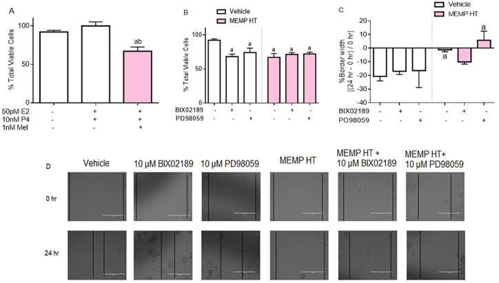
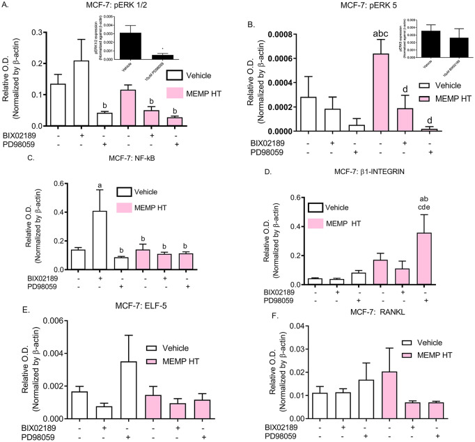
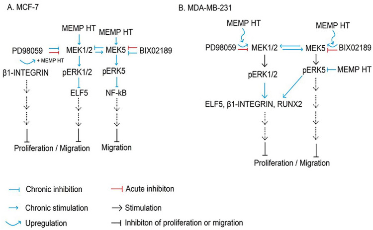
A novel melatonin, estrogen, and progesterone hormone therapy was developed as a safe bio-identical alternative hormone therapy for menopausal women based on the Women's Health Initiative findings that PremPro™ increased breast cancer risk and mortality of all types of breast cancer in postmenopausal women. For HER2 breast cancer, melatonin, estrogen, and progesterone delayed tumor onset and reduced tumor incidence in neu female mice. For other breast cancers, its actions are unknown. In this study, melatonin, estrogen, and progesterone hormone therapy were assessed in human ER+ (MCF-7) and triple negative breast cancer (MDA-MB-231) cells, and found to decrease proliferation and migration of both breast cancer lines. Inhibition of MEK1/2 and 5 using PD98059 and BIX02189, respectively, inhibited proliferation and migration in MDA-MB-231 cells and proliferation in MCF-7 cells; however, when combined with melatonin, estrogen, and progesterone, BIX02189 blocked melatonin, estrogen, and progesterone-mediated inhibition of migration in MCF-7 cells and induced Elf-5. For MDA-MB-231 cells, BIX02189 combined with melatonin, estrogen, and progesterone inhibited proliferation and increased pERK1/2 and β1-INTEGRIN; levels of pERK5 remained low/nearly absent in both breast cancer lines. These findings demonstrate novel anti-cancer actions of melatonin, estrogen, and progesterone in ER+ and triple negative breast cancer cells through intricate MEK1/2- and MEK5-associated signaling cascades that favor anti-proliferation and anti-migration.

Keywords: ERK1/2; ERK5; Elf-5; MCF-7; MDA-MB-231; Melatonin receptors; NF-κB; PremPro; RANKL; RUNX2; breast cancer; estrogen; melatonin; progesterone; β1-INTEGRIN.


Conflict of interest statement

Declaration of Conflicting Interests:The author(s) declared the following potential conflicts of interest with respect to the research, authorship, and/or publication of this article: P.A.W.-E., inventor, Duquesne University, assignee. Combination hormone replacement therapy (HRT) and melatonin to prevent and treat mammary cancer. US Patent 8618083 (2013) and 9370526 (2016). The remaining authors declare that the research was conducted in the absence of any commercial or financial relationships that could be construed as a potential conflict of interest.

Figures

Figure 1.
Figure 1.
Melatonin and estrogen receptor binding in MCF-7 and MDA-MB-231 cells, and the effect of E2, P4, and melatonin on cell proliferation and migration. Total 2-[125I]-iodomelatonin and [3H]-estradiol binding to ERs (A) and melatonin receptors (B), respectively, expressed in MCF-7 and MDA-MB-231 cells. Cells were exposed to E2 (1 pM, 1 nM, 100 nM, 1 µM, and 10 µM), P4 (1 pM, 1 nM, 100 nM, 1 µM, and 10 µM), or Mel (1 pM, 1 nM, 100 nM, 1 µM, and 10 µM) for 24 hours and then counted using trypan blue in MCF-7 (C) and MDA-MB-231 (D) cells. Data represent the mean ± SEM from 3 independent experiments. Data were analyzed by one-way ANOVA followed by Newman-Keuls’ post hoc t-test, where a = P < .05 vs vehicle; b = P < .05 vs 1 pM E2; c = P < .05 vs 1 nM E2; d = P < .05 vs 100 nM E2; e = P < .05 vs 1 µM E2; f = P < .05 vs 1 pM Mel; g = P < .05 vs 1 nM Mel. ANOVA indicates analysis of variance; SEM, standard error of the mean.
Figure 2.
Figure 2.
Effect of MEMP HT, MEK1/2, and MEK5 on MCF-7 cell proliferation and migration. The MEMP HT and inhibitors were used on MCF-7 cell viability (A, B) and migration (C). The representative images of the scratches were given for each treatment 0 and 24 hours (D). The MEK1/2 inhibitor (10 µM PD98059) or the MEK5 inhibitor (10 µM BIX02189) was added to the treatments. For migration assays, % border width ([24 hours – 0 hours] / 0 hours) is represented on the y-axis, and for the proliferation assays, % of living cells relative to the total number of cells counted is represented on the y-axis. Data were analyzed by one-way ANOVA followed by Newman-Keuls’ post hoc t-test where significance was defined as P < .05. Each bar represents the mean ± SEM from 3 independent experiments; a = P < .05 vs vehicle; b = P < .05 vs 50 pM E2 + 10 nM P4. ANOVA indicates analysis of variance; MEMP HT, melatonin, estrogen, and progesterone hormone therapy; SEM, standard error of the mean.
Figure 3.
Figure 3.
Effect of MEMP HT, MEK1/2, and MEK5 on MDA-MB-231 cell proliferation and migration. The MEMP HT and inhibitors were used on MDA-MB-231 cell viability (A, B) and migration (C). The MEK1/2 inhibitor (10 µM PD98059) or the MEK5 inhibitor (10 µM BIX02189) was added to the treatments. The representative images of the scratches were given for each treatment 0 and 24 hours (D). For migration assays, % border width ([24 hours – 0 hours] / 0 hours) is represented on the y-axis, and for the proliferation assays, % of living cells relative to the total number of cells counted is represented on the y-axis. Data were analyzed by one-way ANOVA followed by Newman-Keuls’ post hoc t-test where significance was defined as P < .05. Each bar represents the mean ± SEM from 3 independent experiments; a = P < .05 vs vehicle; b = P < .05 vs 10 µM BIX02189; d = P < .05 vs MEMP HT. ANOVA indicates analysis of variance; MEMP HT, melatonin, estrogen, and progesterone hormone therapy; SEM, standard error of the mean.
Figure 4.
Figure 4.
Treatment effects on pERK1/2, pERK5, NF-κB, β1-INTEGRIN, Elf-5, and RANKL expression in MCF-7 cells. MCF-7 cells were exposed to vehicle or MEMP HT for 24 hours and their effects on pERK1/2 (A), pERK5 (B), NF-κB (C), β1-INTEGRIN (D), Elf-5 (E), or RANKL (F) expression in MCF-7 cells by western blot analysis. The MEK1/2 inhibitor (10 µM PD98059) or the MEK5 inhibitor (10 µM BIX02189) was added to the treatments. Treatment groups are denoted on the x-axis and protein expression (normalized by β-actin) on the y-axis. Data were analyzed by one-way ANOVA followed by Newman-Keuls’ post hoc t-test where significance was defined as P < .05; a = P < .05 vs vehicle; b = P < .05 vs 10 µM BIX02189; c = P < .05 vs 10 µM PD98059; d = P < .05 vs MEMP HT; e = P < .05 vs MEMP HT + 10 µM BIX02189. Each bar represents the mean ± SEM from 3 independent experiments (please see Table 1 for band images). ANOVA indicates analysis of variance; MEMP HT, melatonin, estrogen, and progesterone hormone therapy; SEM, standard error of the mean.
Figure 5.
Figure 5.
Treatment effects on pERK1/2, pERK5, NF-κB, β1-INTEGRIN, Elf-5, RANKL, and RUNX2 expression in MDA-MB-231 cells. MDA-MB-231 cells were exposed to vehicle or MEMP HT for 24 hours and its effects on pERK1/2 (A), pERK5 (B), NF-κB (C), β1-INTEGRIN (D), Elf-5 (E), RANKL (F), or RUNX2 (G) expression in MDA-MB-231 cells by western blot analysis. The MEK1/2 inhibitor (10 µM PD98059) or the MEK5 inhibitor (10 µM BIX02189) was added to the treatments. Treatment groups are denoted on the x-axis and protein expression (normalized by β-actin) on the y-axis. Data were analyzed by one-way ANOVA followed by Newman-Keuls’ post hoc t-test where significance was defined as P < .05; a = P < .05 vs vehicle; b = P < .05 vs 10 µM BIX02189; c = P < .05 vs 10 µM PD98059; d = P < .05 vs MEMP HT; e = P < .05 vs MEMP HT + 10 µM BIX02189. Each bar represents the mean ± SEM from 3 independent experiments (please see Table 2 for band images). ANOVA indicates analysis of variance; MEMP HT, melatonin, estrogen, and progesterone hormone therapy; SEM, standard error of the mean.
Figure 6.
Figure 6.
Summary of mechanisms of action underlying MEMP HT in BC cells. As depicted, MEMP HT inhibits proliferation and migration in MCF-7 cells (A) and MDA-MB-231 BC cells (B). Acute (1 hour) or chronic (24 hours) exposure to the MEK1/2 (PD98059) or MEK5 (BIX02189) inhibitors in MCF-7 cells decreased pERK1/2 and pERK5 (trends), respectively, leading to decreases in NF-κB and increases in Elf-5. In MCF-7 cells, MEMP HT alone increased pERK5 in MCF-7 cells, which was blocked in the presence of 02189 and PD98059, suggesting that both MEK1/2 and 5 modulate MEMP HT’s actions on pERK5. β1-INTEGRIN levels increased only when MEMP HT was combined with PD98059 suggesting that a co-(up)-regulation of β1-INTEGRIN occurred between MEMP HT and MEK1/2. For NF-κB, the BIX02189-mediated up-regulation of NF-κB was blocked in the presence of MEMP HT, suggesting that the MEK5 pathway inhibits NF-κB and MEMP HT reverses this. For MDA-MB-231 cells, acute (1 hour) exposure to the MEK1/2 (PD98059) or MEK5 (02189) inhibitors decreased pERK1/2 and pERK5, respectively; however, chronic (24 hours) exposure to PD98059 alone produced a compensatory up-regulation of MEK1/2 and MEK5 pathways leading to increases in pERK1/2, pERK5, and RUNX2; RUNX2 was blocked when MEMP HT was combined with PD98059. Although MEMP HT alone was without effect on the proteins examined, when it was combined with PD98059 a compensatory up-regulation of pERK1/2 and β1-INTEGRIN occurred and when combined with 02189 a compensatory up-regulation of pERK1/2, β1-INTEGRIN, and Elf-5 occurred suggesting cross-modulation of these signaling pathways is occurring in MDA-MB-231 cells distinct from that of MCF-7 cells. BC indicates breast cancer; MEMP HT, melatonin, estrogen, and progesterone hormone therapy.

Similar articles

Cited by

References

    1. Chlebowski RT, Anderson GL, Gass M, et al. Estrogen plus progestin and breast cancer incidence and mortality in postmenopausal women. JAMA. 2010;304:1684-1692. - PMC - PubMed
    1. Rossouw JE, Anderson GL, Prentice RL, et al. Risks and benefits of estrogen plus progestin in healthy postmenopausal women: principal results From the Women’s Health Initiative randomized controlled trial. JAMA. 2002;288:321-333. - PubMed
    1. Dodda BR, Bondi CD, Hasan M, et al. Co-administering melatonin with an estradiol-progesterone menopausal hormone therapy represses mammary cancer development in a mouse model of HER2-positive breast cancer. Front Oncol. 2019;9:525. - PMC - PubMed
    1. Kubatka P, Zubor P, Busselberg D, et al. Melatonin and breast cancer: evidences from preclinical and human studies. Crit Rev Oncol Hematol. 2018;122:133-143. - PubMed
    1. Hsieh MJ, Lin CW, Su SC, et al. Effects of miR-34b/miR-892a upregulation and inhibition of ABCB1/ABCB4 on melatonin-induced apoptosis in VCR-resistant oral cancer cells. Mol Ther Nucleic Acids. 2020;19:877-889. - PMC - PubMed