

doi: 10.25259/CytoJournal_6_2020.
eCollection 2020.
Cytomorphological diagnosis of rapidly growing, hard, non-tender thyroid lesion
Affiliations
- PMID: 32636921
- PMCID: PMC7332510
- DOI: 10.25259/CytoJournal_6_2020
Item in Clipboard
Cytomorphological diagnosis of rapidly growing, hard, non-tender thyroid lesion
Cytojournal.
.
No abstract available
Conflict of interest statement
The authors declare that they have no competing interests.
Figures
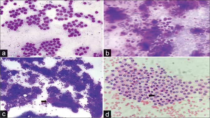
(a) Follicular neoplasm showing presence of repetitive prominent microfollicular pattern in a colloid free background (Giemsa, ×40), (b) medullary carcinoma small cell variant showing singly scattered cells with stippled chromatin in a background of magenta-colored amyloid (Giemsa, ×40), (c) papillary carcinoma showing a cellular smear forming syncytial aggregates and sheets focally with a distinct anatomical border and scanty viscous colloid (depicted by black arrow) also known as chewing gum colloid can be seen (Giemsa, ×10), (d) large, oval, and pale nuclei seen with pale powdered chromatin and presence of intranuclear cytoplasmic inclusion (depicted by black arrow) best appreciated in Pap stain (Papanicolaou, ×40).
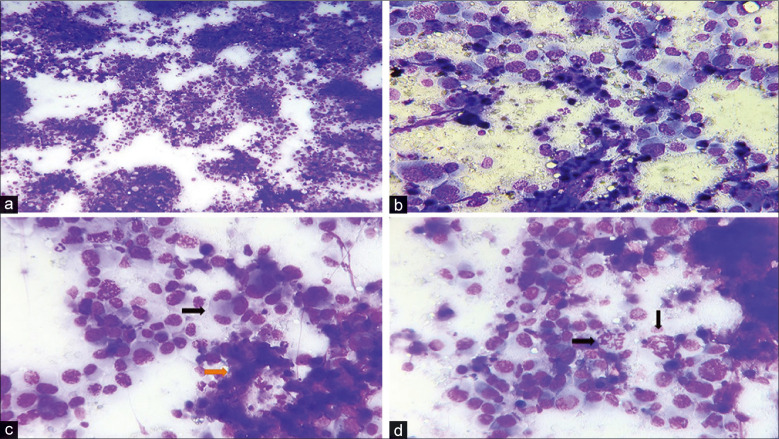
(a) Highly cellular smear with a necrotic background (Giemsa, ×10), (b) giant mononuclear cells present with multilobated nuclei, irregular nuclear membrane, and coarsely clumped chromatin (Giemsa, ×40), (c) multinucleated cells (depicted by black arrow) present along with presence of necrosis (as depicted by orange arrow) (Giemsa, ×40), (d) abnormal mitosis (depicted by black arrow) can be appreciated in a background of highly pleomorphic malignant cells (Giemsa, ×40).
Similar articles
-
Noninvasive follicular thyroid neoplasm with papillary-like nuclear features: An interobserver study of key cytomorphological features from a large academic medical centre.Cytopathology. 2019 Jan;30(1):39-45. doi: 10.1111/cyt.12632. Epub 2018 Nov 8. Cytopathology. 2019. PMID: 30230094
-
Significance of BRAF V600E Mutation and Cytomorphological Features for the Optimization of Papillary Thyroid Cancer Diagnostics in Cytologically Indeterminate Thyroid Nodules.Exp Clin Endocrinol Diabetes. 2019 Apr;127(4):247-254. doi: 10.1055/a-0588-4885. Epub 2018 Mar 22. Exp Clin Endocrinol Diabetes. 2019. PMID: 29566402
-
[Cytomorphological and cytochemical studies of experimentally-induced thyroid tumors in the rat].Zentralbl Pathol. 1991;137(1):29-34. Zentralbl Pathol. 1991. PMID: 2059608 German.
-
The role of fine-needle aspiration in the diagnosis of thyroid lymphoma: a retrospective study of nine cases and review of published series.J Clin Pathol. 2010 Feb;63(2):129-33. doi: 10.1136/jcp.2009.071423. J Clin Pathol. 2010. PMID: 20154034 Review.
-
[Clinical importance of thyroid gland cytology].Pathologe. 2015 Nov;36(6):543-52. doi: 10.1007/s00292-015-0093-0. Pathologe. 2015. PMID: 26462485 Review. German.
References
-
- Dwipayana MP, Yogi P, Semadi S, Wirawan S, Widiana K. Diagnosis and management of an anaplastic thyroid cancer: Case report. Biomed Pharmacol J. 2017;10:1369–77. doi: 10.13005/bpj/1241. - DOI
-
- Chiacchio S, Lorenzoni A, Boni G, Rubello D, Elisei R, Mariani G. Anaplastic thyroid cancer: Prevalence, diagnosis and treatment. Minerva Endocrinol. 2008;33:341–57. - PubMed
LinkOut - more resources
Full Text Sources