

A paravertebral approach for CT-guided percutaneous biopsy of presumably inaccessible, posterior and centrally located pulmonary nodules
- PMID: 32636977
- PMCID: PMC7327127
- DOI: 10.1016/j.radcr.2020.06.004
A paravertebral approach for CT-guided percutaneous biopsy of presumably inaccessible, posterior and centrally located pulmonary nodules
Abstract
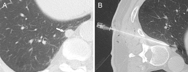
Introduction: To present 3 cases were a paravertebral approach had to be used for the biopsy of posterior and centrally located pulmonary nodules. Case presentation: Three patients underwent percutaneous CT-guided transthoracic biopsy of pulmonary nodules that were initially thought to be inaccessible because of their central, posterior location by a paravertebral approach. The first 2 patients had a history of extra thoracic malignancy and the third patient presented with a bone metastasis and an isolated pulmonary nodule in the right lower lobe, corresponding to potential stage IV lung cancer. Biopsy was feasible in all 3 patients using the paravertebral approach. Pulmonary metastases were confirmed in the first 2 patients, while a TTF-1 positive pulmonary adenocarcinoma was diagnosed in the last patient. No complications occurred. Conclusion: A paravertebral approach is feasible for posterior and centrally located pulmonary nodules.
Keywords: Lung cancer; Paravertebral biopsy; Pulmonary nodule biopsy.
© 2020 The Authors. Published by Elsevier Inc. on behalf of University of Washington.
Figures


Similar articles
-
CT-guided biopsy of pulmonary nodules ≤10 mm: Diagnostic yield based on nodules' lobar and segmental distribution.Clin Imaging. 2020 Oct;66:7-9. doi: 10.1016/j.clinimag.2020.04.040. Epub 2020 May 3. Clin Imaging. 2020. PMID: 32442858
-
Adequacy of Cytologic Samples by Ultrasound-Guided Percutaneous Transthoracic Fine-Needle Aspiration Cytology of Peripheral Pulmonary Nodules for Morphologic Diagnosis and Molecular Evaluations: Comparison With Computed Tomography-Guided Percutaneous Transthoracic Fine-Needle Aspiration Cytology.Arch Pathol Lab Med. 2020 Mar;144(3):361-369. doi: 10.5858/arpa.2018-0346-OA. Epub 2019 Jul 22. Arch Pathol Lab Med. 2020. PMID: 31329477
-
Multiple primary lung cancer versus intrapulmonary metastatic cancer: A case of multiple pulmonary nodules.Thorac Cancer. 2019 Feb;10(2):352-358. doi: 10.1111/1759-7714.12918. Epub 2018 Dec 13. Thorac Cancer. 2019. PMID: 30548923 Free PMC article.
-
[Pulmonary nodule. The surgeon's approach].Zentralbl Chir. 1999;124(2):128-35. Zentralbl Chir. 1999. PMID: 10209847 Review. German.
-
Increased Serum Sedimentation and Positive Tuberculosis Antibody Combined Multiple Pulmonary Nodules in Chest CT in a Middle-Aged Patient Firstly Misdiagnosed as Tuberculosis Proved as Sarcoidosis by CT Guided Percutaneous Lung Puncture Biopsy: a Case Report and Literature Review.Clin Lab. 2019 Sep 1;65(9). doi: 10.7754/Clin.Lab.2019.190325. Clin Lab. 2019. PMID: 31532094 Review.
References
-
- Saji H, Nakamura H, Tsuchida T, Tsuboi M, Kawate N, Konaka C. The incidence and the risk of pneumothorax and chest tube placement after percutaneous CT-guided lung biopsy: the angle of the needle trajectory is a novel predictor. Chest. 2002;121:1521–1526. - PubMed
-
- Hiraki T, Mimura H, Gobara H, Shibamoto K, Inoue D, Matsui Y. Incidence of and risk factors for pneumothorax and chest tube placement after CT fluoroscopy-guided percutaneous lung biopsy: retrospective analysis of the procedures conducted over a 9-year period. AJR Am J Roentgenol. 2010;194:809–814. - PubMed
Publication types
LinkOut - more resources
Full Text Sources
