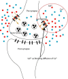
Influence Blocking by Gadolinium in Calcium Diffusion on Synapse Model: A Monte Carlo Simulation Study
- PMID: 32637369
- PMCID: PMC7321394
- DOI: 10.31661/jbpe.v0i0.1155
Influence Blocking by Gadolinium in Calcium Diffusion on Synapse Model: A Monte Carlo Simulation Study
Abstract
Background: Gadolinium (Gd3+) is a chemical element belonging to the lanthanide group and commonly used in magnetic resonance imaging (MRI) as a contrast agent. However, recently, gadolinium has been reported deposition in the body after a patient receives multiple injections. Gadolinium is a potent block and competes with calcium diffusion into the presynaptic. There has not been a precise mechanism of gadolinium blocking calcium channel as a channel of calcium diffusion to presynaptic until now.
Objective: This study aims to investigate the mechanism of calcium influx model and the effect of neurotransmitter release to the synaptic cleft influenced by the presence of Gd3+.
Material and methods: Monte Carlo Cell simulation was used to analyze simulation and also Blender was used to create and visualize the model for synapse. The synapse modeled by a form resembling the actual synapse base on a spherical shape.
Results: The presence of gadolinium around the presynaptic has been disturbing diffusion of calcium influx presynaptic. The result shows that the presence of gadolinium around the presynaptic has caused a decrease in the amount of calcium influx presynaptic. These factors contribute to reducing the establishment of the active membrane, then the amount of synaptic vesicle docking and finally the amount of released neurotransmitter.
Conclusion: Gadolinium and calcium compete with each other across of calcium channel. The presence of gadolinium has caused a chain effect for signal transmission at the chemical synapse, reducing the amount of active membrane, synaptic vesicle docking, and releasing neurotransmitter.
Keywords: Contrast Agents; Diffusion; Gadolinium; Gadolinium Blocking; Monte Carlo Cell; Synapses.
Copyright: © Journal of Biomedical Physics and Engineering.
Figures


References
-
- Lohrke J, Frenzel T, Endrikat J, Alves F C, Grist T M, Law M, et al. 25 Years of Contrast-Enhanced MRI: Developments, Current Challenges and Future Perspectives. Adv Ther. 2016;33:1–28. doi: 10.1007/s12325-015-0275-4. [ PMC Free Article ] - DOI - PMC - PubMed
-
- Aime S, Caravan P. Biodistribution of gadolinium-based contrast agents, including gadolinium deposition. J Magn Reson Imaging. 2009;30:1259–67. doi: 10.1002/jmri.21969. [ PMC Free Article ] - DOI - PMC - PubMed
LinkOut - more resources
Full Text Sources
