

Subcorneal pustular dermatosis: Comprehensive review and report of a case presenting during pregnancy
- PMID: 32637535
- PMCID: PMC7330443
- DOI: 10.1016/j.ijwd.2020.02.003
Subcorneal pustular dermatosis: Comprehensive review and report of a case presenting during pregnancy
Erratum in
-
Erratum regarding previously published articles.Int J Womens Dermatol. 2021 Sep 28;7(5Part B):867. doi: 10.1016/j.ijwd.2021.09.013. eCollection 2021 Dec. Int J Womens Dermatol. 2021. PMID: 35028405 Free PMC article.
Abstract
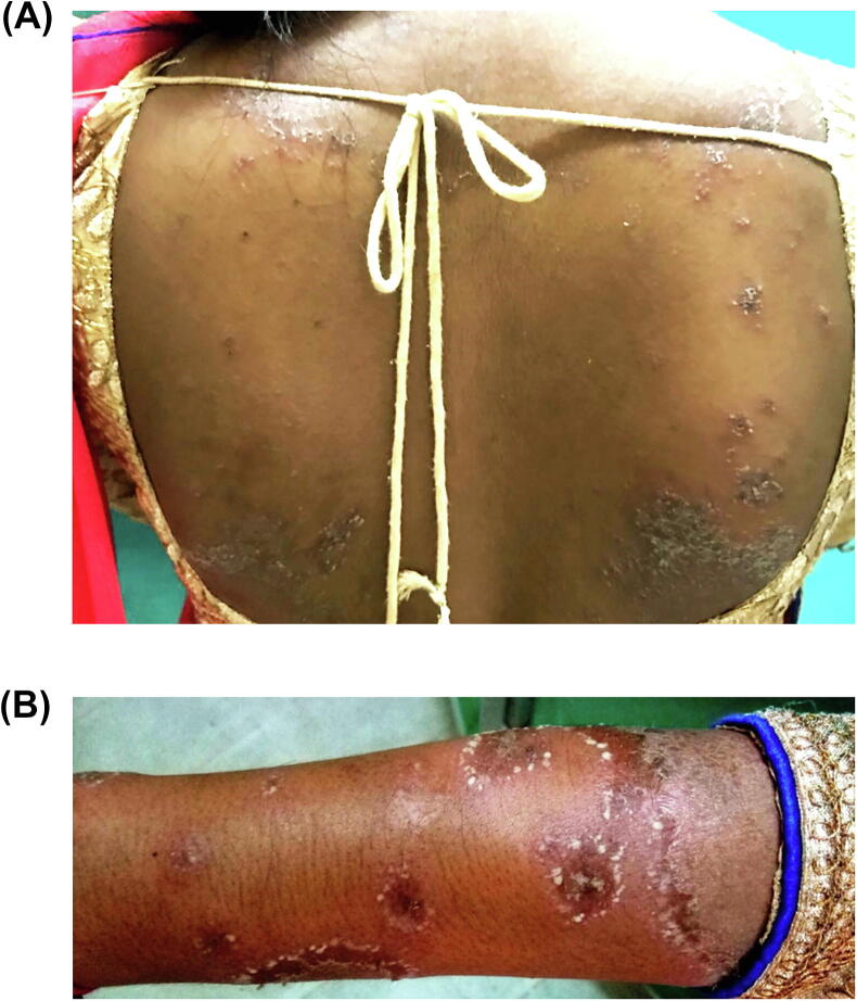
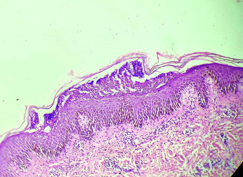
Subcorneal pustular dermatosis (SPD), also known as Sneddon-Wilkinson disease, is a rare, relapsing, sterile pustular eruption of unknown etiology that develops most commonly in middle-aged or mature women. This article reviews the presentation, associations, and management of the condition and highlights advances in pathophysiology. Onset of SPD during pregnancy has not been reported. Herein, we report a case of SPD that developed during pregnancy. The patient was treated with dapsone without complications for her or the fetus. An association between T helper (Th) 17 and Th2 environments in the development of SPD has been advocated. Pregnancy is characterized by a predominance of Th2 responses and increased interleukin-17 levels and thus may favor the development of the condition.
Keywords: Dapsone; Gestation; Neutrophilic dermatosis; Pregnancy; Sneddon-Wilkinson disease; Subcorneal pustular dermatosis.
© 2020 Published by Elsevier Inc. on behalf of Women's Dermatologic Society.
Figures
References
-
- Bauwens M., De Coninck A., Roseeuw D. Subcorneal pustular dermatosis treated with PUVA therapy. A case report and review of the literature. Dermatology. 1999;198(2):203–205. - PubMed
-
- Berk D.R., Hurt M.A., Mann C., Sheinbein D. Sneddon-Wilkinson disease treated with etanercept: Report of two cases. Clin Exp Dermatol. 2009;34(3):347–351. - PubMed
-
- Bohelay G., Duong T.A., Ortonne N., Chosidow O., Valeyrie-Allanore L. Subcorneal pustular dermatosis triggered by Mycoplasma pneumoniae infection: a rare clinical association. J Eur Acad Dermatol Venereol. 2015;29(5):1022–1025. - PubMed
-
- Boms S., Gambichler T. Review of literature on amicrobial pustulosis of the folds associated with autoimmune disorders. Am J Clin Dermatol. 2006;7(6):369–374. - PubMed
Publication types
LinkOut - more resources
Full Text Sources
