Brainstem ischemic syndrome in juvenile NF2
- PMID: 32637630
- PMCID: PMC7323477
- DOI: 10.1212/NXG.0000000000000446
Brainstem ischemic syndrome in juvenile NF2
Abstract
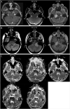
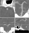
Objective: A new case of brainstem ischemic necrosis in a young woman with de novo neurofibromatosis type 2 (NF2) is reported, and given notable similarities to 7 prior cases of brainstem stroke in the literature, features defining a possible syndrome were sought.
Methods: Case review including detailed clinical assessment, neuroimaging analysis, genetic testing, and brain biopsy, followed by a multicase analysis.
Results: Brainstem ischemia in juvenile NF2 typically occurs in teenagers without previously known NF2 as an acute, monophasic presentation with restricted diffusion in the midbrain or pons following a recent hypoperfusion event, normal vascular imaging, obvious intracranial imaging features of NF2, typical inactivating NF2 alterations, biopsy showing necrosis without small vessel pathology, and subsequent aggressive NF2 lesion progression.
Conclusions: Brainstem ischemia in juvenile NF2 is a rare syndrome of unclear etiology, possibly reflecting an unknown underlying vascular abnormality; a digenic effect is not excluded.
Copyright © 2020 The Author(s). Published by Wolters Kluwer Health, Inc. on behalf of the American Academy of Neurology.
Figures


References
-
- Anand G, Vasallo G, Spanou M, et al. . Diagnosis of sporadic neurofibromatosis type 2 in the paediatric population. Arch Dis Child 2018;103:463–469. - PubMed
-
- Gaudioso C, Listernick R, Fisher MJ, Campen CJ, Paz A, Gutmann DH. Neurofibromatosis 2 in children presenting during the first decade of life. Neurology 2019;93:e964–e967. - PubMed
-
- Ng J, Mordekar SR, Connolly DJA, Baxter P. Stroke in a child with neurofibromatosis type 2. Eur J Paediatr Neurol 2009;13:77–79. - PubMed
-
- Sreedher G, Panigrahy A, Ramos-Martinez SY, Abdel-Hamid HZ, Zuccoli G. Brachium pontis stroke revealing neurofibromatosis type-2. J Neuroimaging 2013;23:132–134. - PubMed
LinkOut - more resources
Full Text Sources
Research Materials
Miscellaneous
