Review
. 2020 May 13;5(2):62-80.
doi: 10.1016/j.synbio.2020.04.001. eCollection 2020 Jun.

Site directed mutagenesis as a precision tool to enable synthetic biology with engineered modular polyketide synthases

Affiliations
Review

Site directed mutagenesis as a precision tool to enable synthetic biology with engineered modular polyketide synthases

Erin E Drufva et al. Synth Syst Biotechnol. .

Erratum in

Abstract

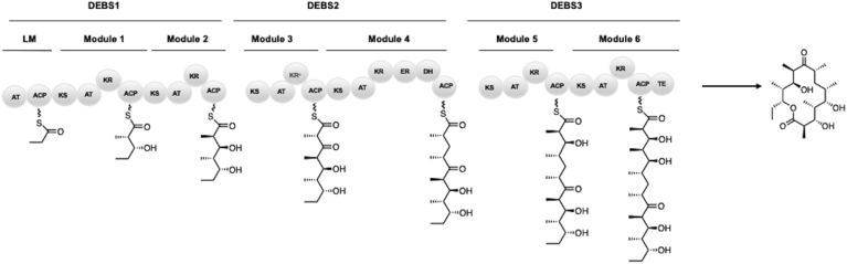
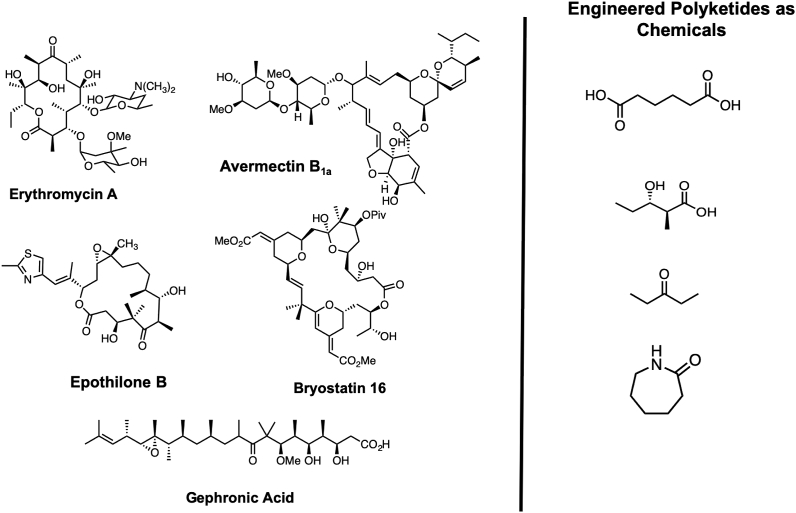
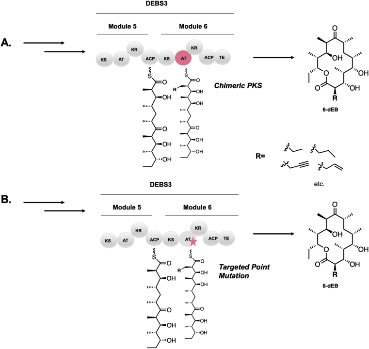
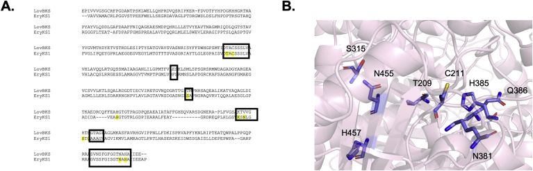
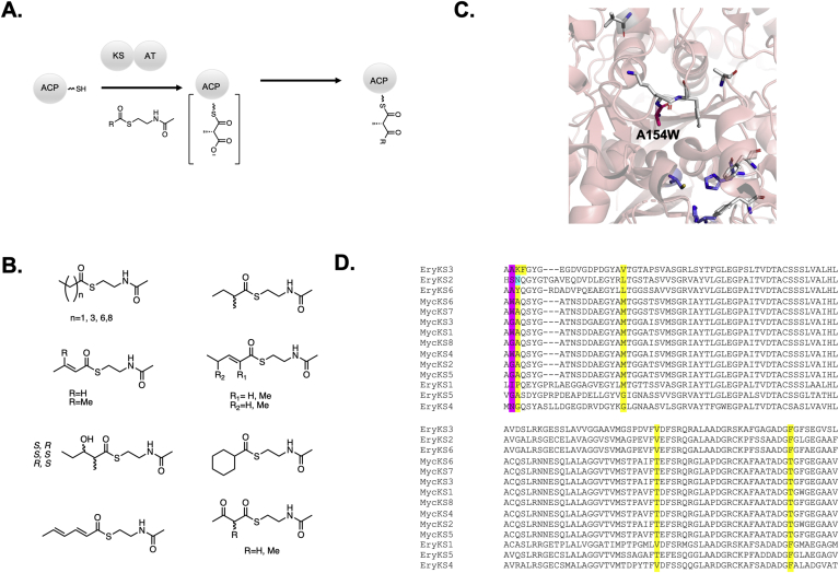
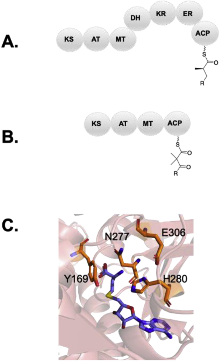
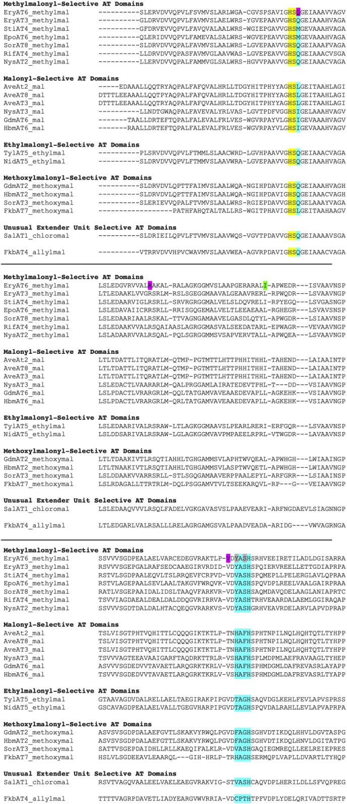
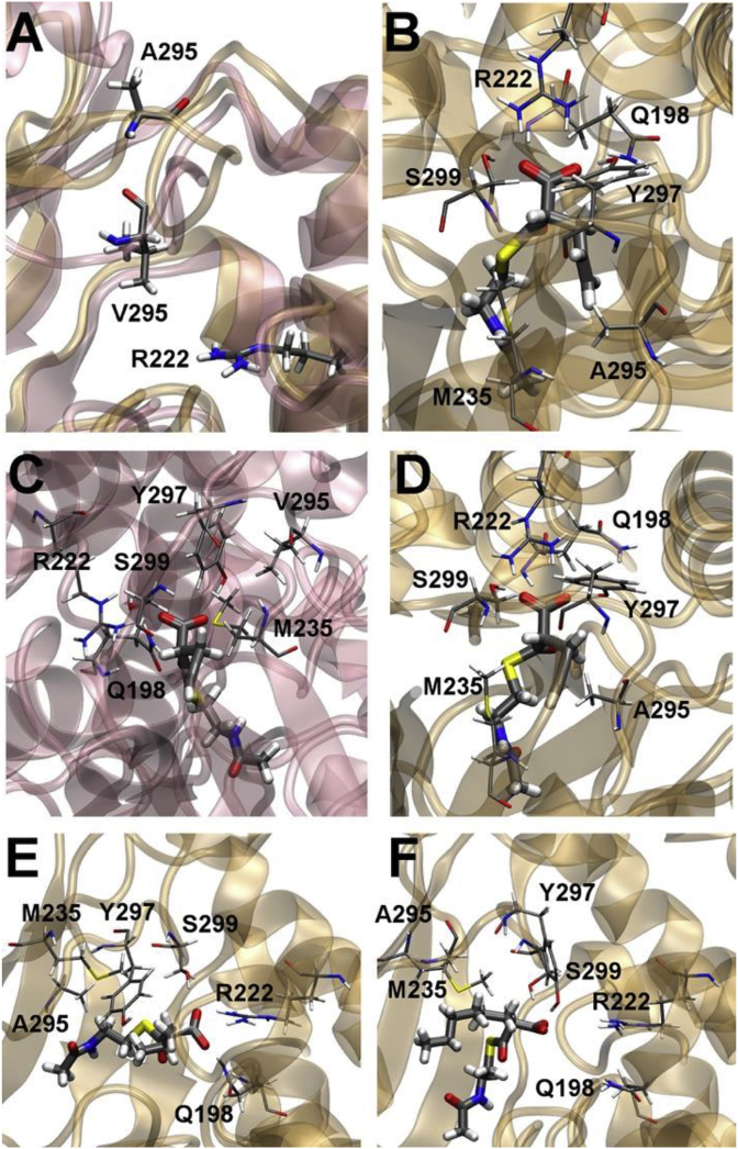
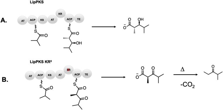
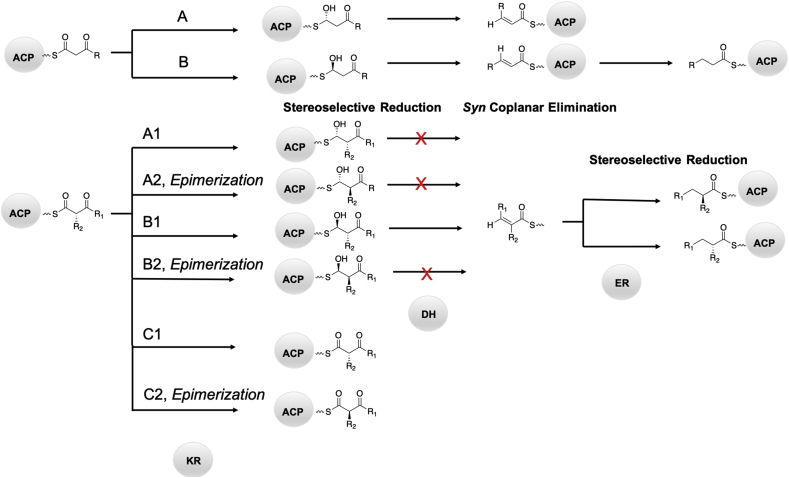
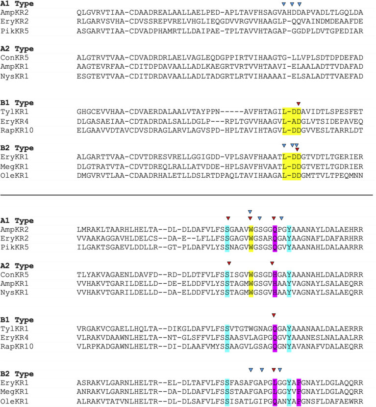
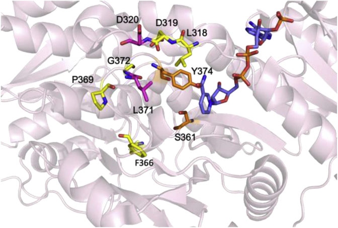
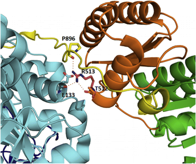
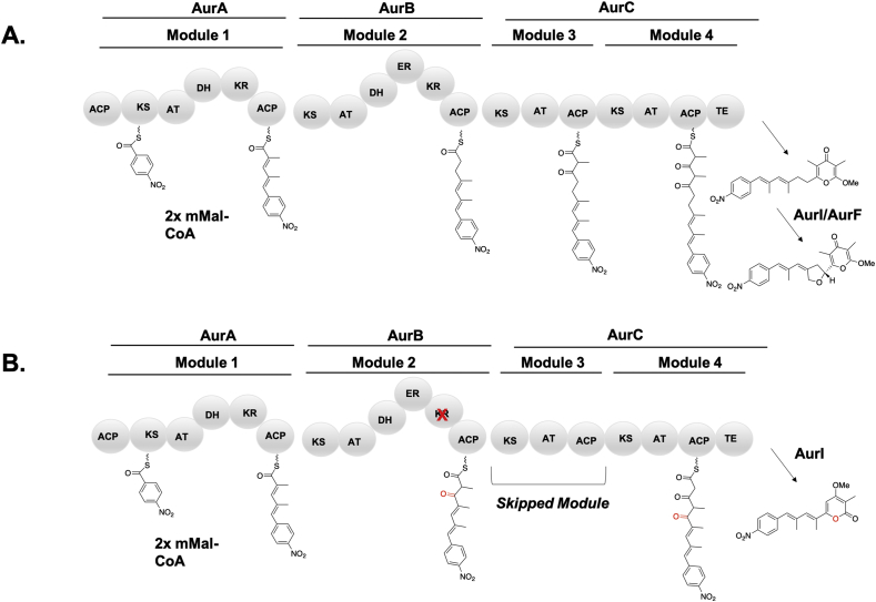
Modular polyketide synthases (PKSs) are a multidomain megasynthase class of biosynthetic enzymes that have great promise for the development of new compounds, from new pharmaceuticals to high value commodity and specialty chemicals. Their colinear biosynthetic logic has been viewed as a promising platform for synthetic biology for decades. Due to this colinearity, domain swapping has long been used as a strategy to introduce molecular diversity. However, domain swapping often fails because it perturbs critical protein-protein interactions within the PKS. With our increased level of structural elucidation of PKSs, using judicious targeted mutations of individual residues is a more precise way to introduce molecular diversity with less potential for global disruption of the protein architecture. Here we review examples of targeted point mutagenesis to one or a few residues harbored within the PKS that alter domain specificity or selectivity, affect protein stability and interdomain communication, and promote more complex catalytic reactivity.

Keywords: ACP, acyl carrier protein; AT, acyltransferase; DEBS, 6-deoxyerthronolide B synthase; DH, dehydratase; EI, enoylisomerase; ER, enoylreductase; KR, ketoreductase; KS, ketosynthase; LM, loading module; MT, methyltransferase; Mod, module; PKS, polyketide synthase; PS, pyran synthase; Polyketide synthase; Protein engineering; Rational design; SNAC, N-acetyl cysteamine; Saturation mutagenesis; Site directed mutagenesis; Synthetic biology.


Figures

Fig. 1
Fig. 1
The 6-deoxyerythronolide B synthase (DEBS) which produces the aglycone precursor to erythromycin, 6-dEB through priming with a loading module (LM) and undergoing six successive decarboxylative Claisen condensations followed by reductive processing to the β-carbon.
Fig. 2
Fig. 2
Comparison of natural product scaffolds and petrochemically derived feedstocks such as adipic acid [3], 3-hydroxy acids [161,162], short chain ketones [97], and caprolactam [163] explored by Keasling and coworkers.
Fig. 3
Fig. 3
Comparison of A) a domain swapping approach and B) a site directed mutagenesis approach to alter the selectivity of EryAT6.
Fig. 4
Fig. 4
A) Alignment of LovB and EryKS1 used in the mutagenesis experiment reported by Khosla and coworkers to identify conserved residues between modular and iterative PKSs essential for catalysis. Residues mutated in the report are indicated in yellow and residues found within 10 Å of the active site cysteine are in boxes. The alignment was performed with MUSCLE. B) Homology Model of EryKS1 with the residues mutated by Khosla and coworkers highlighted in blue. The homology model was made with Swiss PDB using EryKS3 as a template.
Fig. 5
Fig. 5
A) Overview of the ability of KS1 to extend unnatural N-acetyl cysteamine substrates. B) Panel of substrates tested. C) Structure of EryKS3 (pdb code 2QO3) with the residues mutated to explore the sequence space divergence in the mycolactone KSs. D) Sequence alignment of the KSs within DEBS and the those within the mycolactone PKS. The residue found by our laboratory in a common assembly of DEBS1-TE is highlighted (mutated to histidine to introduce a restriction site). The alignment was performed with MUSCLE.
Fig. 6
Fig. 6
A) Example of a monomethylating MT domain harbored within in a PKS module. Frequently, other β-carbon processing domains are found, for example, a full reductive loop shown above. B) Example of a gem-dimethylating MT domain. C) Active site of CurMT3 (pdb code: 5THZ) from the curacin A pathway. Residues targeted for mutation are shown in orange and the SAH cofactor is shown in blue.
Fig. 7
Fig. 7
Sequence alignment of AT domains. Signature motifs including YASH for methylmalonyl-CoA, HAFH for malonyl-CoA, TAGH for ethylmalonyl-CoA, and less conserved motifs for rarer extender units as well as the diagnostic Q versus I/L motif indicating methylmalonyl versus malonyl-CoA are highlighted in cyan. The active site histidine/serine dyad is highlighted in yellow. Mutations introduced by Schulz and coworkers in EryAT6 are indicated in magenta [76,77,115], those introduced by Williams and coworkers [72] are indicated in green, and those explored by both the Williams and Schulz groups are indicated in grey. The alignment was performed with MUSCLE.
Fig. 8
Fig. 8
Snapshots of molecular dynamics simulations of the wild type EryAT6 (rose) and EryAT6 V295A (ochre). A) Depiction of an alanine mutation which allows for greater flexibility of the binding pocket compared to valine. B) Allylmalonyl-SNAC placed in A295A EryAT6 which can be accommodated by the increased binding pocket of the alanine mutation. C) Propargylmalonyl-SNAC does not readily fit into the active site of wild type EryAT6. D) Propargylmalonyl-SNAC fits better in the V295A mutant than the wild type. E) V295A with isopropylmalonyl-SNAC. F) Hexanoylmalonyl-SNAC. Reproduced with permission from ref. [76].
Fig. 9
Fig. 9
Example of strategic inactivation of KR residues to generate keto moieties. A) LipPKS, composed of the Lip1 gene (LM and Mod1) fused to EryTE. B) Inactivation of the KR to strategically generate short chain ketones [97].
Fig. 10
Fig. 10
Schematic of chemical fates of the elongating polyketide by β-carbon processing domains.
Fig. 11
Fig. 11
Sequence alignment indicating diagnostic motifs in the KR domain. Active site residues are shown in cyan. Residues that are fingerprints for hydroxy stereochemistry are indicated in yellow (for A type KRs a W and for B type KRs an LDD motif). Residues associated with α stereochemistry are indicated in magenta. Residues mutated by Leadlay and coworkers are indicated by blue arrows [104,105]. Residues mutated by Keatinge-Clay and coworkers are indicated by red arrows [22,25,100]. The alignment was performed with MUSCLE.
Fig. 12
Fig. 12
Active site of EryKR1 (pdb code 2FR1). The active site tyrosine and serine are indicated in orange. The NADP+ cofactor is indicated in blue. Residues mutated by the Keatinge-Clay group are indicated in magenta [100]. Residues mutated by the Leadlay group are indicated in yellow [104,105]. D320 was mutated by both Keatinge-Clay and coworkers and Leadlay and coworkers.
Fig. 13
Fig. 13
Native β-keto acyl-ACP and truncated small molecule substrate mimics for in vitro studies of decreasing complexity used by standalone KR domains.
Fig. 14
Fig. 14
Types of reactions catalyzed by domains with DH double hot dog folds including A) cyclization to form a pyran and B) isomerization of the olefin.
Fig. 15
Fig. 15
A) Sequence alignment of ER types by stereochemical outcome. The diagnostic tyrosine and valine are indicated in yellow. Other residues that correlate with stereochemical outcome but do not appear to have any impact in mutagenesis studies are indicated in yellow. The alignment was performed with MUSCLE. B) Homology model of EryER4 indicating a hypothesis for the role of the diagnostic tyrosine, which provides steric crowding and prevents approach from the re face. C) Homology model of RapER13 indicating a hypothesis for the role of the diagnostic valine, which sterically affords space for reduction from the re face. Panels B–C reproduced with permission from ref. , .
Fig. 16
Fig. 16
Stereoisomers biocatalytically generated by the wild type and S148C mutant of PikTE through a biocatalytic extension of model substrates via methylmalonyl-SNAC [155,156].
Fig. 17
Fig. 17
A) Energy diagram of and B) transition state calculations of wild type and S148C with the two epimeric substrates. Reproduced with permission from Ref. [156].
Fig. 18
Fig. 18
Critical arginine salt bridge mediating contact between the KS-AT linker. Reproduced with permission from Ref. [157].
Fig. 19
Fig. 19
Example of domain skipping by a single KR inactivation in the aureothin pathway [160].

References

    1. Barajas J.F. Engineered polyketides: synergy between protein and host level engineering. Synth Syst Biotechnol. 2017;2:147–166. - PMC - PubMed
    1. Yuzawa S. Commodity chemicals from engineered modular type I polyketide synthases. 2018;608 - PubMed
    1. Hagen A. Engineering a polyketide synthase for in vitro production of adipic acid. ACS Synth Biol. 2016;5:21–27. - PubMed
    1. Menzella H.G. Rational design and assembly of synthetic trimodular polyketide synthases. Chem Biol. 2007;14:143–151. - PubMed
    1. Donadio S. Modular organization of genes required for complex polyketide biosynthesis. Science. 1991;252:675–679. - PubMed

LinkOut - more resources