. 2020 Jun 21;5(4):768-778.
doi: 10.1016/j.bioactmat.2020.05.008. eCollection 2020 Dec.

Biofilm-inspired adhesive and antibacterial hydrogel with tough tissue integration performance for sealing hemostasis and wound healing

Affiliations

Biofilm-inspired adhesive and antibacterial hydrogel with tough tissue integration performance for sealing hemostasis and wound healing

Wei Han et al. Bioact Mater. .

Abstract

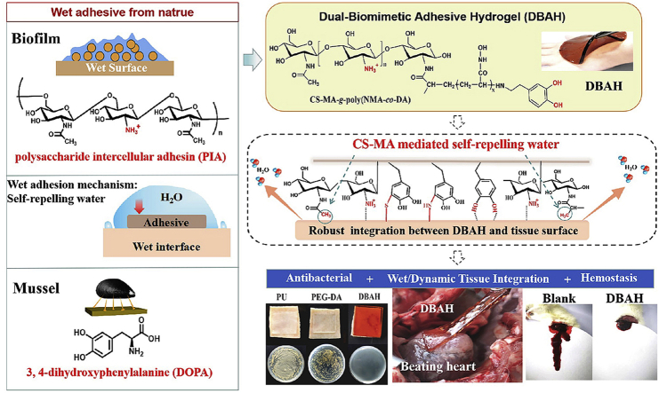
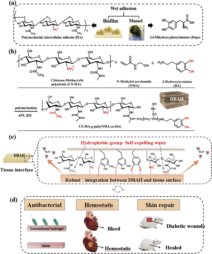
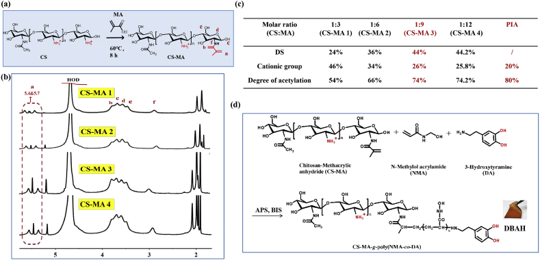
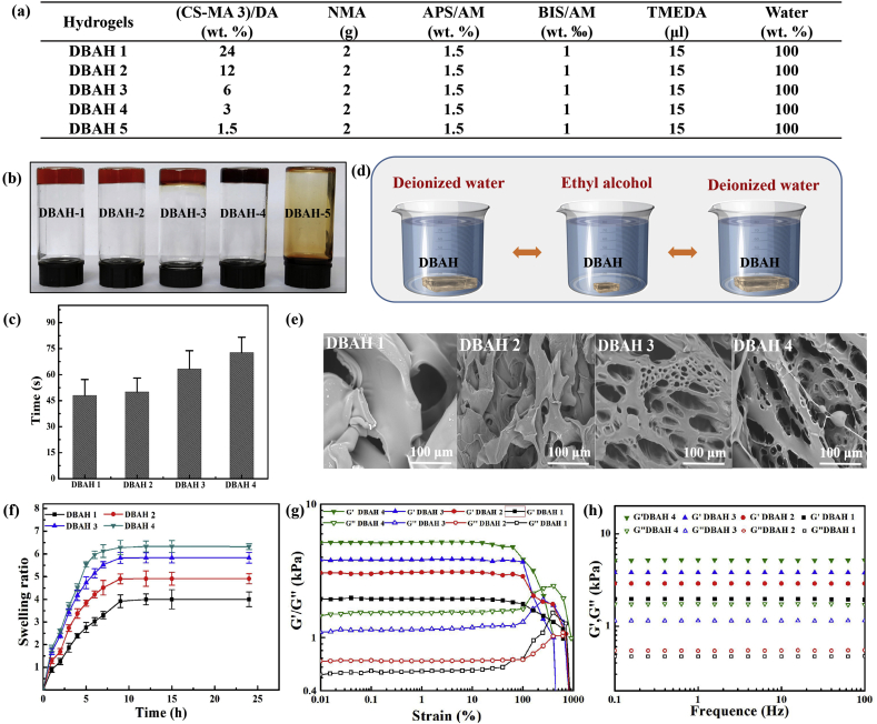
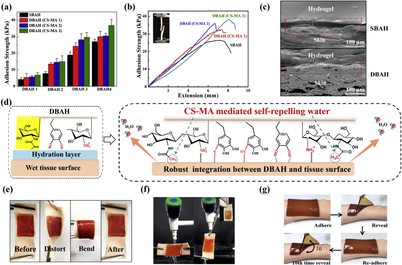
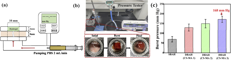
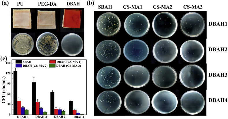
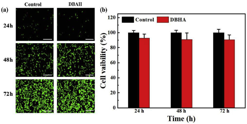
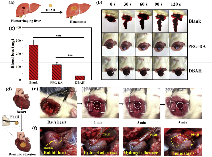
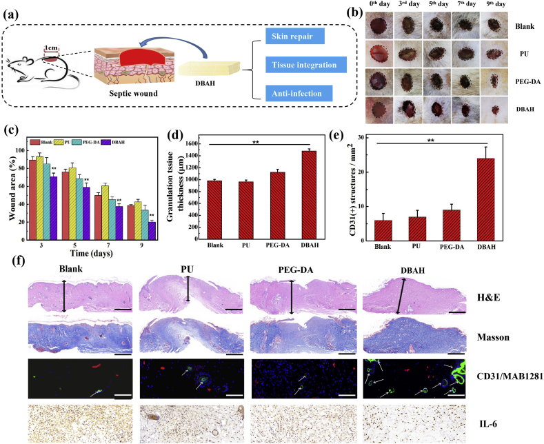
Uncontrolled bleeding and infection can cause significant increases in mortalities. Hydrogel sealants have attracted extensive attention for their ability to control bleeding. However, because interfacial water is a formidable barrier to strong surface bonding, a challenge remains in finding a product that offers robust tissue adhesion combined with anti-infection properties. Inspired by the strong adhesive mechanism of biofilm and mussels, we report a novel dual bionic adhesive hydrogel (DBAH) based on chitosan grafted with methacrylate (CS-MA), dopamine (DA), and N-hydroxymethyl acrylamide (NMA) via a facile radical polymerization process. CS-MA and DA were simultaneously included in the adhesive polymer for imitating the two key adhesive components: polysaccharide intercellular adhesin (PIA) of staphylococci biofilm and 3,4-dihydroxy-l-phenylalanine (Dopa) of mussel foot protein, respectively. DBAH presented strong adhesion at 34 kPa even upon three cycles of full immersion in water and was able to withstand up to 168 mm Hg blood pressure, which is significantly higher than the 60-160 mm Hg measured in most clinical settings. Most importantly, these hydrogels presented outstanding hemostatic capability under wet and dynamic in vivo movements while displaying excellent antibacterial properties and biocompatibility. Therefore, DBAH represents a promising class of biomaterials for high-efficiency hemostasis and wound healing.

Keywords: Adhesive; Antibacterial; Biofilm; Dual-biomimetic; Hemostasis; Hydrogel; Mussel.


Conflict of interest statement

There are no conflict of interest for our manuscript.

Figures

Image 1
Graphical abstract
Scheme 1
Scheme 1
Schematic illustration of design strategy of an engineered biofilm and mussel inspired dual-bionic adhesive hydrogels (DBAH), and its application for Sealing Hemostasis and Wound Healing. (a) The structure of polysaccharide intercellular adhesin (PIA) derived from biofilm and DOPA derived from mussel protein that play a key role in wet adhesion; (b) A biometic biopolymer, chitosan grafted with methacrylate (CS-MA) from PIA, and Dopamine, a catecholamine containing a catechol group of DOPA, was conjugated with NMA for hydrogel formation; (c) Schematic illustration of strong underwater bioinspired adhesion base on the self-repelling water function of CS-MA. (d) The multifunctional properties and potential application in in vivo hemorrhage and diabetic wound healing with antibacterial performance.
Fig. 1
Fig. 1
Synthesis and structure characterization of CS-MA. (a) Schematic illustration of the synthesis of CS-MA; (b) The 1H NMR spectra of CS-MA with different grafting density of MA (CS-MA1, CS-MA2, CS-MA3, CS-MA4); (c) Synthesis of CS-MA with different degrees of substitution (DS) of MA: CS-MA3 with 26% of the monomers are deacetylated, which is comparable to PIA. (d) The formation of DBAH via free radical polymerization reaction based on CS-MA, DA, and NMA.
Fig. 2
Fig. 2
Physical and mechanical characterization of DBAH. (a) Synthesis of DBAH with different ratio of (CS-MA 3)/DA (wt.%). (b) Hydrogel formation of DBAH with different (CS-MA)/DA (wt. %) (DBAH1: (CS-MA)/DA = 24%, DBAH2: (CS-MA)/DA = 12%, DBAH3: (CS-MA)/DA = 6%, DBAH4: (CS-MA)/DA = 3%). Suitable (CS-MA)/DA (wt. %) ((CS-MA)/DA ≥ 3 wt%) resulted in solid hydrogels. Lower CS-MA contents ((CS-MA)/DA = 1.5 wt%) resulted in viscous solution; (c) Gelation time of different hydrogels; (d) Purification of hydrogels with a three cycles of deionized water and ethanol. (e) Morphologies, (f) Swelling ratio, and (g), (h) Rheological analysis of DBAH.
Fig. 3
Fig. 3
(a) Adhesive strengths and (b) Lap shear curve of DBAH containing different kind of CS-MA on two pieces of porcine skin. The single biometic PDA-PAM hydrogel free of CS-MA as control; (c) Scanning electron micrographs of the interface between the hydrogel and the porcine skin; (d) Schematic illustration showing a potential mechanism of the tissue adhesion of DBAH. (e, f) Photographs of the DBAH adhering to the tissue. No detachment was observed between the hydrogel and tissue regardless of bending, distorting, and even under water flushing. (g) Photos of the multiple times repeatable adhering–peeling process of DBAH on the author's arm while no irritability.
Fig. 4
Fig. 4
Burst pressure of DBAH. (a) Schematic diagram; (b) Experimental setup and specimen dimensions for the burst pressure measurement. PBS was pumped into the specimen chamber under a constant flow rate of 2 mL/min, while the pressure was recorded with a pressure gauge; (c) The observed burst pressure of DBAH containing different kind of CS-MA and SBAH.
Fig. 5
Fig. 5
Antibacterial activity analysis of DBAH. (a) Photos of bacteria clones at the interface of pigskin coverd 12 h with PU dressing, PEG-DA hydrogel and DBAH, respectively; (b) Macroscopical images of bacteria clones and (c) Quantitative bacerial viability of specimen treated by DBAH containing different kind of CS-MA and SBAH.
Fig. 6
Fig. 6
In vitro cell compatibility. (a) CLSM images of the cells stained with the LIVE/DEAD assay kit after 24, 48 and 72-hour culture on DBAH's extract media. A normal culture medium was used as the control.(b) Cell viability of NIH-3T3 cells after 24, 48, and 72-hour culture in mediums that were conditioned with DBAH. Scale bars = 100 μm.
Fig. 7
Fig. 7
Hemostatic performance of the DBAH. (a) A schematic illustration of the mouse liver hemorrhage model treated by DBAH; (b) Pictures showing hemostatic efficacy of the damaged SD rats liver treated by DBAH, PEGDA hydrogel (positive control), and untreated (negative control) within 120 s; (c) The accumulated blood loss from liver bleeding treated with different dressing as mentioned above at 120 s (“***” means p < 0.001); (d) A schematic illustration of the rabbit's heart bleeding model treated by DBAH; (e) In vivo test on a beating SD rats heart with blood exposure within 5 min; (f) In vivo test on a beating rabbit's heart with blood exposure within 5 min.
Fig. 8
Fig. 8
Evaluation of DBAH in vivo skin repair promotion at wound of SD rat dorsal skin. (a) Schematic illustrations of wound healing process; (b) Photographs of wounds treated with DBAH, PEGDA hydrogel and PU dressing (positive control), and untreated (Blank, negative control) at 0th, 3rd, 5th, 7th and 9th day; (c) Mean wound area and (d) Quantitative analysis of granulation tissue thickness of wounds at 9th day; (e) Quantification of CD31 labeled structures; (f) Hematoxylin–eosin and Masson staining, immunofluorescence staining of neovascularization, and immunostaining of IL-6 in granulation tissues of wounds at 9th day. Scale bars: H&E, Masson = 1 mm, CD31/MAB1281, IL-6 = 100 μm.

References

    1. Hong Y., Zhou F., Hua Y., Zhang X., Ni C., Pan D., Zhang Y., Jiang D., Yang L., Lin Q., Zou Y., Yu D., Arnot D.E., Zou X., Zhu L., Zhang S., Ouyang H. Nat. Commun. 2019;10:1. - PMC - PubMed
    1. Pfeifer R., Tarkin I.S., Rocos B., Pape H.-C. Injury. 2009;40:907. - PubMed
    1. Zhao X., Guo B., Wu H., Liang Y., Ma P.X. Nat. Commun. 2018;9:1. - PMC - PubMed
    1. Gan D., Xu T., Xing W., Ge X., Fang L., Wang K., Ren F., Lu X. Adv. Funct. Mater. 2019;29:1805964.
    1. Katzenell U., Ash N., Tapia A.L., Campino G.A., Glassberg E. Mil. Med. 2012;177:1065. - PubMed

LinkOut - more resources