[Preprint]. 2020 Jun 29:2020.06.29.174623.
doi: 10.1101/2020.06.29.174623.

SARS-CoV-2 infection of primary human lung epithelium for COVID-19 modeling and drug discovery

Affiliations

SARS-CoV-2 infection of primary human lung epithelium for COVID-19 modeling and drug discovery

A Mulay et al. bioRxiv. .

Update in

Abstract

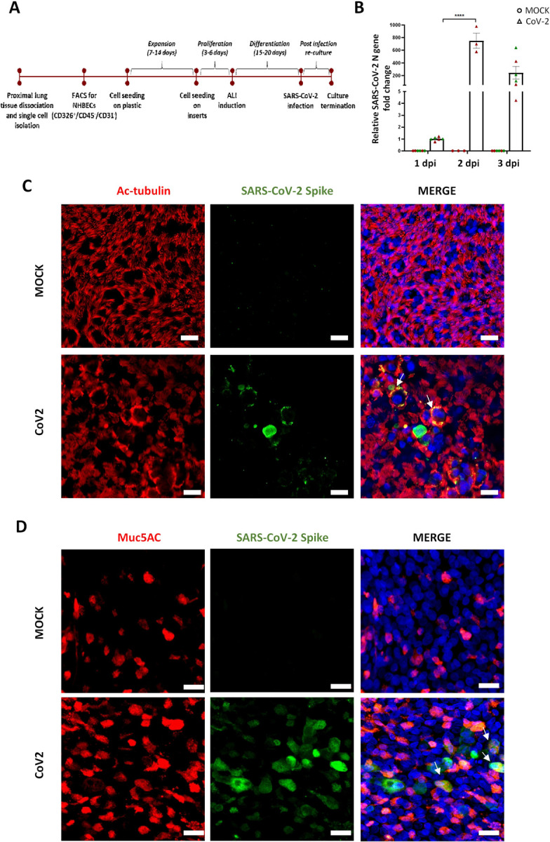
Coronavirus disease 2019 (COVID-19) is the latest respiratory pandemic resulting from zoonotic transmission of severe acute respiratory syndrome-related coronavirus 2 (SARS-CoV-2). Severe symptoms include viral pneumonia secondary to infection and inflammation of the lower respiratory tract, in some cases causing death. We developed primary human lung epithelial infection models to understand responses of proximal and distal lung epithelium to SARS-CoV-2 infection. Differentiated air-liquid interface cultures of proximal airway epithelium and 3D organoid cultures of alveolar epithelium were readily infected by SARS-CoV-2 leading to an epithelial cell-autonomous proinflammatory response. We validated the efficacy of selected candidate COVID-19 drugs confirming that Remdesivir strongly suppressed viral infection/replication. We provide a relevant platform for studying COVID-19 pathobiology and for rapid drug screening against SARS-CoV-2 and future emergent respiratory pathogens.

One sentence summary: A novel infection model of the adult human lung epithelium serves as a platform for COVID-19 studies and drug discovery.


Conflict of interest statement

Competing interests: Authors declare no competing interests.

Figures

Fig. 1.
Fig. 1.. SARS-CoV-2 infects and replicates within normal human proximal airway cells.
(A) Workflow for establishment of human proximal airway ALI cultures and their infection with SARS-CoV-2. (B) ALI cultures of proximal airway epithelial cells are susceptible to SARS-CoV-2 infection which peaked at 2dpi; n= 3 to 6 cultures from 2 independent donors. Circles indicate mock cultures and triangles indicate SARS-CoV-2 infected cultures. Red and green colors indicate cultures from separate donors. Data were analyzed using Two-Way ANOVA with Sidak’s post-hoc correction and represented as fold change for individual cultures ± SEM relative to mean N gene expression at 1dpi ****p<0.0001. 2 days after SARS-CoV-2 infection, viral spike protein (green) was found to heterogeneously co-localize with (C) acetylated tubulin positive ciliated cells (red) and (D) a proportion of Muc5AC positive goblet cells (red). Scale bar = 20μm. Arrows indicate colocalization of markers.
Fig. 2.
Fig. 2.. SARS-CoV-2 infects and replicates within normal human distal alveolar organoids.
(A) Workflow for establishment of human distal alveolar organoid cultures and their infection with SARS-CoV-2. (B) Enzymatic and mechanical disruption of alveolar organoids to expose the apical surface was essential for robust infection, as evaluated by detection of SARS-CoV-2 N gene abundance; n = 3 to 4 organoid cultures established from 2 different donor samples for each condition. (C) Viral infection levels peaked at 2 days post infection; n=3 organoid cultures. Data were analyzed using Two-Way ANOVA with Sidak’s post-hoc correction and represented as fold change for individual cultures ± SEM relative to mean N gene expression at 1dpi. ****p=0.0002 for (B) and ***p=0.0009 ****p<0.0001 for (C). (D) Viral infection was assessed at 2dpi by antibody against SARS-CoV-2 “spike” protein (green) and colocalized with AT2 cell marker HTII-280 (red) (E) Infected alveolar organoids (red) also demonstrated signs of cellular apoptosis at 3dpi, indicated by positive staining for cleaved caspase-3 (green); scale bar= 20μm.
Fig. 3.
Fig. 3.. Primary human lung epithelial models for study of SARS-CoV-2 induced host response and drug validation.
(A) Heatmap showing RNA seq analysis of differentially expressed genes at 2 dpi. (B) Volcano plot of gene expression changes in SARS-CoV-2 infected vs mock cultures defined by p-value and >2-fold change. Several viral genes such as Virus_N, virus_ORF1ab, virus_ ORF3a were detected in the infected samples. Cytokines such as IFNB1, and antiviral response genes OAS1, OAS2, ISG15 and MX1 were significantly upregulated. (C) The most upregulated canonical transcriptional pathway in SARS-CoV-2 infected alveolar cultures was IFN signaling pathway. In addition to TLR and NFKB signaling. (D) Downregulated canonical transcriptional pathways include antigen presentation and Th1 and Th2 activation pathways. (E) Pre-treatment of alveolar organoid cultures with IFNB1, Hydroxychloroquine and Remdesivir significantly reduced viral replication. The effect of Remdesivir on viral replication was more pronounced than that of IFNB1 or hydroxychloroquine. Data are represented as log2fold change for individual cultures ± SEM, normalized to mean infection and analyzed using One-Way ANOVA with Tukey’s post-hoc test **p=0.0012 for IFNB1, **p=0.0044 for HCQ and ****p<0.0001 for Remdesivir. The 3 different colors indicate cultures from different biological replicates. (F) Pre-treatment of proximal ALI cultures with Remdesivir significantly reduced viral infection/replication. Pre-treatment with Hydroxychloroquine and IFNB did not have an effect on viral replication; n= 3–4 independent cultures. Data are represented as log2fold change for individual cultures, normalized to mean infection and analyzed using One-Way ANOVA plus Tukey’s posthoc test ****p<0.0001.

References

    1. Zhou P. et al. , A pneumonia outbreak associated with a new coronavirus of probable bat origin. Nature 579, 270–273 (2020). - PMC - PubMed
    1. Tay M. Z., Poh C. M., Rénia L., MacAry P. A., Ng L. F. P., The trinity of COVID-19: immunity, inflammation and intervention. Nat Rev Immunol 20, 363–374 (2020). - PMC - PubMed
    1. Cui J., Li F., Shi Z. L., Origin and evolution of pathogenic coronaviruses. Nat Rev Microbiol 17, 181–192 (2019). - PMC - PubMed
    1. Vijaykrishna D. et al. , Reassortment of pandemic H1N1/2009 influenza A virus in swine. Science 328, 1529 (2010). - PMC - PubMed
    1. Barkauskas C. E. et al. , Type 2 alveolar cells are stem cells in adult lung. J Clin Invest 123, 3025–3036 (2013). - PMC - PubMed

Publication types