[Preprint]. 2020 Jul 1:2020.07.01.182659.
doi: 10.1101/2020.07.01.182659.

High affinity binding of SARS-CoV-2 spike protein enhances ACE2 carboxypeptidase activity

Affiliations

High affinity binding of SARS-CoV-2 spike protein enhances ACE2 carboxypeptidase activity

Jinghua Lu et al. bioRxiv. .

Update in

Abstract

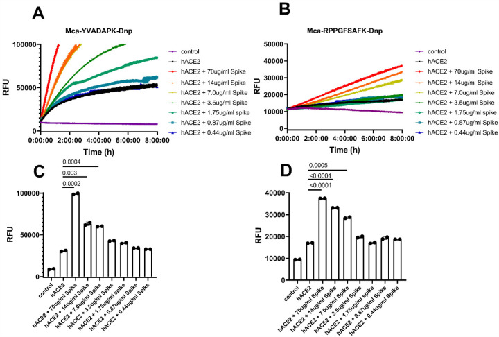
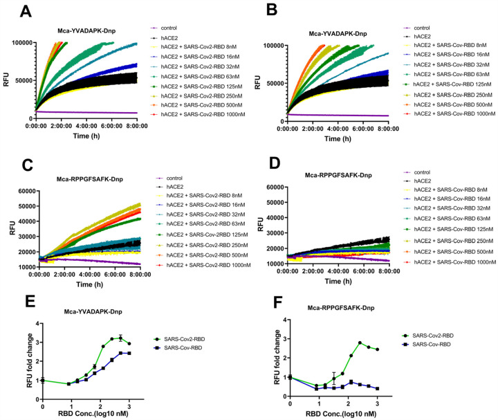
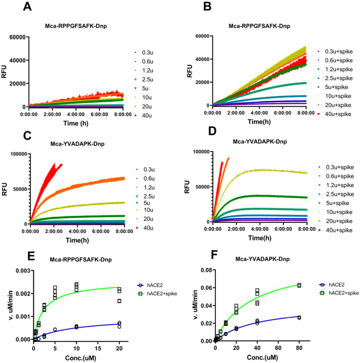
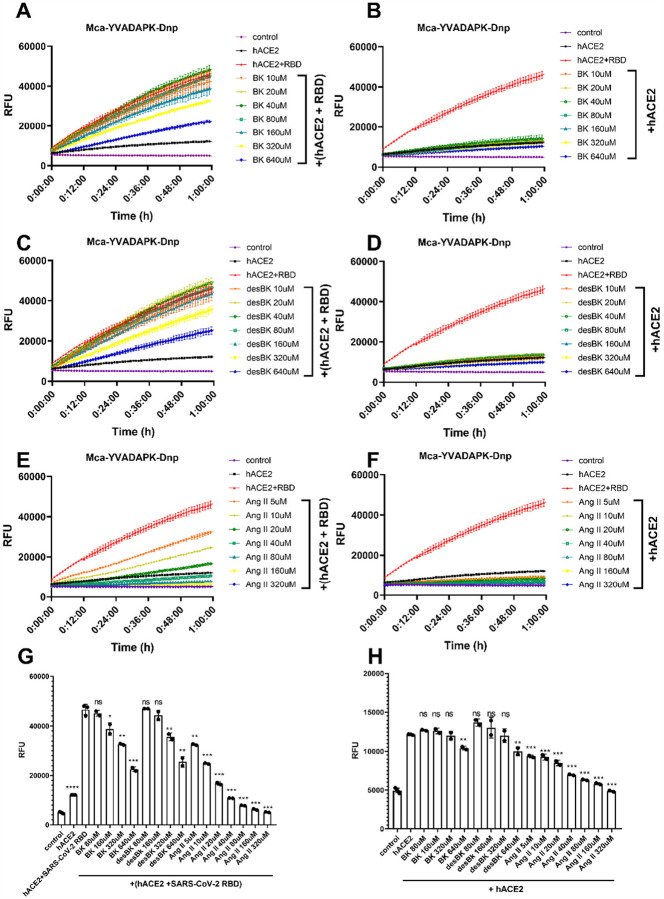
A novel coronavirus (SARS-CoV-2) has emerged to a global pandemic and caused significant damages to public health. Human angiotensin-converting enzyme 2(ACE2) was identified as the entry receptor for SARS-CoV-2. As a carboxypeptidase, ACE2 cleaves many biological substrates besides Ang II to control vasodilatation and permeability. Given the nanomolar high affinity between ACE2 and SARS-CoV-2 spike protein, we wonder how this interaction would affect the enzymatic activity of ACE2. Surprisingly, SARS-CoV-2 trimeric spike protein increased ACE2 proteolytic activity ~3-10 fold when fluorogenic caspase-1 substrate and Bradykinin-analog peptides were used to characterize ACE2 activity. In addition, the enhancement was mediated by ACE2 binding of RBD domain of SARS-CoV-2 spike. These results highlighted the altered activity of ACE2 during SARS-CoV-2 infection and would shed new lights on the pathogenesis of COVID-19 and its complications for better treatments.


Conflict of interest statement

Disclosure

The authors declare that no competing interests exist.

Figures

Figure 1
Figure 1
SARS-CoV-2 spike protein enhanced ACE2 cleavage of fluorogenic caspase-1 substrate and bradykinin analog in a concentration dependent manner. A) and B) Kinetic reading of relative fluorescence units (RFU) monitoring the hydrolysis of Mca-YVADAPK-Dnp(20uM) and Mca-RPPGFSAFK-Dnp (20uM) in the presence of SARS-CoV-2 spike protein at indicated final concentrations in the enzymatic assays. The maximum reading is limited to 100000 RFU on Synergy H1 plate reader. C) and D) Comparison of RFU generation during the cleavage of Mca-YVADAPK-Dnp and Mca-RPPGFSAFK-Dnp at 1.5h and 8 h, respectively.
Figure 2
Figure 2
SARS-CoV-2 RBD but not SARS-CoV RBD enabled ACE2 to cleave bradykinin analog. A) and B) hydrolysis of Mca-YVADAPK-Dnp(20uM) in the presence of SARS-CoV-2 RBD and SARS-CoV RBD proteins at indicated concentrations. C) and D) hydrolysis of Mca-RPPGFSAFK-Dnp (20uM) in the presence of SARS-CoV-2 RBD and SARS-CoV RBD proteins at indicated concentrations. E) Fold change of RFU during Mca-YVADAPK-Dnp cleavage in the presence of SARS-CoV-2 RBD or SARS-CoV RBD at the time point of 1.5h due to instrument overflow. F) Fold change of RFU during Mca-RPPGFSAFK-Dnp cleavage in the presence of SARS-CoV-2 RBD or SARS-CoV RBD at time point of 8h. All RFU readings at different concentrations of RBD proteins were normalized to that of ACE2 cleavage without RBD proteins correspondingly.
Figure 3
Figure 3
Measurement of kinetic constants for hydrolysis of Mca-RPPGFSAFK-Dnp and Mca-YVADAPK-Dnp in the absence or presence of SARS-CoV-2 spike protein (14ug/ml). A) and B) background subtracted RFU kinetic readings during ACE2 hydrolysis of Mca-RPPGFSAFK-Dnp at different concentrations in the absence (A) and presence (B) of SARS-CoV-2 spike protein. C) and D) background subtracted RFU kinetic readings during ACE2 hydrolysis of Mca-YVADAPK-Dnp at different concentrations in the absence (C) and presence (D) of SARS-CoV-2 spike protein. E) and F) Michaelis plots for ACE2 hydrolysis of Mca-RPPGFSAFK-Dnp and Mca-YVADAPK-Dnp in the absence (blue) or presence (green)of SARS-CoV-2 spike protein. The initial velocity conditions were limited to 30min for Mca-YVADAPK-Dnp and 60min for Mca-RPPGFSAFK-Dnp due to different cleavage rate. All determinations were repeated with duplicates.
Figure 4
Figure 4
Competition of Bradykinin(BK), des-Arg9-BK, and Ang II peptide with Mca-YVADAPK-Dnp (10uM) for ACE2 hydrolysis. A),C) and E) panels showing the substrates competition in the presence of SARS-CoV-2 RBD protein at different concentrations. B),D) and F) panels showing the substrates competition in the absence of SARS-CoV-2 RBD protein at different concentrations. G) Comparison of RFU during ACE2 hydrolysis of Mca-YVADAPK-Dnp in the presence of competitive substrate at time 1h. The pairwise p-value statistics were calculated between (hACE2 + SARS-CoV-2 RBD) and corresponding concentrations of competitive peptides. H) Comparison of RFU during ACE2 hydrolysis of Mca-YVADAPK-Dnp in the absence of competitive substrate at time 1h. The pairwise p-value statistics were calculated between hACE2 and corresponding concentrations of competitive peptides.
Figure 5
Figure 5
Conformational changes of ACE2 upon SARS-CoV-2 binding. A) apo structure of ACE2 (PDB ID: 1R42).The N-terminal subdomain was colored in cyan with the secondary structures of SARS-CoV-2 spike binding sites highlighted in blue. The C-terminal domain of apo ACE2 was colored in wheat. All subsequent structural superpositions were based the alignment of ACE2 residues 20–84 that formed the first 2 helix for RBD domain interaction. The ACE2 inhibitor MLN-4760 (purple) and Angiotensin II (yellow) were positioned in the substrate pocket based on the structural alignment. B) ACE2 structure with inhibitor MLN-4760 binding (PDB ID: 1R4L). The C-terminal domain was highlighted in orange. C) hACE in complex with Ang II(PDB ID: 4APH). The C-terminal domain was highlighted in orange. D)ACE2 structure in complex with SARS-CoV RBD(PDB ID: 2AJF). The C-terminal domain was highlighted in yellow. E) ACE2 structure in complex with SARS-CoV-2 RBD (PDB ID: 6M0J). The C-terminal domain was highlighted in lime green. F) ACE2 structure in complex with a chimeric SARS-CoV-2 RBD domain (PDB ID: 6VW1). The C-terminal domain was highlighted in green. G) Enlarged view of ACE2 substrate binding pocket. One additional ACE2-SARS-CoV-2 RBD complex(PDB ID: 6LZG) was included. The residue color scheme was listed in the bottom panel.

References

    1. Wu F. et al. A new coronavirus associated with human respiratory disease in China. Nature 579, 265–269 (2020). - PMC - PubMed
    1. Zhou P. et al. A pneumonia outbreak associated with a new coronavirus of probable bat origin. Nature 579, 270–273 (2020). - PMC - PubMed
    1. Zhu N. et al. A Novel Coronavirus from Patients with Pneumonia in China, 2019. N Engl J Med 382, 727–733 (2020). - PMC - PubMed
    1. Huang C. et al. Clinical features of patients infected with 2019 novel coronavirus in Wuhan, China. Lancet 395, 497–506 (2020). - PMC - PubMed
    1. Wichmann D. et al. Autopsy Findings and Venous Thromboembolism in Patients With COVID-19. Ann Intern Med (2020). - PubMed

Publication types

LinkOut - more resources