Review
. 2020 Jul 22;107(2):219-233.
doi: 10.1016/j.neuron.2020.06.032. Epub 2020 Jul 1.

COVID-19 and the Chemical Senses: Supporting Players Take Center Stage

Affiliations
Review

COVID-19 and the Chemical Senses: Supporting Players Take Center Stage

Keiland W Cooper et al. Neuron. .

Abstract

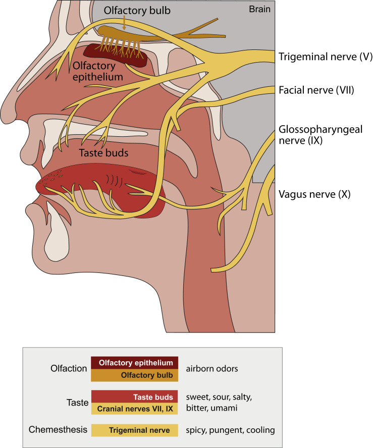
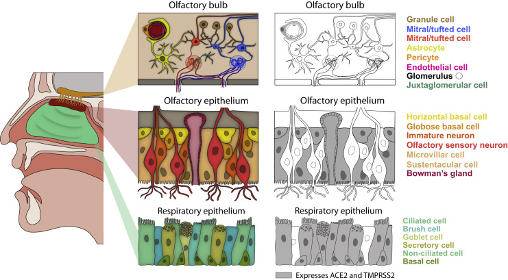
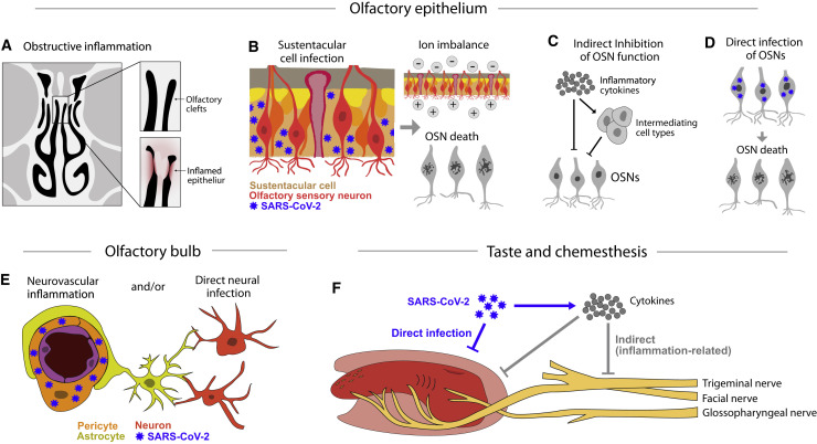
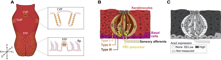
The main neurological manifestation of COVID-19 is loss of smell or taste. The high incidence of smell loss without significant rhinorrhea or nasal congestion suggests that SARS-CoV-2 targets the chemical senses through mechanisms distinct from those used by endemic coronaviruses or other common cold-causing agents. Here we review recently developed hypotheses about how SARS-CoV-2 might alter the cells and circuits involved in chemosensory processing and thereby change perception. Given our limited understanding of SARS-CoV-2 pathogenesis, we propose future experiments to elucidate disease mechanisms and highlight the relevance of this ongoing work to understanding how the virus might alter brain function more broadly.


Figures

Figure 1
Figure 1
Chemosensory Anatomy Defines the Potential Attack Surface for SARS-CoV-2 Chemosensation occurs in sensory epithelia in the nose and mouth. Multiple cranial nerves relay the senses of smell, taste, and chemesthesis to the brain. Airborne odors are detected by olfactory sensory neurons that reside in the olfactory epithelium; their axons pierce the bony cribriform plate to enter the olfactory bulb in the brain. Taste buds on the tongue are innervated by sensory afferents from the facial nerve (VII) and glossopharyngeal nerve (IX). The vagus nerve (X) also innervates taste buds residing in the pharynx. The detection of pungent chemicals, also known as chemesthesis, is mediated by both oral and nasal afferents of the trigeminal nerve (V). Although deficits in smell are most commonly reported in COVID-19, all three chemosensory modalities have been reported to be affected.
Figure 2
Figure 2
ACE2-Positive Cells in the Nasal Respiratory Epithelium, Olfactory Epithelium, and the Olfactory Bulb Schematic of a sagittal view of the human head, in which respiratory and olfactory epithelium as well as the olfactory bulb in the brain are colored (left). For each tissue, a schematic of the anatomy and known major cell types are shown (middle). The nose contains both respiratory and olfactory epithelia. The olfactory epithelium is restricted to medial portion of the superior turbinate and the superior portion of the nasal septum, whereas the respiratory epithelium is continuous with the upper airway (Dahl and Mygind, 1998). Olfactory sensory neurons within the olfactory epithelium are responsible for detecting odors, and in mice and humans they are continuously regenerated from globose progenitors throughout life (Durante et al., 2020; Schwob et al., 2017). The olfactory epithelium also contains other support and non-neuronal cell types, as well as reserve horizontal basal stem cells that respond to injury and can reconstitute olfactory epithelial cell types (Choi and Goldstein, 2018). In the respiratory epithelium, basal progenitor cells generate all epithelial cell types, including ciliated and secretory cells. In the olfactory bulb in the brain (tan), the axons from olfactory sensory neurons coalesce into glomeruli, and mitral/tufted cells innervate these glomeruli and send olfactory projections to downstream olfactory areas. ACE2-positive cell types are indicated in gray (right). Four recent reports have all concluded that ACE2 is not expressed in olfactory sensory neurons (Brann et al., 2020; Chen et al., 2020b; Fodoulian et al., 2020; Ziegler et al., 2020). Pericyte ACE2 expression in the olfactory bulb is inferred from the mouse data; there are currently no available human olfactory bulb sequencing datasets. Figure modified from Brann et al. (2020).
Figure 3
Figure 3
Possible Mechanisms of Chemosensory Disturbances (A) COVID-19 patients have reported olfactory loss in the absence of features common to upper respiratory infections like widespread nasal inflammation or obstruction. Such symptoms are consistent with recent CT imaging suggesting that SARS-CoV-2 infection may cause inflammation that is localized to the olfactory clefts (Eliezer et al., 2020). (B) Sustentacular cells, Bowman’s gland cells, and microvillar cells in the olfactory epithelium express both ACE2 and TMPRSS2 and may be directly infected by SARS-CoV-2. Support cell infection may cause changes in the olfactory mucus or ion imbalances that can inhibit olfactory signaling; such changes may be rapidly reversible. The loss of these support cells in animal models can also result in the death of olfactory sensory neurons. (C) Inflammatory cytokines may also directly or indirectly inhibit olfactory sensory neuron function. (D) Although current data suggest that sustentacular and other support cells are the primary targets of SARS-CoV-2, a remaining possibility is that SARS-CoV-2 directly infects OSNs. (E) Immunostaining for ACE2 protein in the mouse olfactory bulb suggests that ACE2 expression is restricted to vascular pericytes (Brann et al., 2020), which may directly (via perfusion changes) or indirectly (via inflammation) affect chemosensory perception in the brain. (F) Taste and chemesthetic disturbances may result from the direct infection of cells in the tongue, the secondary consequences of obstruction due to inflammation, or damage following the release of inflammatory cytokines.
Figure 4
Figure 4
Organization of the Tongue and Taste Buds (A) In the front of the tongue, taste buds are situated in fungiform papillae (FFP). FFP are distributed throughout the anterior lingual epithelium, which is covered by mechanosensory filiform papillae (flp). In the posterior tongue, large complex papillae (circumvallate [CVP] and foliate [FolP]) are epithelial invaginations that house hundreds of buds each; rodents have a single CVP, while humans possess 8–12 CVPs in an inverted V arrangement across the posterior tongue (not shown). Modified from Barlow (2015). (B) Each bud is a collection of ∼100 taste receptor cells (TRCs) categorized into three morphological types (I for support or glial-like, II for sweet, bitter, umami, and III for sour). All TRCs are continuously renewed from basal stem cells adjacent to taste buds (“Basal cells”) (Gaillard et al., 2015; Liu et al., 2013; Okubo et al., 2009). Taste bud-fated daughters generated by basal cells exit the cell cycle and enter buds as postmitotic TRC precursors, which differentiate into each of the TRC types (Miura et al., 2006, 2014). Basal cells also give rise to keratinocytes of the non-taste lingual epithelium including those surrounding taste buds. Individual TRCs have a brief lifespan ranging from 1 to 6 weeks (Beidler and Smallman 1965; Hamamichi et al., 2006; Perea-Martinez et al., 2013). Type III TRCs make conventional presynaptic contacts, while type II TRCs have specialized contacts with sensory afferents to transmit taste information to the CNS (Finger et al., 2005; Romanov et al., 2018; Roper and Chaudhari, 2017; Taruno et al., 2013). (C) To date, RNA profiling of taste relevant cell populations has been obtained primarily from murine CVP, with two additional datasets for general anterior tongue epithelium in mice and human (see text). A summary of approximate ACE2 expression levels extracted from these studies is depicted here as discussed in the text. Of note, type I TRCs and precursor TRCs have yet to be profiled, and ACE2 is not detected in sensory neuron afferents.

References

    1. Adamczyk K., Herman M., Fraczek J., Piec R., Szykula-Piec B., Zaczynski A., Wojtowicz R., Bojanowski K., Rusyan E., Krol Z., et al. Sensitivity and specifity of prediction models based on gustatory disorders in diagnosing COVID-19 patients: a case-control study. medRxiv. 2020 doi: 10.1101/2020.05.31.20118380. - DOI
    1. Åkerlund A., Bende M., Murphy C. Olfactory threshold and nasal mucosal changes in experimentally induced common cold. Acta Otolaryngol. 1995;115:88–92. - PubMed
    1. Ansari M.A., Marchi E., Ramamurthy N., Aschenbrenner D., Hackstein C.-P., Bowden R., Sharma E., Pedergnana V., Venkateswaran S., Kugathasan S., et al. A gene locus that controls expression of ACE2 in virus infection. medRxiv. 2020 doi: 10.1101/2020.04.26.20080408. - DOI
    1. Armulik A., Genové G., Betsholtz C. Pericytes: developmental, physiological, and pathological perspectives, problems, and promises. Dev. Cell. 2011;21:193–215. - PubMed
    1. Bader M. Tissue renin-angiotensin-aldosterone systems: Targets for pharmacological therapy. Annu. Rev. Pharmacol. Toxicol. 2010;50:439–465. - PubMed

Publication types

MeSH terms