Review
. 2020 Jul 6;7(3):71.
doi: 10.3390/bioengineering7030071.

Prospects and Challenges of Translational Corneal Bioprinting

Affiliations
Review

Prospects and Challenges of Translational Corneal Bioprinting

Matthias Fuest et al. Bioengineering (Basel). .

Abstract

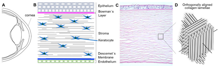
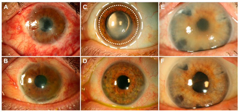
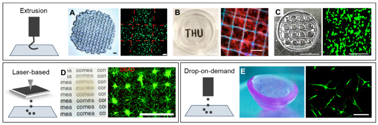
Corneal transplantation remains the ultimate treatment option for advanced stromal and endothelial disorders. Corneal tissue engineering has gained increasing interest in recent years, as it can bypass many complications of conventional corneal transplantation. The human cornea is an ideal organ for tissue engineering, as it is avascular and immune-privileged. Mimicking the complex mechanical properties, the surface curvature, and stromal cytoarchitecure of the in vivo corneal tissue remains a great challenge for tissue engineering approaches. For this reason, automated biofabrication strategies, such as bioprinting, may offer additional spatial control during the manufacturing process to generate full-thickness cell-laden 3D corneal constructs. In this review, we discuss recent advances in bioprinting and biomaterials used for in vitro and ex vivo corneal tissue engineering, corneal cell-biomaterial interactions after bioprinting, and future directions of corneal bioprinting aiming at engineering a full-thickness human cornea in the lab.

Keywords: 3D; bioprinting; cell-biomaterial interaction; corneal tissue engineering; hydrogel.


Conflict of interest statement

The authors declare no conflict of interest.

Figures

Figure 1
Figure 1
Bioarchitecture of the human cornea. Schematic illustration of (A) the cross-section of the human eye ball, (B) cornea, and (C) histological section of a human cornea showing the ultrastructure of the three main layers (epithelium, stroma, and endothelium) as well as the Bowman’s layer and the Descemet’s membrane (courtesy of Matthias Fuest, MD, RWTH Aachen University). (D) Illustration of orthogonally aligned collagen lamellae present in the human corneal stroma.
Figure 2
Figure 2
Eyes with diseases of the three different layers of the cornea before and after treatment. The crucial circular limbal area, host to most corneal stem cells, is located between the dotted white circles shown in (C). Patient (A) suffered a chemical burn to the surface of the eye that killed all epithelial stem cells, leading to a conjunctivalization of the cornea. The patient was treated with a limbal transplant from the healthy collateral eye ((B), 3 months post-surgery). Patient (C) had a stromal corneal scar following Herpes keratitis and underwent full thickness corneal transplantation ((D), 4 months post-surgery). Corneal endothelial cells of patient (E) died after a complicated cataract surgery, which led to swelling of the cornea and impaired vision. The eye underwent a lamellar endothelial corneal transplantation (Descemet membrane endothelial keratoplasty (DMEK), (F), 6 months post-surgery). All images are courtesy of Matthias Fuest, MD, RWTH Aachen University.
Figure 3
Figure 3
Cellular morphology in bioprinted corneal stromal tissue-like structures strongly depends on the material used as bioink. (A) Extrusion bioprinting of human corneal stromal keratocytes (CSKs) in 15% GelMa. Fluorescent staining shows round cells after bioprinting. Scale bars represent 1 mm (left) and 100 µm (right) [88]. (B) Extrusion bioprinting of rat limbal stromal stem cells in PEG-PCL reinforced 15% GelMa. Fluorescent staining shows cellular filopodial extensions up to 50 µm. Scale bars represent 10 mm (left) and 100 µm (right) [87]. (C) Extrusion bioprinting of human turbinate-derived mesenchymal stem cells cultured in CSK differentiation media and embedded in a cornea-derived decellularized ECM bioink. Cells show filopodial extension up to 50 µm. Scale bars represent 5 mm (left) and 200 µm (right) [89]. (D) Laser-based bioprinting of limbal epithelial cells and adipose-derived stem cells in a Matrigel-collagen type-I-based bioink. After bioprinting, cells can spread in the printed substrate. Scale bar represents 1 mm [84]. (E) Drop-on-demand bioprinting of human CSKs in 0.5% agarose–0.2% collagen type-I hybrid bioink. Cells are able to extend their filopodia up to 100 µm. Scale bar represents 100 µm [86].

References

    1. DelMonte D.W., Kim T. Anatomy and physiology of the cornea. J. Cataract Refract. Surg. 2011;37:588–598. doi: 10.1016/j.jcrs.2010.12.037. - DOI - PubMed
    1. Nakamura T., Inatomi T., Sotozono C., Koizumi N., Kinoshita S. Ocular surface reconstruction using stem cell and tissue engineering. Prog. Retin. Eye Res. 2016;51:187–207. doi: 10.1016/j.preteyeres.2015.07.003. - DOI - PubMed
    1. O’Callaghan A.R., Daniels J.T. Concise Review: Limbal Epithelial Stem Cell Therapy: Controversies and Challenges. Stem Cells. 2011;29:1923–1932. doi: 10.1002/stem.756. - DOI - PubMed
    1. Yam G.H.F., Riau A.K., Funderburgh M.L., Mehta J.S., Jhanji V. Keratocyte biology. Exp. Eye Res. 2020;196:108062. doi: 10.1016/j.exer.2020.108062. - DOI - PubMed
    1. Doughty M.J., Zaman M.L. Human corneal thickness and its impact on intraocular pressure measures: A review and meta-analysis approach. Surv. Ophthalmol. 2000;44:367–408. doi: 10.1016/S0039-6257(00)00110-7. - DOI - PubMed

LinkOut - more resources