Kidney Involvement in Hypocomplementemic Urticarial Vasculitis Syndrome-A Case-Based Review
- PMID: 32640739
- PMCID: PMC7408727
- DOI: 10.3390/jcm9072131
Kidney Involvement in Hypocomplementemic Urticarial Vasculitis Syndrome-A Case-Based Review
Abstract
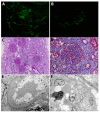
Hypocomplementemic urticarial vasculitis syndrome (HUVS), or McDuffie syndrome, is a rare small vessel vasculitis associated with urticaria, hypocomplementemia and positivity of anti-C1q antibodies. In rare cases, HUVS can manifest as an immune-complex mediated glomerulonephritis with a membranoproliferative pattern of injury. Due to the rarity of this disorder, little is known about the clinical manifestation, pathogenesis, treatment response and outcome of such patients. We describe here three cases of HUVS with severe renal involvement. These patients had a rapidly progressive form of glomerulonephritis with severe nephrotic syndrome against a background of a membranoproliferative pattern of glomerular injury with extensive crescent formation. Therefore, these patients required aggressive induction and maintenance immunosuppressive therapy, with a clinical and renal response in two patients, while the third patient progressed to end-stage renal disease. Because of the rarity of this condition, there are few data regarding the clinical presentation, pathology and outcome of such patients. Accordingly, we provide an extensive literature review of cases reported from 1976 until 2020 and place them in the context of the current knowledge of HUVS pathogenesis. We identified 60 patients with HUVS and renal involvement that had adequate clinical data reported, out of which 52 patients underwent a percutaneous kidney biopsy. The most frequent renal manifestation was hematuria associated with proteinuria (70% of patients), while one third had abnormal kidney function on presentation (estimated glomerular filtration (GFR) below 60 mL/min/1.73 m2). The most frequent glomerular pattern of injury was membranoproliferative (35%), followed by mesangioproliferative (21%) and membranous (19%). Similar to other systemic vasculitis, renal involvement carries a poorer prognosis, but the outcome can be improved by aggressive immunosuppressive treatment.
Keywords: anti-C1q antibodies; crescentic glomerulonephritis; hypocomplementemic urticarial vasculitis syndrome (HUVS); kidney involvement; nephrotic syndrome.
Conflict of interest statement
The authors declare no conflict of interest.
Figures


References
-
- Schwartz H.R., McDuffie F.C., Black L.F., Schroeter A.L., Conn D.L. Hypocomplementemic urticarial vasculitis. Association with chronic obstructive pulmonary disease. Mayo Clin. Proc. 1982;57:231–238. - PubMed
-
- Loricera J., Calvo-Río V., Mata C., Ortiz-Sanjuán F., González-López M.A., Alvarez L., González-Vela M.C., Armesto S., Fernández-Llaca H., Rueda-Gotor J., et al. Urticarial vasculitis in Northern Spain: Clinical study of 21 cases. Medicine. 2014;93:53–60. doi: 10.1097/MD.0000000000000013. - DOI - PMC - PubMed
-
- Jachiet M., Flageul B., Deroux A., Le Quellec A., Maurier F., Cordoliani F., Godmer P., Abasq C., Astudillo L., Belenotti P., et al. The clinical spectrum and therapeutic management of hypocomplementemic urticarial vasculitis: Data from a french nationwide study of fifty-seven patients. Arthritis Rheumatol. 2015;67:527–534. doi: 10.1002/art.38956. - DOI - PubMed
-
- McDuffie F.C., Sams W.M., Maldonado J.E., Andreini P.H., Conn D.L., Samayoa E.A. Hypocomplementemia with cutaneous vasculitis and arthritis. Possible immune complex syndrome. Mayo Clin. Proc. 1973;48:340–348. - PubMed
Publication types
LinkOut - more resources
Full Text Sources

