Review
. 2020 Jul;245(13):1163-1174.
doi: 10.1177/1535370220938741. Epub 2020 Jul 8.

The role of extracellular matrix in normal and pathological pregnancy: Future applications of microphysiological systems in reproductive medicine

Affiliations
Review

The role of extracellular matrix in normal and pathological pregnancy: Future applications of microphysiological systems in reproductive medicine

Blakely B O'Connor et al. Exp Biol Med (Maywood). 2020 Jul.

Abstract

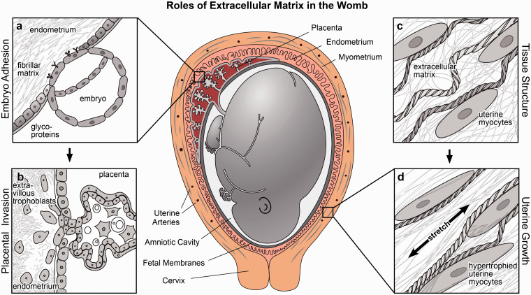
Extracellular matrix in the womb regulates the initiation, progression, and completion of a healthy pregnancy. The composition and physical properties of extracellular matrix in the uterus and at the maternal-fetal interface are remodeled at each gestational stage, while maladaptive matrix remodeling results in obstetric disease. As in vitro models of uterine and placental tissues, including micro-and milli-scale versions of these organs on chips, are developed to overcome the inherent limitations of studying human development in vivo, we can isolate the influence of cellular and extracellular components in healthy and pathological pregnancies. By understanding and recreating key aspects of the extracellular microenvironment at the maternal-fetal interface, we can engineer microphysiological systems to improve assisted reproduction, obstetric disease treatment, and prenatal drug safety.

Keywords: Extracellular matrix; maternal–fetal interface; obstetric diseases; placenta; pregnancy.


Figures

Figure 1.
Figure 1.
Roles of extracellular matrix in the womb. Extracellular provides structural support in the womb and mediates important cellular processes including adhesion, migration, invasion, and mechanical signaling. (a) Extracellular matrix regulates embryonic adhesion by defining a window of receptivity for implantation. (b) Extracellular matrix guides placental invasion and is dynamically remodeled when extravillous trophoblasts invade the endometrium. (c–d) Extracellular matrix forms a fibrillar scaffold to reinforce tissue strength and extensibility, propagating mechanical signals that induce stretch-mediated hypertrophy and uterine contractile activation at the onset of labor.
Figure 2.
Figure 2.
Integrin and matrix metalloproteinase gene expression in placental cell types. (a) First trimester placental villi stained for Collagen IV and counterstained with hematoxylin and simplified illustration (right). Arrow indicates basement membrane staining and box represents globular Col IV in extravillous column (reprinted with permission from Oefner et al.). (b–e) Heatmaps of alpha (panel b) and beta (panel c) integrin and matrix metalloproteinase (panel d) and tissue inhibitors of matrix metalloproteinases (panel e) transcription levels in placental cells (RNA sequencing data were extracted from a previously published dataset) For clarity, only genes with more than 3.6 fragments per kilobase of transcript per million mapped reads (FPKM) in at least one cell type are included.
Figure 3.
Figure 3.
In vitro models of the maternal–fetal interface and embryogenesis. (a) Primary endometrial cells suspended in matrigel self-organize into organoids in chemically defined medium (adapted from Turco et al.). (b) Human trophoblast stem cells form three-dimensional syncytiotrophoblasts aggregates that produced human chorionic gonadotropin (CGB) and syndecan-1 (SDC) a trophoblast marker (adapted from Okae et al.). (c) Mouse embryonic stem cells and extra-embryonic trophoblast stem cells create embryo-like structures in three-dimensional matrigel scaffolds (adapted from Harrison et al.). (d) Microphysiological model of the placental barrier composed of trophoblasts and endothelial cells on a fibronectin-coated membrane, stained with E-cadherin and VE-cadherin, respectively (adapted from Blundell et al.). (e) Microfluidic model of trophoblast invasion, with primary extravillous trophoblasts (EVTs) embedded in Matrigel between two channels that create a chemokine gradient (adapted from Abbas et al. under Creative Commons CC BY 4.0).

References

    1. Napso T, Yong HEJ, Lopez-Tello J, Sferruzzi-Perri AN. The role of placental hormones in mediating maternal adaptations to support pregnancy and lactation. Front Physiol 2018; 9:1091 - PMC - PubMed
    1. Knöfler M. Critical growth factors and signalling pathways controlling human trophoblast invasion. Int J Dev Biol 2010; 54:269–80 - PMC - PubMed
    1. Muiznieks LD, Keeley FW. Molecular assembly and mechanical properties of the extracellular matrix: a fibrous protein perspective. Biochim Biophys Acta 2013; 1832:866–75 - PubMed
    1. Yanagishita M. Function of proteoglycans in the extracellular matrix. Acta Pathol Jpn 1993; 43:283–93 - PubMed
    1. Piez KA. History of extracellular matrix: a personal view. Matrix Biol 1997; 16:85–92 - PubMed

Publication types