. 2020 Sep;40(9):2212-2226.
doi: 10.1161/ATVBAHA.120.314729. Epub 2020 Jul 9.

Fibulin-1 Integrates Subendothelial Extracellular Matrices and Contributes to Anatomical Closure of the Ductus Arteriosus

Affiliations

Fibulin-1 Integrates Subendothelial Extracellular Matrices and Contributes to Anatomical Closure of the Ductus Arteriosus

Satoko Ito et al. Arterioscler Thromb Vasc Biol. 2020 Sep.

Abstract

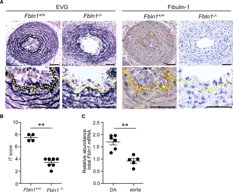
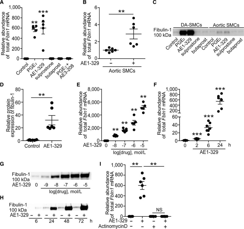
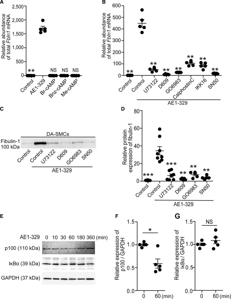
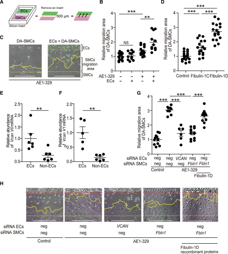
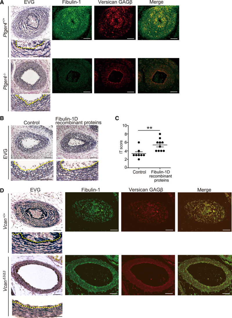
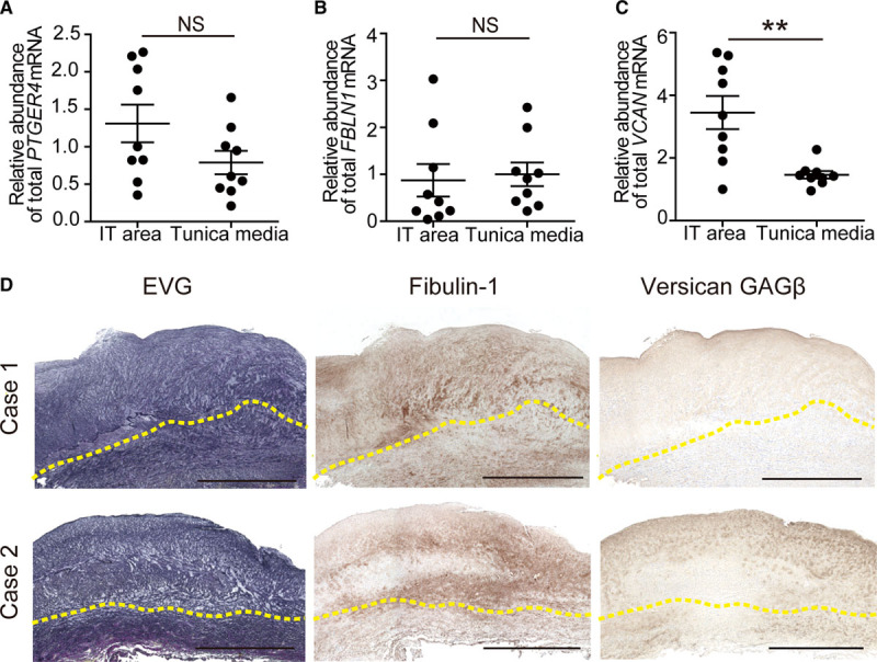
Objective: The ductus arteriosus (DA) is a fetal artery connecting the aorta and pulmonary arteries. Progressive matrix remodeling, that is, intimal thickening (IT), occurs in the subendothelial region of DA to bring anatomic DA closure. IT is comprised of multiple ECMs (extracellular matrices) and migrated smooth muscle cells (SMCs). Because glycoprotein fibulin-1 binds to multiple ECMs and regulates morphogenesis during development, we investigated the role of fibulin-1 in DA closure. Approach and Results: Fibulin-1-deficient (Fbln1-/-) mice exhibited patent DA with hypoplastic IT. An unbiased transcriptome analysis revealed that EP4 (prostaglandin E receptor 4) stimulation markedly increased fibulin-1 in DA-SMCs via phospholipase C-NFκB (nuclear factor κB) signaling pathways. Fluorescence-activated cell sorting (FACS) analysis demonstrated that fibulin-1 binding protein versican was derived from DA-endothelial cells (ECs). We examined the effect of fibulin-1 on directional migration toward ECs in association with versican by using cocultured DA-SMCs and ECs. EP4 stimulation promoted directional DA-SMC migration toward ECs, which was attenuated by either silencing fibulin-1 or versican. Immunofluorescence demonstrated that fibulin-1 and versican V0/V1 were coexpressed at the IT of wild-type DA, whereas 30% of versican-deleted mice lacking a hyaluronan binding site displayed patent DA. Fibulin-1 expression was attenuated in the EP4-deficient mouse (Ptger4-/-) DA, which exhibits patent DA with hypoplastic IT, and fibulin-1 protein administration restored IT formation. In human DA, fibulin-1 and versican were abundantly expressed in SMCs and ECs, respectively.

Conclusions: Fibulin-1 contributes to DA closure by forming an environment favoring directional SMC migration toward the subendothelial region, at least, in part, in combination with EC-derived versican and its binding partner hyaluronan.

Keywords: ductus arteriosus; extracellular matrix; fibulin; mice; neointima; vascular remodeling; versicans.


Conflict of interest statement

None.

Figures

Figure 1.
Figure 1.
Patency of the ductus arteriosus (DA) in neonatal Fbln1−/− mice. A, Elastica van Gieson (EVG) staining and immunohistochemistry for fibulin-1 in the DA tissues from Fbln1+/+ and Fbln1−/− neonates (day 0). Dotted yellow lines indicate internal elastic lamina. Scale bars, 50 µm. B, Scoring of the degree of intimal thickening (IT) in the DAs of Fbln1+/+ and Fbln1−/ mice. n=4–7, **P<0.01. Data are presented as mean±SEM. C, Total fibulin-1 (fibulin-1C and -1D) mRNA expression in the DA and aortic tissues of rats on gestational day 21. n=5–6, **P<0.01. Data are presented as mean±SEM.
Figure 2.
Figure 2.
Fibulin-1 expression was increased by PGE2 (prostaglandin E2)-EP4 (prostaglandin E receptor 4) signaling pathways. A, Expression of total fibulin-1mRNA in rat ductus arteriosus (DA)–smooth muscle cell (SMCs) treated for 24 h by PGE2 (1 μmol/L), ONO-AE1-329 (AE1-329: EP4 agonist, 1 μmol/L), ONO-AE3-208 (AE3-208: EP4 antagonist, 1 μmol/L), sulprostone (EP1/3 agonist, 1 μmol/L), or butaprost (EP2 agonist, 1 μmol/L). n=5–9, **P<0.01, ***P<0.001 vs control. Data are presented as mean±SEM. B, Relative expression of total fibulin-1 (fibulin-1C and-1D) mRNA in aortic SMCs stimulated with ONO-AE1-329 (AE1-329, 1 μmol/L) for 24 h. n=6, **P<0.01. Data are presented as mean±SEM. C, Protein expression of fibulin-1 in the supernatant of rat DA-SMCs and aortic SMCs cultured with each drug as indicated for 72 h. The same dose as (A) was used for each drug. D, Quantification of C. n=6, **P<0.01. Data are presented as mean±SEM. E, Dose-dependent total fibulin-1 mRNA expression after 24 h stimulation with ONO-AE1-329. n=5, **P<0.01 vs control. Data are presented as mean±SEM. F, Time-dependent fibulin-1 mRNA expression after stimulation by ONO-AE1-329 (AE1-329, 1 μmol/L). n=6–9, ***P<0.001 vs control. Data are presented as mean±SEM. G, Dose-dependent fibulin-1 protein production in supernatant of DA-SMCs treated for 72 h by ONO-AE1-329. H, Time-dependent fibulin-1 protein production in supernatant of DA-SMCs treated with ONO-AE1-329 (AE1-329, 1 μmol/L). I, EP4-induced total fibulin-1 mRNA expression in rat DA-SMCs treated with or without actinomycin D (5 μg/mL) for 24 h. n=6, **P<0.01. Data are presented as mean±SEM. NS indicates not significant.
Figure 3.
Figure 3.
The PLC (phospholipase C)-PKC (protein kinase C)–noncanonical NFκB (nuclear factor κB) signaling pathway plays a primary role in the EP4 (prostaglandin E receptor 4)-mediated increase in fibulin-1. A, Fibulin-1 mRNA expression in ductus arteriosus (DA)–smooth muscle cells (SMCs) treated for 24 h with ONO-AE1-329 (AE1-329, 1 μmol/L) or cAMP analogs (50 μmol/L): 8-bromo-cAMP (Br-cAMP), nonselective; N6-benzoyladenosine-cAMP (Bnz-cAMP), PKA (protein kinase A)-selective; and 8-p-methoxyphenylthon-2’-O-methyl-cAMP (Me-cAMP), Epac (exchange protein activated by a cAMP)-selective cAMP analogs. n=5–6, **P<0.01. Not significant (NS) vs control. Data are presented as mean±SEM. B, Fibulin-1 mRNA expression in DA-SMCs after 24-h stimulation by ONO-AE1-329 (AE1-329, 1 μmol/L) with or without PLC inhibitors, U73122 (5 μmol/L) or D609 (50 μmol/L); PKC inhibitors, GO6983 (10 μmol/L) or Calphostin C (0.2 μmol/L); or NFκB inhibitors, IKK (IκB kinase) 16 (3 μmol/L) or SN50 (20 μmol/L). n=5, **P<0.01 vs control with ONO-AE1-329. Data are presented as mean±SEM. C, Protein expression of fibulin-1 in supernatant of rat DA-SMCs treated for 72 h by ONO-AE1-329 (AE1-329, 1 μmol/L) combined with PLC inhibitors, U73122 (5 μmol/L) or D609 (50 μmol/L); PKC inhibitor, GO6983 (10 μmol/L); or NFκB inhibitor, SN50 (20 μmol/L). D, Quantification of (C). n=5-8, **P<0.01, ***P<0.001 vs control with ONO-AE1-329. Data are presented as mean±SEM. E, Protein expression of p100, IκBα (nuclear factor kappa-B inhibitor, alpha), and GAPDH at 0–360 min in DA-SMCs stimulated by ONO-AE1-329 (AE1-329, 1 μmol/L). F and G, Quantification of E at 0 min and 60 min. n=5, *P<0.05. Data are presented as mean±SEM.
Figure 4.
Figure 4.
Fibulin-1 in cooperation with versican promoted directional smooth muscle cell (SMC) migration toward endothelial cells (ECs). A, A method of coculture using a silicon-insert is shown as a schematic representation. EA. hy926 cells (ECs) were labeled with GFP (green fluorescence protein) or PKH67 Green Fluorescent Cell Linker Midi Kit. Migration areas for 96 h were quantified. Dotted red lines, boundary of ECs; dotted yellow lines, start line of ductus arteriosus (DA)-SMCs. B, Quantification of DA-SMC migration area. DA-SMCs were pretreated with or without ONO-AE1-329 (AE1-329, 1 μmol/L) before removal of the silicon-insert. DA-SMCs were cultured with or without ECs. n=11–19, **P<0.01, ***P<0.001. Data are presented as mean±SEM. C, Representative images of B. Dotted red lines, boundary of ECs; dotted yellow lines, start line of DA-SMCs; solid yellow lines, 96-h migrated lines of DA-SMCs. D, Quantification of DA-SMC migration area. DA-SMCs were pretreated with fibulin-1–targeted siRNA (small interfering RNA) and cultured with ECs under stimulation of fibulin-1C or fibulin-1D recombinant proteins for 24 h. The amounts of fibulin-1C and fibulin-1D were equal (1.10 μg/mL). n=16–18, ***P<0.001. Data are presented as mean±SEM. E and F, Expression of versican V0 and V1 mRNAs in rat DA-ECs and non-ECs that were isolated by fluorescence-activated cell sorting (FACS). n=5–6, **P<0.01. Data are presented as mean±SEM. G, Migration areas of DA-SMCs treated with or without Fbln1-targeted siRNA. EA. hy926 cells (ECs) were treated with or without VCAN-targeted siRNA. DA-SMCs were treated with or without ONO-AE1-329 (AE1-329, 1 μmol/L) after siRNA transfection. Fibulin-1D recombinant proteins were added after silencing of Fbln1 in DA-SMCs. n=6-12, ***P<0.001. Data are presented as mean±SEM. H, Representative images of G. Dotted red lines, boundary of ECs; dotted yellow lines, start line of DA-SMCs; and solid yellow lines, 96-h migrated lines of DA-SMCs. Scale bars, 500 μm. Neg indicates negative control siRNA; and NS, not significant.
Figure 5.
Figure 5.
Fibulin-1-versican complex and fibulin-1-versican–hyaluronic acid complex promote intimal thickening (IT) formation of the ductus arteriosus (DA) in mice. A, Elastica van Gieson staining (EVG) and immunofluorescent staining for fibulin-1 (green) and versican glycosaminoglycan β (GAGβ) domain (red) in the DA of EP4 (prostaglandin E receptor 4)-deficient (Ptger4−/−) and wild-type (Ptger4+/+) neonatal mice (day 0). Dotted yellow lines indicate internal elastic lamina. Scale bars, 50 µm. B, Effect of fibulin-1D recombinant proteins (1.10 μg/mL) on IT formation of organ-cultured Ptger4−/− DAs. Dotted yellow lines show internal elastic lamina. Scale bars, 50 µm. C, IT formation of B was evaluated by IT score. n=8-10, **P<0.01. Data are presented as mean±SEM. D, EVG and immunofluorescent staining for fibulin-1 (green) and versican GAGβ domain (red) in the DAs from versican G1 A-subdomain deleted (VcanΔ3/Δ3) and wild-type (Vcan+/+) neonatal mice (day 0). Dotted yellow lines indicate internal elastic lamina. Scale bars, 50 µm.
Figure 6.
Figure 6.
Human ductus arteriosus (DA) tissues show a similar distribution to rodents in fibulin-1 and versican. A–C, Expression of EP4 (prostaglandin E receptor 4; PTGER4), total fibulin-1 (FBLN1), and total versican (VCAN) mRNAs in the intimal thickening (IT) area and the tunica media of human DA tissues. n=9, **P<0.01. Data are presented as mean±SEM. D, Elastica van Gieson (EVG) staining and immunohistological analysis of fibulin-1 and versican glycosaminoglycan β (GAGβ) domain in human DA tissues. Dotted yellow lines show internal elastic lamina. Case 1, patient no. 10; case 2, patient no. 11 in Table II in the Data Supplement. Scale bars, 1 mm. NS indicates not significant.

Similar articles

Cited by

References

    1. Yokoyama U. Prostaglandin E-mediated molecular mechanisms driving remodeling of the ductus arteriosus. Pediatr Int 201557820–827doi: 10.1111/ped.12769 - PubMed
    1. Mitra S, Florez ID, Tamayo ME, Mbuagbaw L, Vanniyasingam T, Veroniki AA, Zea AM, Zhang Y, Sadeghirad B, Thabane L. Association of placebo, indomethacin, ibuprofen, and acetaminophen with closure of hemodynamically significant patent ductus arteriosus in preterm infants: a systematic review and meta-analysis. JAMA 20183191221–1238doi: 10.1001/jama.2018.1896 - PMC - PubMed
    1. Ito S, Matsuda T, Usuda H, Watanabe S, Kitanishi R, Hanita T, Watanabe T, Adachi O. Surgical ligation for patent ductus arteriosus in extremely premature infants: strategy to reduce their risk of neurodevelopmental impairment. Tohoku J Exp Med 20162407–13doi: 10.1620/tjem.240.7 - PubMed
    1. Noori S, McCoy M, Friedlich P, Bright B, Gottipati V, Seri I, Sekar K. Failure of ductus arteriosus closure is associated with increased mortality in preterm infants. Pediatrics 2009123e138–e144doi: 10.1542/peds.2008-2418 - PubMed
    1. Clyman RI. Ibuprofen and patent ductus arteriosus. N Engl J Med 2000343728–730doi: 10.1056/NEJM200009073431009 - PubMed

Publication types

MeSH terms