

Extracellular vesicles: novel communicators in lung diseases
- PMID: 32641036
- PMCID: PMC7341477
- DOI: 10.1186/s12931-020-01423-y
Extracellular vesicles: novel communicators in lung diseases
Abstract
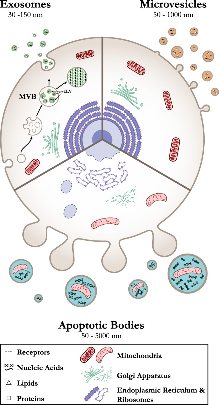
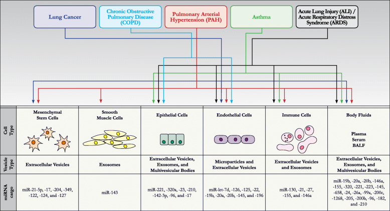
The lung is the organ with the highest vascular density in the human body. It is therefore perceivable that the endothelium of the lung contributes significantly to the circulation of extracellular vesicles (EVs), which include exosomes, microvesicles, and apoptotic bodies. In addition to the endothelium, EVs may arise from alveolar macrophages, fibroblasts and epithelial cells. Because EVs harbor cargo molecules, such as miRNA, mRNA, and proteins, these intercellular communicators provide important insight into the health and disease condition of donor cells and may serve as useful biomarkers of lung disease processes. This comprehensive review focuses on what is currently known about the role of EVs as markers and mediators of lung pathologies including COPD, pulmonary hypertension, asthma, lung cancer and ALI/ARDS. We also explore the role EVs can potentially serve as therapeutics for these lung diseases when released from healthy progenitor cells, such as mesenchymal stem cells.
Conflict of interest statement
The authors declare that they have no competing interests.
Figures
References
-
- Lai FW, Lichty BD, Bowdish DM. Microvesicles: ubiquitous contributors to infection and immunity. J Leukoc Biol. 2015;97:237–245. - PubMed
-
- Johnstone RM. The Jeanne Manery-Fisher Memorial Lecture 1991. Maturation of reticulocytes: formation of exosomes as a mechanism for shedding membrane proteins. Biochem Cell Biol. 1992;(70):179–90. - PubMed
-
- Wolf P. The nature and significance of platelet products in human plasma. Br J Haematol. 1967;13:269–288. - PubMed
Publication types
MeSH terms
Substances
Grants and funding
LinkOut - more resources
Full Text Sources
Medical
