. 2020 Aug 21;295(34):12203-12213.
doi: 10.1074/jbc.RA120.012732. Epub 2020 Jul 8.

In vivo two-photon microscopy reveals the contribution of Sox9+ cell to kidney regeneration in a mouse model with extracellular vesicle treatment

Affiliations

In vivo two-photon microscopy reveals the contribution of Sox9+ cell to kidney regeneration in a mouse model with extracellular vesicle treatment

Kaiyue Zhang et al. J Biol Chem. .

Abstract

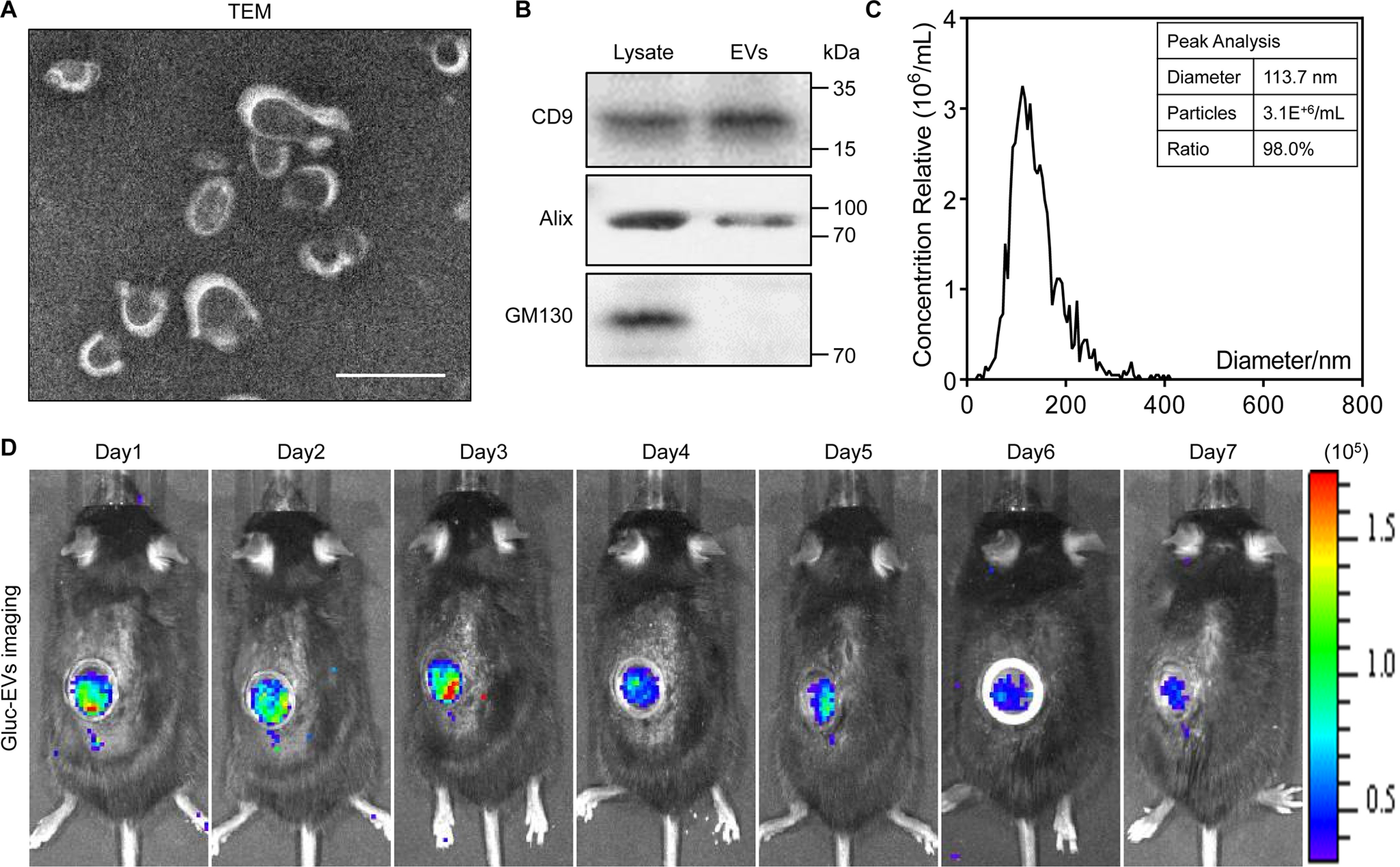
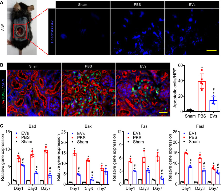
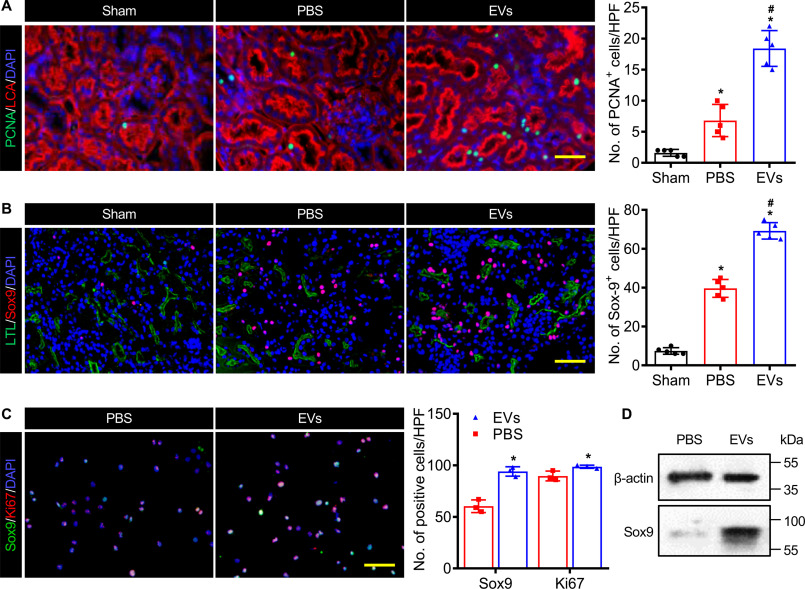
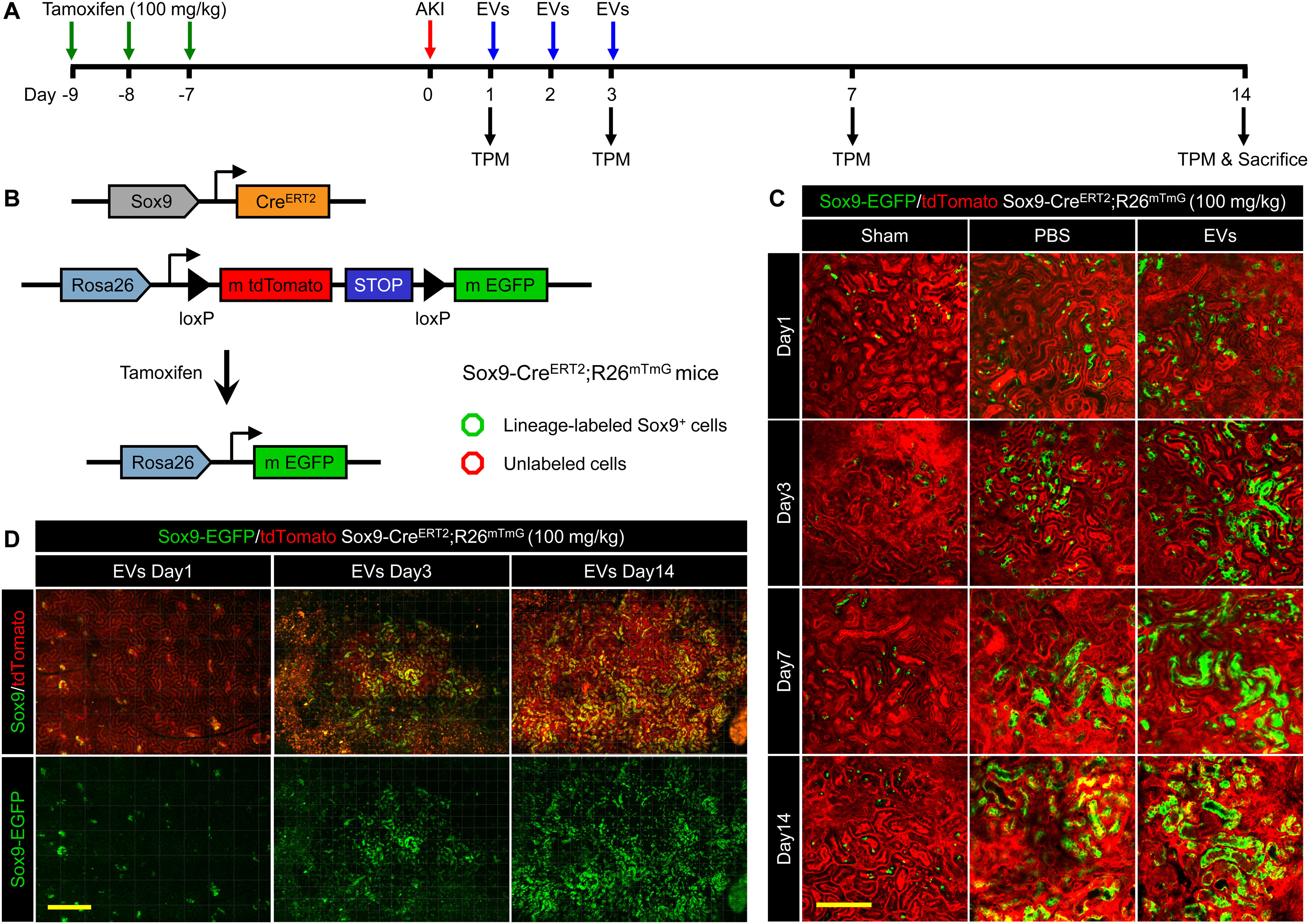
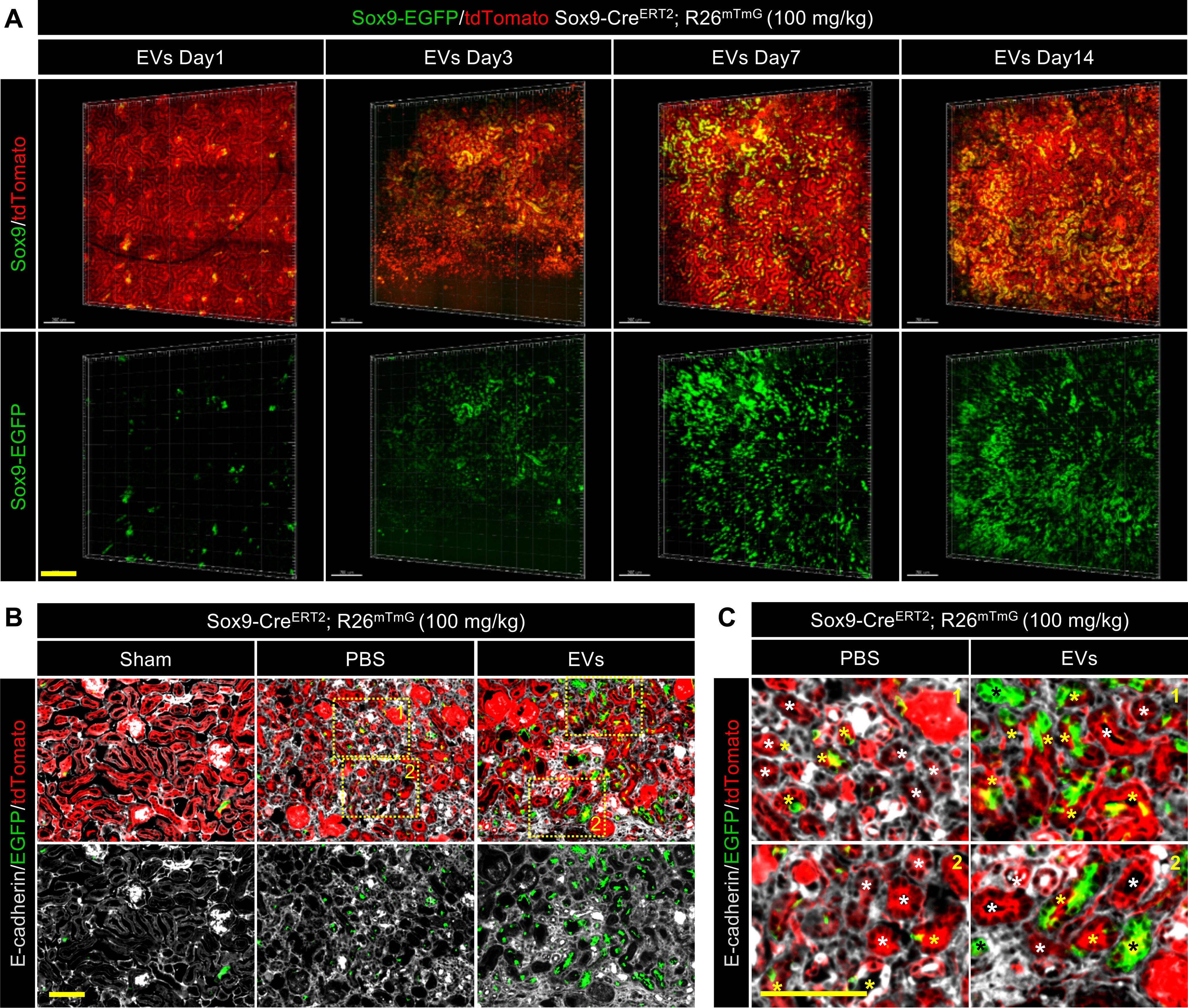
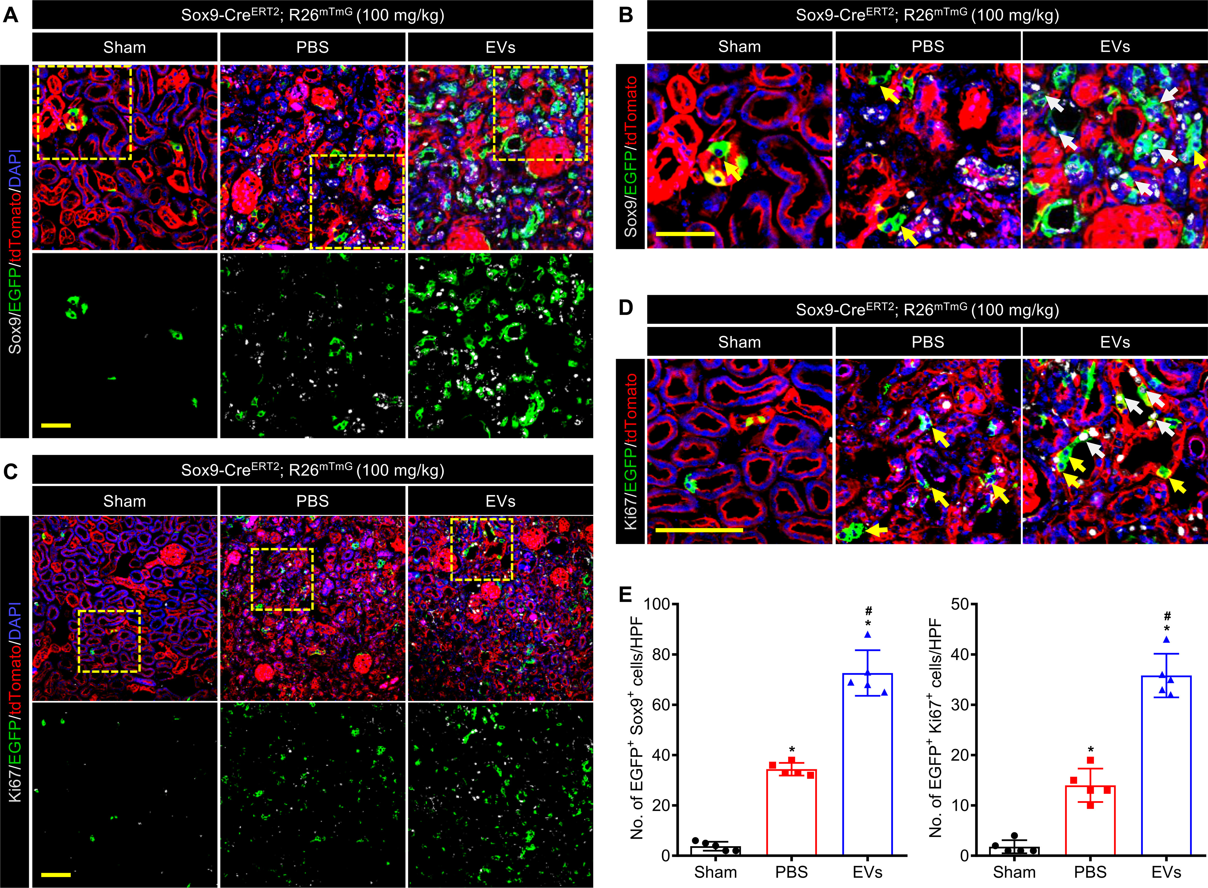
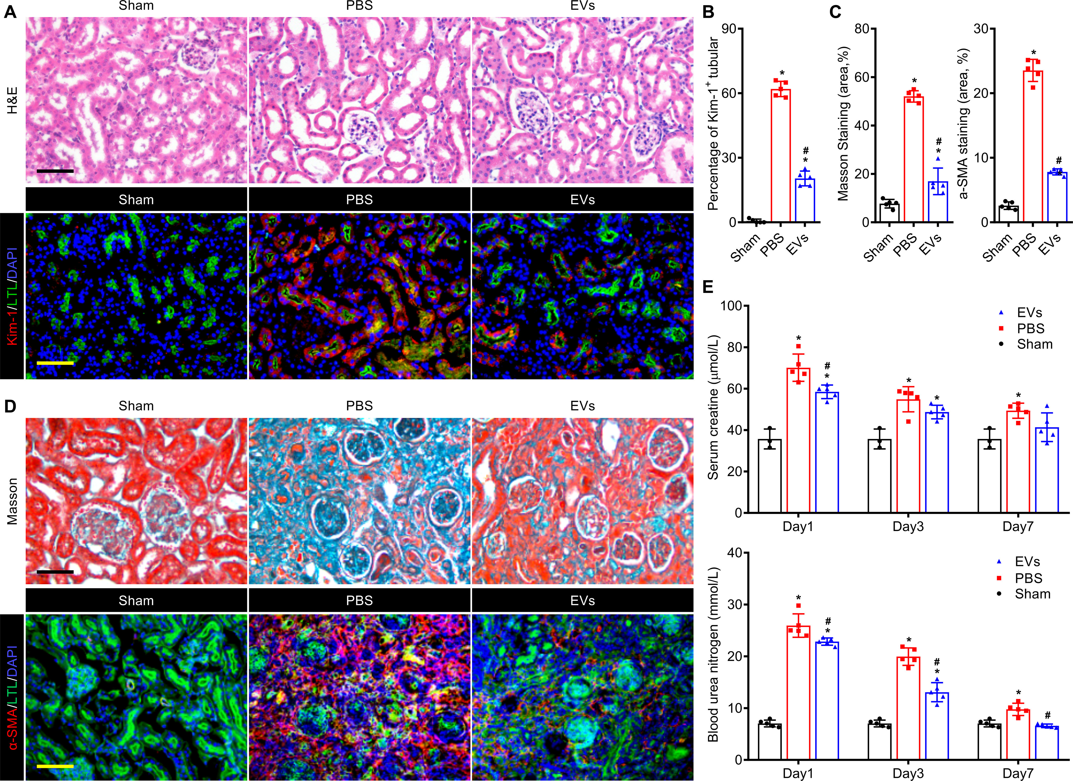
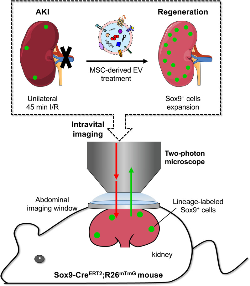
Mesenchymal stem cell (MSC)-derived extracellular vesicles (EVs) have been shown to stimulate regeneration in the treatment of kidney injury. Renal regeneration is also thought to be stimulated by the activation of Sox9+ cells. However, whether and how the activation mechanisms underlying EV treatment and Sox9+ cell-dependent regeneration intersect is unclear. We reasoned that a high-resolution imaging platform in living animals could help to untangle this system. To test this idea, we first applied EVs derived from human placenta-derived MSCs (hP-MSCs) to a Sox9-CreERT2; R26mTmG transgenic mouse model of acute kidney injury (AKI). Then, we developed an abdominal imaging window in the mouse and tracked the Sox9+ cells in the inducible Sox9-Cre transgenic mice via in vivo lineage tracing with two-photon intravital microscopy. Our results demonstrated that EVs can travel to the injured kidneys post intravenous injection as visualized by Gaussia luciferase imaging and markedly increase the activation of Sox9+ cells. Moreover, the two-photon living imaging of lineage-labeled Sox9+ cells showed that the EVs promoted the expansion of Sox9+ cells in kidneys post AKI. Histological staining results confirmed that the descendants of Sox9+ cells contributed to nephric tubule regeneration which significantly ameliorated the renal function after AKI. In summary, intravital lineage tracing with two-photon microscopy through an embedded abdominal imaging window provides a practical strategy to investigate the beneficial functions and to clarify the mechanisms of regenerative therapies in AKI.

Keywords: Sox9; acute kidney injury; extracellular vesicles; intravital microscopy; kidney; lineage tracing; mesenchymal stem cells (MSCs); microscopic imaging; regenerative medicine; two-photon microscopy.


Conflict of interest statement

Conflict of interest—The authors declare that they have no conflicts of interest with the contents of this article.

Figures

Figure 1.
Figure 1.
Characteristics and bioluminescence imaging of EVs. A, transmission electron microscope (TEM) image of EVs. Scale bar, 100 nm. B, Western blot analysis confirmed the three categories of exosomal markers: CD9, Alix, and GM130. C, nanoparticle tracking analysis indicates the peak diameter of EVs is 113.7 nm. D, the biodistribution of EVs was traced in vivo by Gaussia luciferase (Gluc) imaging through the AIW.
Figure 2.
Figure 2.
EVs suppressed apoptosis of the kidney after ischemia-reperfusion injury. A, fluorescence stereomicroscope imaging of the injured kidney stained by Hoechst 33342 through the embedded AIW. Scale bar, 100 μm. B, the TUNEL staining (green) of the kidney sections (rhodamine-labeled LCA, red, proximal tubular) on day 3. Scale bar, 50 μm. C, real-time qPCR analysis of apoptosis-related genes in injured kidneys on days 1, 3, and 7 after IRI. Relative gene expression was normalized to Gapdh. The experiments were performed in triplicate. Data are expressed as scatter plots with mean ± S.D. *, P < 0.05 versus Sham; #, P < 0.05 versus PBS.
Figure 3.
Figure 3.
EVs improved the Sox9 activation in the injured kidney. A, anti-PCNA immunostaining (green) of proximal tubules (rhodamine-labeled LCA, red) on day 3 post IRI. Scale bar, 50 μm. B, representative images and analysis of anti-Sox9 immunostaining (red) in kidney (FITC-labeled LTL, green, proximal tubules) at 3 days after IRI. Scale bar, 100 μm. C, immunostaining analysis for Sox9 (green) and Ki67 (red) in HK2 cells treated with EVs for 24 h. Scale bar, 100 μm. DAPI demarcates nuclei. D, Western blot analysis of Sox9 expression in HK2 cells treated with EVs for 24 h. Data are expressed as scatter plots with mean ± S.D. *, P < 0.05 versus Sham; #, P < 0.05 versus PBS.
Figure 4.
Figure 4.
Two-photon living imaging of Sox9+ cells expansion following EV treatment. A, schematic illustration for two-photon living imaging of lineage tracing marked Sox9+ cells after renal IRI and EV treatment. Sox9-CreERT2; R26mTmG mice were injected intraperitoneally with tamoxifen once a day for 3 continuous days to label the Sox9+ cells. 7 days after the final tamoxifen injection, the renal ischemia-reperfusion injury was performed in these mice with simultaneous implantation of AIW. The EVs or PBS were intravenously injected once a day for 3 continuous days post IRI. The two-photon living imaging was carried out on days 1, 3, 7, and 14 after IRI. The kidney tissues were collected on day 14 after the injury. B, the scheme of generation of a tamoxifen-inducible Sox9-CreERT2; R26mTmG mice for genetic lineage tracing. C, the representative images of two-photon intravital tracing showed Sox9+ cell-derived cells abundantly expanded with the administration of EVs. Scale bar, 200 μm. D, whole-vision scans of the injured kidney from the same mouse treated with EVs through the embedded AIW by using a two-photon microscope on days 1, 3, and 14 post IRI. Scale bars, 500 μm.
Figure 5.
Figure 5.
EVs promoted the formation of functional renal tubules by descendants of the Sox9+ cells. A, 3D reconstruction of the dynamic variation in the injured kidney treated with EVs by using two-photon living imaging. Scale bar, 200 μm. B and C, representative images (B) and local zoom images (C) for co-localization analysis of anti-E-cadherin immunostaining (gray) and Sox9-CreERT2–activated EGFP fluorescence in kidneys at 14 days post injury. White asterisks highlighted the tubules formed by descendants of Sox9 cells. Yellow asterisks highlighted the tubules formed by descendants of both Sox9+ cells and Sox9 cells. Black asterisks highlighted the tubules formed by descendants of only Sox9+ cells. Scale bar, 100 μm.
Figure 6.
Figure 6.
EVs activated Sox9 expression and Sox9+ cells proliferation during renal regeneration. A and B, confocal images (A) and local zoom images (B) for co-localization analysis of anti-Sox9 immunostaining (gray) and Sox9-CreERT2 activated EGFP fluorescence in kidneys at 14 days post injury. White arrowheads highlighted Sox9+/EGFP co-labeled cells, and yellow arrowheads highlighted the Sox9 but EGFP-labeled cells. Scale bar, 50 μm. C and D, confocal images (C) and local zoom images (D) for co-localization analysis of anti-Ki67 immunostaining (gray) and Sox9-CreERT2–activated EGFP fluorescence in kidneys at 14 days post injury. White arrowheads highlighted Ki67+/EGFP co-labeled cells, and yellow arrowheads highlighted the Ki67 but EGFP-labeled cells. Scale bar, 100 μm. E, quantification of Sox9+/EGFP co-labeled cells (A) and Ki67+/EGFP co-labeled cells (C) in the kidneys of the mice administrated with PBS or EVs on day 14 post IRI. Data are expressed as scatter plots with mean ± S.D. *, P < 0.05 versus Sham; #, P < 0.05 versus PBS.
Figure 7.
Figure 7.
EVs attenuated renal injury and promoted kidney regeneration in renal IRI mouse model. A, histological analysis of kidney injury by H&E staining and anti–Kim-1 immunostaining (red) on day 3 post IRI. The proximal tubules were co-stained by FITC-labeled LTL (green). Scale bar, 100 μm. B, quantitative assessments of Kim-1–positive injured tubules. C, quantitative assessments of fibrotic area in Masson staining and anti–α-SMA immunostaining. D, representative images of Masson staining and anti–α-SMA immunostaining (red) for renal tissues harvested on day 28 post IRI. The proximal tubules were co-stained by FITC-labeled LTL (green). Scale bar, 100 μm. E, serum creatinine and blood urea nitrogen levels of each group were measured on days 1, 3, and 7 post IRI. Data are expressed as scatter plots with mean ± S.D. *, P < 0.05 versus Sham; #, P < 0.05 versus PBS.
Figure 8.
Figure 8.
Schematic diagram of two-photon living imaging of dynamic Sox9-dependent renal regeneration activated by EVs. Intravital lineage tracing with two-photon intravital microscopy through an abdominal imaging window provides a practical strategy for investigating Sox9-dependent EVs treatment at a high resolution in Sox9-CreERT2; R26mTmG mouse.

References

    1. Lameire N. H., Bagga A., Cruz D., De Maeseneer J., Endre Z., Kellum J. A., Liu K. D., Mehta R. L., Pannu N., Van Biesen W., and Vanholder R. (2013) Acute kidney injury: An increasing global concern. Lancet 382, 170–179 10.1016/S0140-6736(13)60647-9 - DOI - PubMed
    1. Hoste E. A. J., Kellum J. A., Selby N. M., Zarbock A., Palevsky P. M., Bagshaw S. M., Goldstein S. L., Cerdá J., and Chawla L. S. (2018) Global epidemiology and outcomes of acute kidney injury. Nat. Rev. Nephrol. 14, 607–625 10.1038/s41581-018-0052-0 - DOI - PubMed
    1. Kumar S., Liu J., Pang P., Krautzberger A. M., Reginensi A., Akiyama H., Schedl A., Humphreys B. D., and McMahon A. P. (2015) Sox9 activation highlights a cellular pathway of renal repair in the acutely injured mammalian kidney. Cell Rep. 12, 1325–1338 10.1016/j.celrep.2015.07.034 - DOI - PubMed
    1. Kang H. M., Huang S., Reidy K., Han S. H., Chinga F., and Susztak K. (2016) Sox9-positive progenitor cells play a key role in renal tubule epithelial regeneration in mice. Cell Rep. 14, 861–871 10.1016/j.celrep.2015.12.071 - DOI - PMC - PubMed
    1. Agrahari V., Agrahari V., Burnouf P. A., Chew C. H., and Burnouf T. (2019) Extracellular microvesicles as new industrial therapeutic frontiers. Trends Biotechnol. 37, 707–729 10.1016/j.tibtech.2018.11.012 - DOI - PubMed

Publication types

MeSH terms

Substances