

Cerebrovascular reactivity in cerebral amyloid angiopathy, Alzheimer disease, and mild cognitive impairment
- PMID: 32641520
- PMCID: PMC7538216
- DOI: 10.1212/WNL.0000000000010201
Cerebrovascular reactivity in cerebral amyloid angiopathy, Alzheimer disease, and mild cognitive impairment
Abstract
Objective: To assess cerebrovascular reactivity in response to a visual task in participants with cerebral amyloid angiopathy (CAA), Alzheimer disease (AD), and mild cognitive impairment (MCI) using fMRI.
Methods: This prospective cohort study included 40 patients with CAA, 22 with AD, 27 with MCI, and 25 healthy controls. Each participant underwent a visual fMRI task using a contrast-reversing checkerboard stimulus. Visual evoked potentials (VEPs) were used to compare visual cortex neuronal activity in 83 participants. General linear models using least-squares means, adjusted for multiple comparisons with the Tukey test, were used to estimate mean blood oxygen level-dependent (BOLD) signal change during the task and VEP differences between groups.
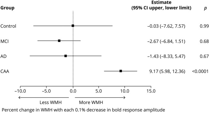
Results: After adjustment for age and hypertension, estimated mean BOLD response amplitude was as follows: CAA 1.88% (95% confidence interval [CI] 1.60%-2.15%), AD 2.26% (1.91%-2.61%), MCI 2.15% (1.84%-2.46%), and control 2.65% (2.29%-3.00%). Only patients with CAA differed from controls (p = 0.01). In the subset with VEPs, group was not associated with prolonged latencies or lower amplitudes. Lower BOLD amplitude response was associated with higher white matter hyperintensity (WMH) volumes in CAA (for each 0.1% lower BOLD response amplitude, the WMH volume was 9.2% higher, 95% CI 6.0%-12.4%) but not other groups (p = 0.002 for interaction) when controlling for age and hypertension.
Conclusions: Mean visual BOLD response amplitude was lowest in participants with CAA compared to controls, without differences in VEP latencies and amplitudes. This suggests that the impaired visual BOLD response is due to reduced vascular reactivity in CAA. In contrast to participants with CAA, the visual BOLD response amplitude did not differ between those with AD or MCI and controls.
© 2020 American Academy of Neurology.
Figures


Comment in
-
Impaired occipital cerebrovascular reactivity as a biomarker for vascular β-amyloid.Neurology. 2020 Sep 8;95(10):415-416. doi: 10.1212/WNL.0000000000010207. Epub 2020 Jul 8. Neurology. 2020. PMID: 32641536 No abstract available.
References
-
- Weller R, Boche D, Nicoll JR. Microvasculature changes and cerebral amyloid angiopathy in Alzheimer's disease and their potential impact on therapy. Acta Neuropathol 2009;118:87–102. - PubMed
-
- Cupino T, Zabel M. Alzheimer's silent partner: cerebral amyloid angiopathy. Transl Stroke Res 2013;5:330–337. - PubMed
-
- Shin HK, Jones PB, Garcia-Alloza M, et al. . Age-dependent cerebrovascular dysfunction in a transgenic mouse model of cerebral amyloid angiopathy. Brain 2007;130:2310–2319. - PubMed
Publication types
MeSH terms
Grants and funding
LinkOut - more resources
Full Text Sources
Medical