

Time course and diagnostic utility of NfL, tau, GFAP, and UCH-L1 in subacute and chronic TBI
- PMID: 32641529
- PMCID: PMC7455355
- DOI: 10.1212/WNL.0000000000009985
Time course and diagnostic utility of NfL, tau, GFAP, and UCH-L1 in subacute and chronic TBI
Erratum in
-
Time Course and Diagnostic Utility of Nfl, Tau, GFAP, and UCH-L1 in Subacute and Chronic TBI.Neurology. 2021 Mar 23;96(12):593. doi: 10.1212/WNL.0000000000011717. Neurology. 2021. PMID: 33753530 Free PMC article. No abstract available.
Abstract
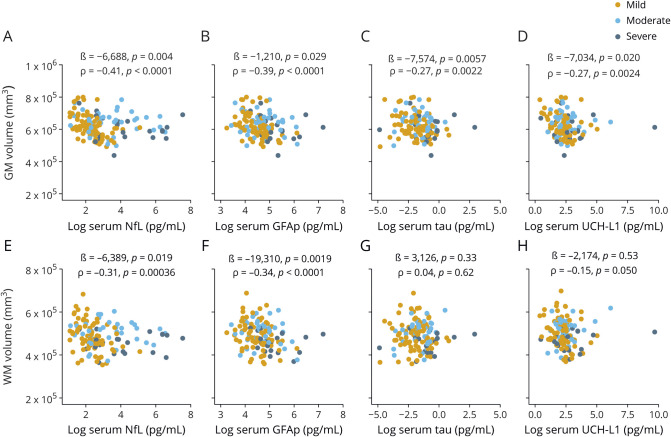
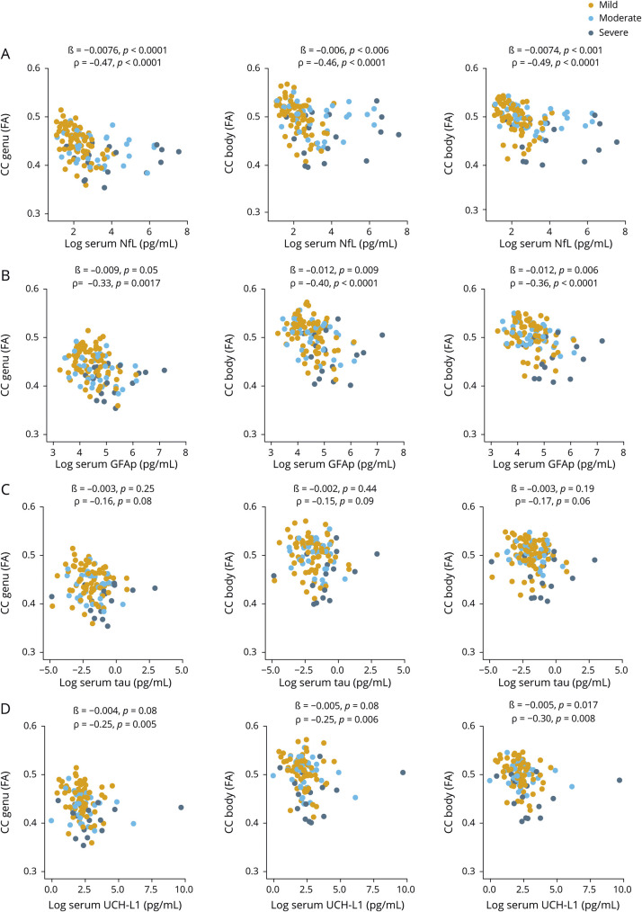
Objective: To determine whether neurofilament light (NfL), glial fibrillary acidic protein (GFAP), tau, and ubiquitin C-terminal hydrolase-L1 (UCH-L1) measured in serum relate to traumatic brain injury (TBI) diagnosis, injury severity, brain volume, and diffusion tensor imaging (DTI) measures of traumatic axonal injury (TAI) in patients with TBI.
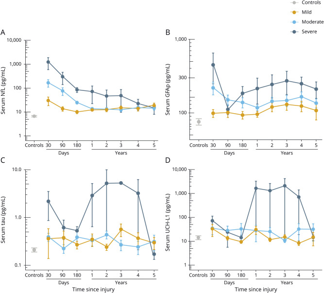
Methods: Patients with TBI (n = 162) and controls (n = 68) were prospectively enrolled between 2011 and 2019. Patients with TBI also underwent serum, functional outcome, and imaging assessments at 30 (n = 30), 90 (n = 48), and 180 (n = 59) days, and 1 (n = 84), 2 (n = 57), 3 (n = 46), 4 (n = 38), and 5 (n = 29) years after injury.
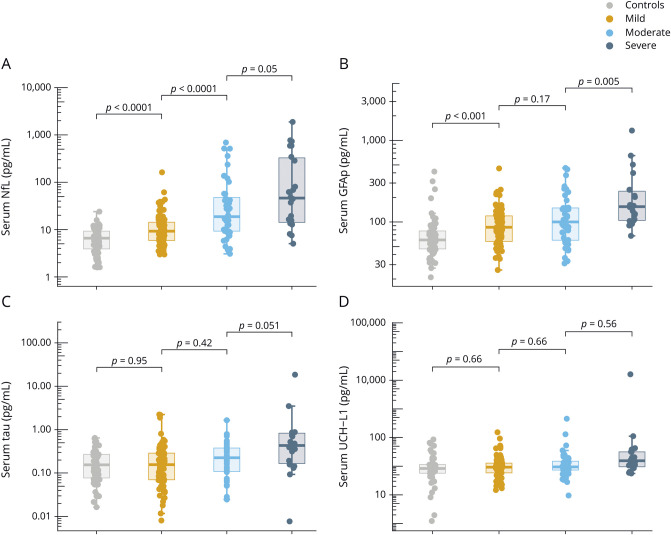
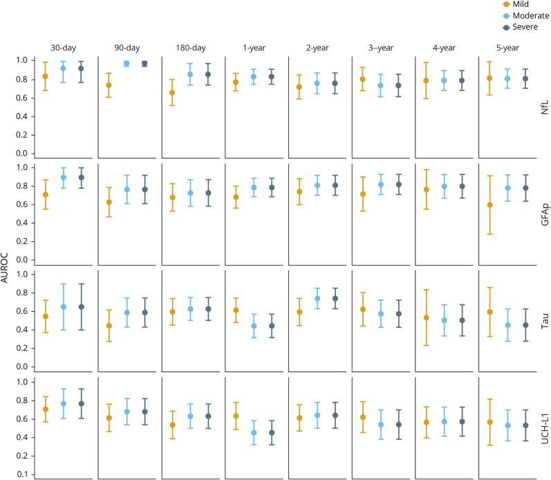
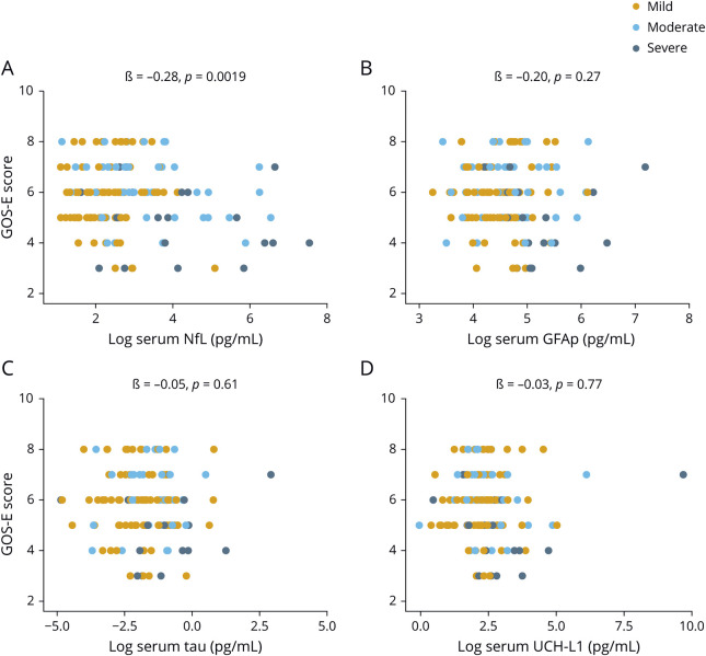
Results: At enrollment, patients with TBI had increased serum NfL compared to controls (p < 0.0001). Serum NfL decreased over the course of 5 years but remained significantly elevated compared to controls. Serum NfL at 30 days distinguished patients with mild, moderate, and severe TBI from controls with an area under the receiver-operating characteristic curve (AUROC) of 0.84, 0.92, and 0.92, respectively. At enrollment, serum GFAP was elevated in patients with TBI compared to controls (p < 0.001). GFAP showed a biphasic release in serum, with levels decreasing during the first 6 months of injury but increasing over the subsequent study visits. The highest AUROC for GFAP was measured at 30 days, distinguishing patients with moderate and severe TBI from controls (both 0.89). Serum tau and UCH-L1 showed weak associations with TBI severity and neuroimaging measures. Longitudinally, serum NfL was the only biomarker that was associated with the likely rate of MRI brain atrophy and DTI measures of progression of TAI.
Conclusions: Serum NfL shows greater diagnostic and prognostic utility than GFAP, tau, and UCH-L1 for subacute and chronic TBI.
Classification of evidence: This study provides Class III evidence that serum NfL distinguishes patients with mild TBI from healthy controls.
© 2020 American Academy of Neurology.
Figures
Comment in
-
Progress in the diagnosis of traumatic brain injury.Neurology. 2020 Aug 11;95(6):235-236. doi: 10.1212/WNL.0000000000009992. Epub 2020 Jul 8. Neurology. 2020. PMID: 32641522 No abstract available.
References
-
- Smith DH, Chen XH, Pierce JE, et al. Progressive atrophy and neuron death for one year following brain trauma in the rat. J Neurotrauma 1997;14:715–727. - PubMed
Publication types
MeSH terms
Substances
LinkOut - more resources
Full Text Sources
Medical
Research Materials
Miscellaneous