. 2020 Jul 8;10(1):11226.
doi: 10.1038/s41598-020-66959-x.

Sevoflurane induces neuronal activation and behavioral hyperactivity in young mice

Affiliations

Sevoflurane induces neuronal activation and behavioral hyperactivity in young mice

Lei Yang et al. Sci Rep. .

Abstract

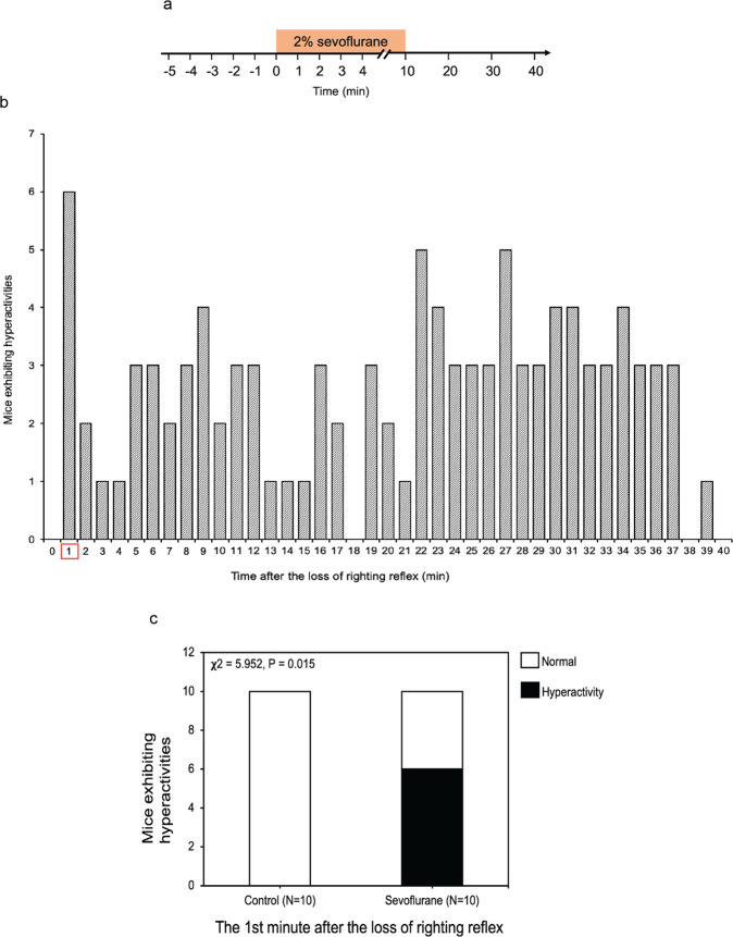
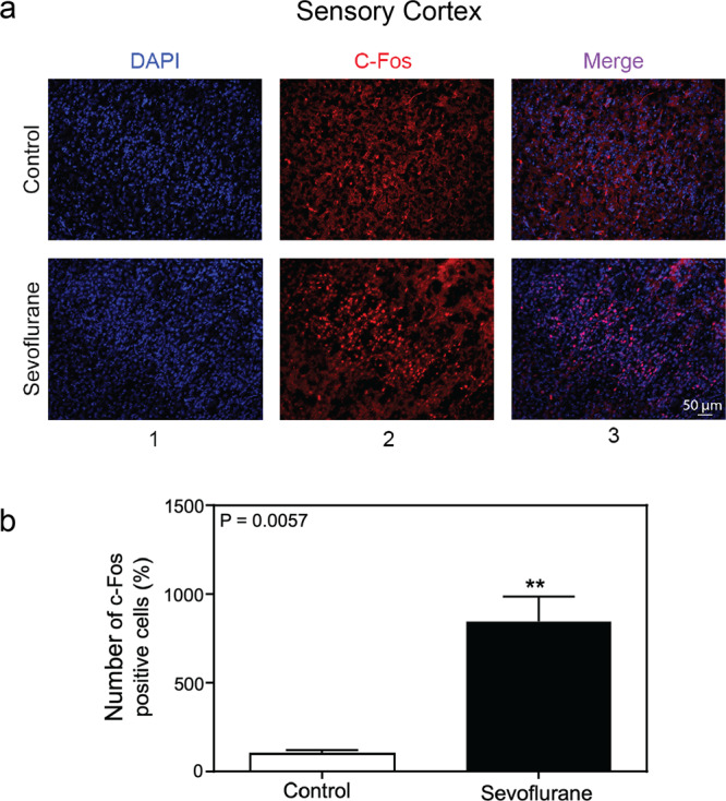
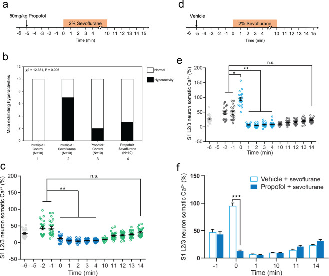
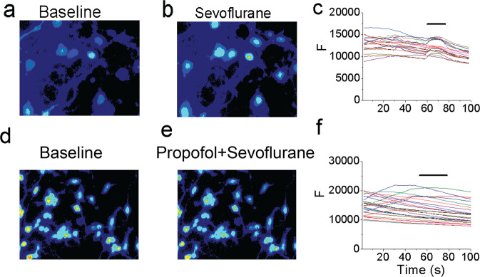
Sevoflurane, a commonly used anesthetic, may cause agitation in patients. However, the mechanism underlying this clinical observation remains largely unknown. We thus assessed the effects of sevoflurane on neuronal activation and behaviors in mice. Ten-day-old mice received 2% sevoflurane, 1% isoflurane, or 6% desflurane for 10 minutes. The behavioral activities were recorded and evaluated at one minute after the loss of righting reflex in the mice, which was about two minutes after the anesthetic administration. The neuronal activation was evaluated by c-Fos expression and calcium imaging at one minute after the anesthetic administration. Propofol, which reduces neuronal activation, was used to determine the cause-and-effect of sevoflurane. We found that sevoflurane caused an increase in neuronal activation in primary somatosensory cortex of young mice and behavioral hyperactivity in the mice at one minute after the loss of righting reflex. Desflurane did not induce behavioral hyperactivity and isoflurane only caused behavioral hyperactivity with borderline significance. Finally, propofol attenuated the sevoflurane-induced increase in neuronal activation and behavioral hyperactivity in young mice. These results demonstrate an unexpected sevoflurane-induced increase in neuronal activation and behavioral hyperactivity in young mice. These findings suggest the potential mechanisms underlying the sevoflurane-induced agitation and will promote future studies to further determine whether anesthetics can induce behavioral hyperactivity via increasing neuronal activation.


Conflict of interest statement

The authors declare no competing interests.

Figures

Figure 1
Figure 1
Sevoflurane induced behavioral hyperactivity in young mice. (a) Timeline of the experiment. The activity of each of P10 mice was recorded before, during, and after the administration of 2% anesthetic sevoflurane for 10 minutes (indicated by the box). (b) The number of mice exhibiting hyperactivity after the loss of righting reflex. (c) At the first minute after the loss of righting reflex (induced by the administration of 2% sevoflurane), 60% of the young mice exhibited hyperactivity (χ2 = 5.952, P = 0.015, n = 10, χ2 test).
Figure 2
Figure 2
Isoflurane and desflurane did not induce behavioral hyperactivity in young mice. (a) Timeline of the experiment. The activity of each of P10 mice was recorded before, during, and after the administration of 6% desflurane for 10 minutes (indicated by the box). (b) At the first minute after the loss of righting reflex, 30% of the young mice (n = 10) exhibited hyperactivity in the 6% desflurane group (χ2 = 1.569, P = 0.210). (c) Timeline of the experiment. The activity of each of P10 mice was recorded before, during, and after the administration of 1% isoflurane for 10 minutes (indicated by the box). (d) At the first minute after the loss of righting reflex, 40% of the young mice (n = 10) exhibited hyperactivity in the 1% isoflurane group (χ2 = 5.000, P = 0.087).
Figure 3
Figure 3
Sevoflurane increased the number of c-Fos-positive cells in the somatosensory cortex of young mice. (a) Immunohistochemistry staining of c-Fos in sensory cortex (20×). Column 1 is the image of nuclei (blue), column 2 is the image of c-Fos (red), and column 3 is the merged image. The top row represents the brain tissues of mice following the control condition, and the bottom row represents the brain tissues of mice treated with 2% sevoflurane (20 ×). (b) Quantification of the immunohistochemistry staining showed that the anesthetic sevoflurane increased the number of the c-Fos-positive cells as compared to the control condition in the sensory cortex of the mice (847.8% versus 100%, P = 0.0057, n = 3, t test).
Figure 4
Figure 4
Sevoflurane increased neuronal activity in the primary somatosensory cortex of young mice. (a) Timeline of the experiment. The neuronal activity in P10 mice was recorded before, during, and after the administration of 2% sevoflurane for 10 minutes (indicated by the orange box). (b) Schematic diagram showing in vivo two-photon imaging in layer 2/3 (L2/3) of the primary somatosensory cortex (S1). (c) Representative images of L2/3 somata in S1 expressing GCaMP6s at the indicated time points. Scale bar: 20 µm. (d) Representative calcium fluorescence traces from 3 cells in one mouse at the indicated time points. (e) Summary quantification of the neuronal calcium activity averaged over 30 seconds at each time point (-2 minute: 37.7 ± 5.4%; −1 minute: 31.4 ± 3.9%; 0 minute: 94.7 ± 14.5%; 1 minute: 1.1 ± 2.3%; 2 minute: 5.2 ± 1.9%; 3 minute: 5.9 ± 1.9%; 4 minute: 8.3 ± 1.8%; 10 minute: 11.9 ± 7.1%; 11 minute: 14.1 ± 2.3%; 12 minute: 13.3 ± 2.2%; 13 minute: 12.8 ± 3.1%; 14 minute: 27.7 ± 5.2%, 32 cells from 4 mice; ***P < 0.001, n.s. P > 0.05, one-way ANOVA). Summary data are presented as mean ± s.e.m. Each circle represents an individual cell.
Figure 5
Figure 5
Propofol attenuated the sevoflurane-induced behavioral and neuronal hyperactivity in young mice. (a) Timeline of the experiment. 50 mg/kg propofol was injected intraperitoneally 5 minutes before the administration of 2% sevoflurane. Mice were exposed to 2% sevoflurane for 10 minutes. Afterward, mice were left in the chamber to recover for 5 minutes. The behavior of each mouse was recorded for 20 minutes. (b) At the first minute after the administration of 2% sevoflurane plus intralipid (the vehicle of propofol), 70% of young mice exhibited hyperactivity (bar 2). 20% of young mice exhibited hyperactivity after the administration of intravenous anesthetic propofol (bar 3). Finally, 30% of young mice exhibited hyperactivity following the administration of propofol plus sevoflurane (bar 4) (χ2 = 12.381, P = 0.006, n = 10, χ2 test). These data demonstrated that propofol attenuated the sevoflurane-induced hyperactivity in young mice. (c) In vivo calcium imaging was performed in the S1 of P10 mice that received an intraperitoneal injection of propofol (at −5 minute) and then sevoflurane (at 0 minute). Light grey is baseline without any treatment. Green is propofol only. Dark blue is propofol + sevoflurane. (d) Timeline of vehicle (intralipid) experiment. In vivo calcium imaging was performed in the S1 of P10 mice that received an intraperitoneal injection of intralipid (vehicle of propofol, indicated by the arrow, at −5 minute). Five minutes after the administration of the vehicle, the mice received the administration of 2% sevoflurane (at 0 minute). (e) Summary quantification of neuronal calcium activity averaged over 30 seconds at each time point. Light grey is baseline without any treatment. Dark grey (−2 minute, −1 minute) is vehicle. Light blue is vehicle + sevoflurane. (f) Comparison of neuronal calcium activity in P10 mice between propofol plus sevoflurane treatment and vehicle plus sevoflurane treatment (t-test). Summary data are presented as mean ± s.e.m. Each circle represents an individual cell. *P < 0.05, **P < 0.01, ***P < 0.001, n.s. P > 0.05.
Figure 6
Figure 6
Propofol attenuated the sevoflurane-induced increase in the number of c-Fos-positive cells in the somatosensory cortex of young mice. (a) Immunohistochemistry staining of c-Fos (magnification 20 ×). Column 1 is the image of nuclei (blue), column 2 is the image of c-Fos (red), and column 3 is the merged image. Top row represents the brain tissues of mice following the intralipid plus control condition, the second row represents the brain tissues of mice treated with intralipid plus 2% sevoflurane, the third row represents the brain tissues of mice treated with 50 mg/kg propofol, and the fourth row represents the brain tissues of mice treated with 50 mg/kg propofol plus 2% sevoflurane (20 ×). (b) Quantification of the immunohistochemistry staining showed that sevoflurane increased the number of c-Fos-positive cells as compared to the control condition. Propofol attenuated the sevoflurane-induced increase in the number of c-Fos-positive cells in the somatosensory cortex of the young mice (F = 51.80, P < 0.0001, n = 3, one-way ANOVA).
Figure 7
Figure 7
Propofol attenuated the sevoflurane-induced increase in intracellular calcium levels in primary neurons. (a) The baseline intracellular calcium imaging in the neurons. (b) The intracellular calcium imaging in the neurons treated by 0.45 mM (2%) sevoflurane. (c) The quantification of the intracellular calcium imaging of a and b. (d) The baseline intracellular calcium imaging in the neurons. (e) The intracellular calcium imaging in the neurons treated by 0.45 mM (2%) sevoflurane plus propofol (10 µM). (f) The quantification of the intracellular calcium imaging of d and e. F means fluorescence intensity.

References

    1. Meara JG, et al. Global Surgery 2030: evidence and solutions for achieving health, welfare, and economic development. The Lancet. 2015;386:569–624. - PubMed
    1. Gibert S, et al. Epileptogenic Effect of SevofluraneDetermination of the Minimal Alveolar Concentration of Sevoflurane Associated with Major Epileptoid Signs in Children. Anesthesiology: The Journal of the American Society of Anesthesiologists. 2012;117:1253–1261. - PubMed
    1. Jääskeläinen SK, Kaisti K, Suni L, Hinkka S, Scheinin H. Sevoflurane is epileptogenic in healthy subjects at surgical levels of anesthesia. Neurology. 2003;61:1073. - PubMed
    1. Campagna JA, Miller KW, Forman SA. Mechanisms of actions of inhaled anesthetics. The New England journal of medicine. 2003;348:2110–2124. - PubMed
    1. Zhang B, et al. The effects of isoflurane and desflurane on cognitive function in humans. Anesthesia and analgesia. 2012;114:410–415. doi: 10.1213/ANE.0b013e31823b2602. - DOI - PMC - PubMed

Publication types

MeSH terms