. 2020 Sep;585(7825):414-419.
doi: 10.1038/s41586-020-2457-8. Epub 2020 Jul 8.

Envelope protein ubiquitination drives entry and pathogenesis of Zika virus

Affiliations

Envelope protein ubiquitination drives entry and pathogenesis of Zika virus

Maria I Giraldo et al. Nature. 2020 Sep.

Abstract

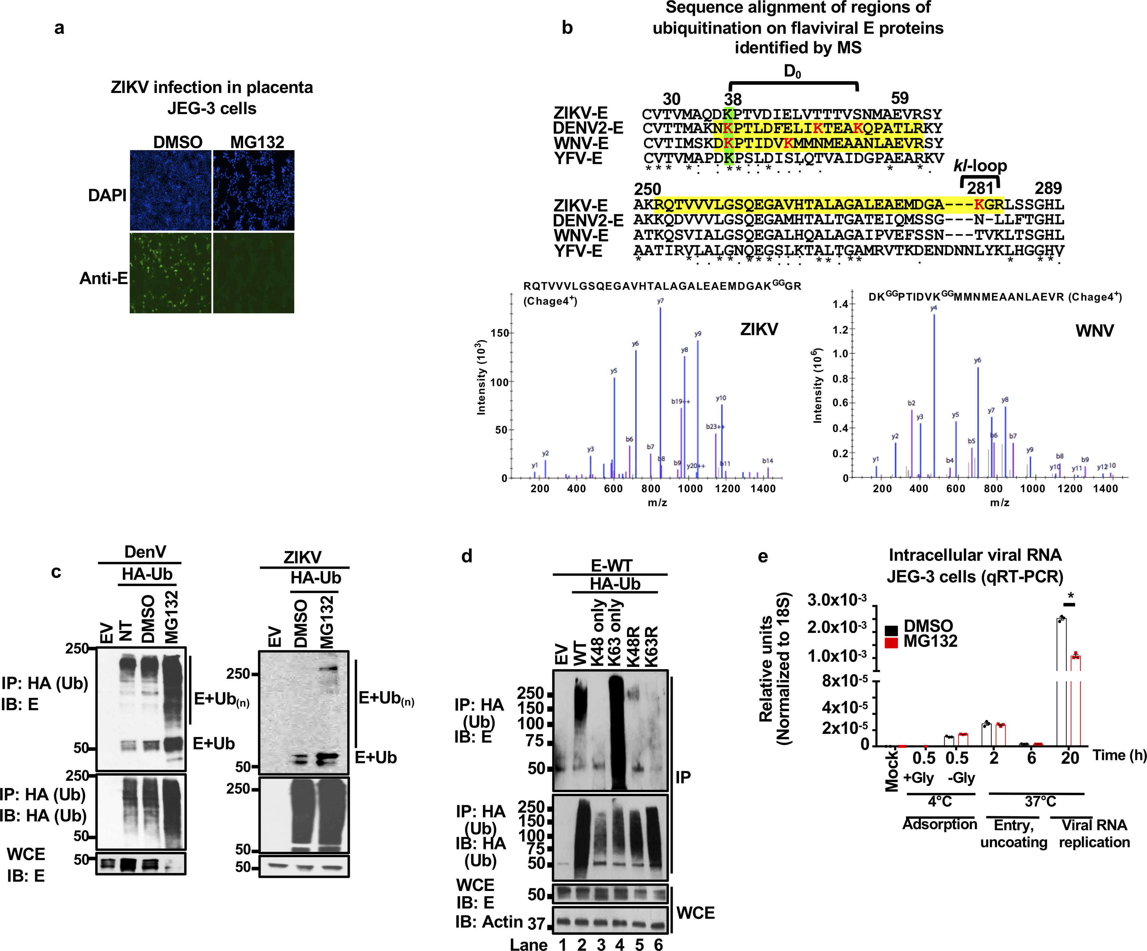
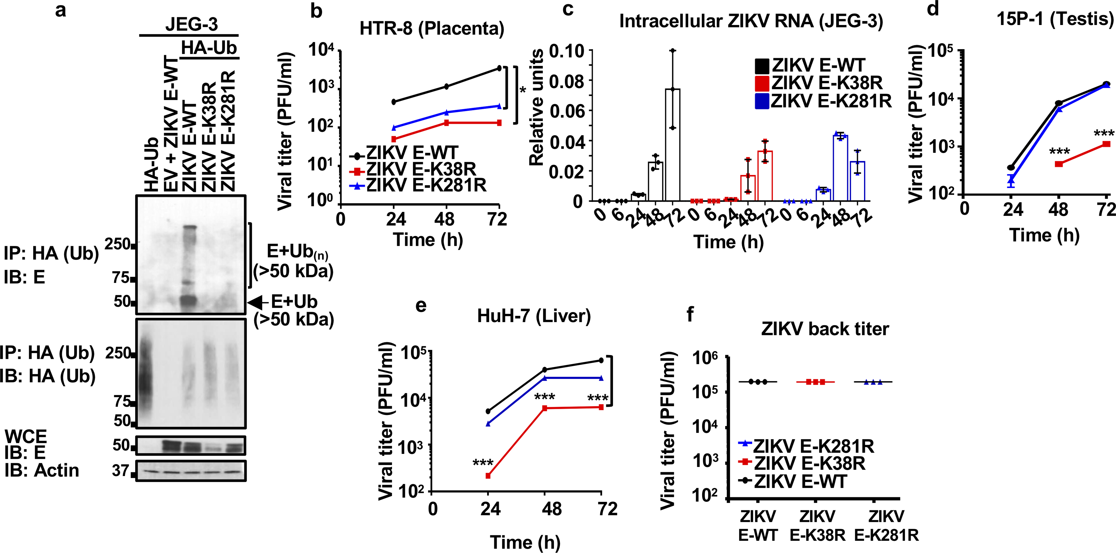
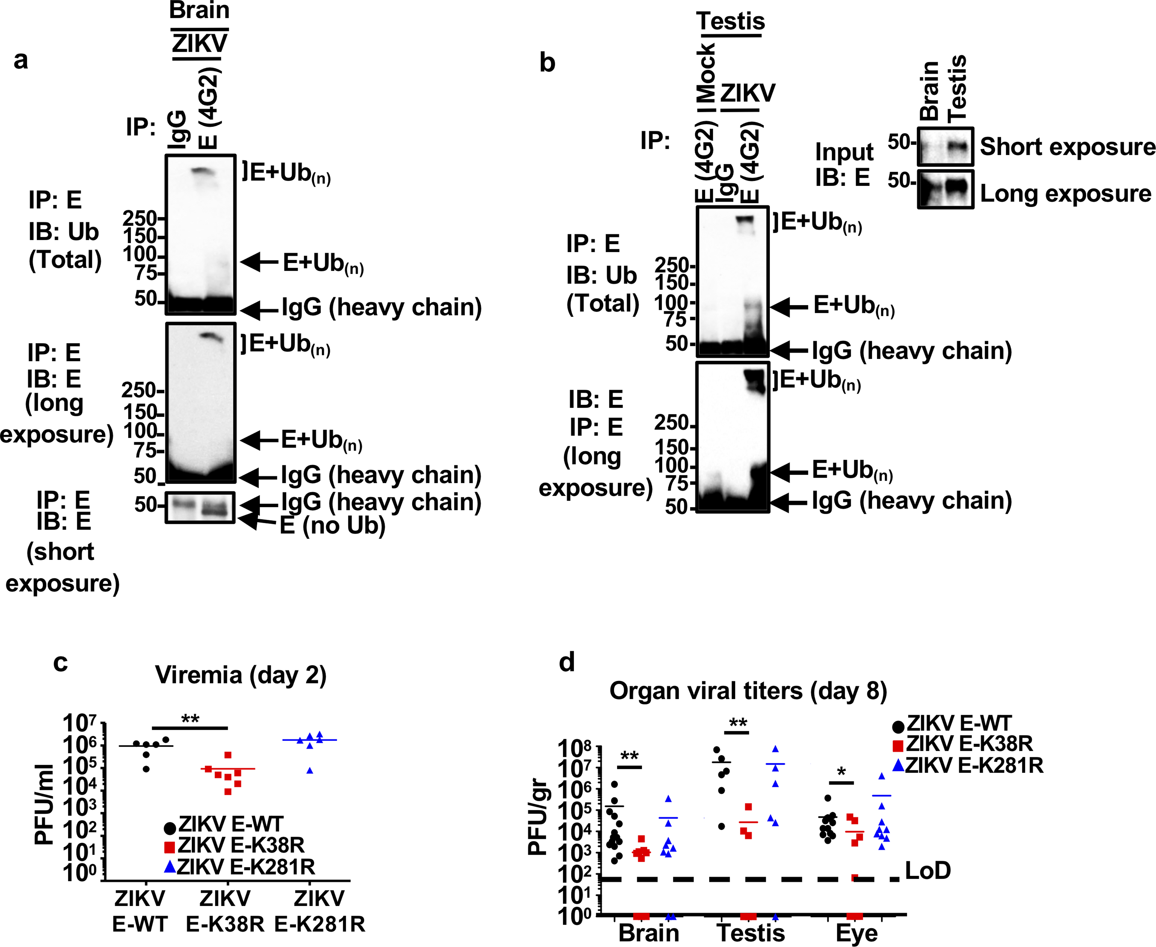
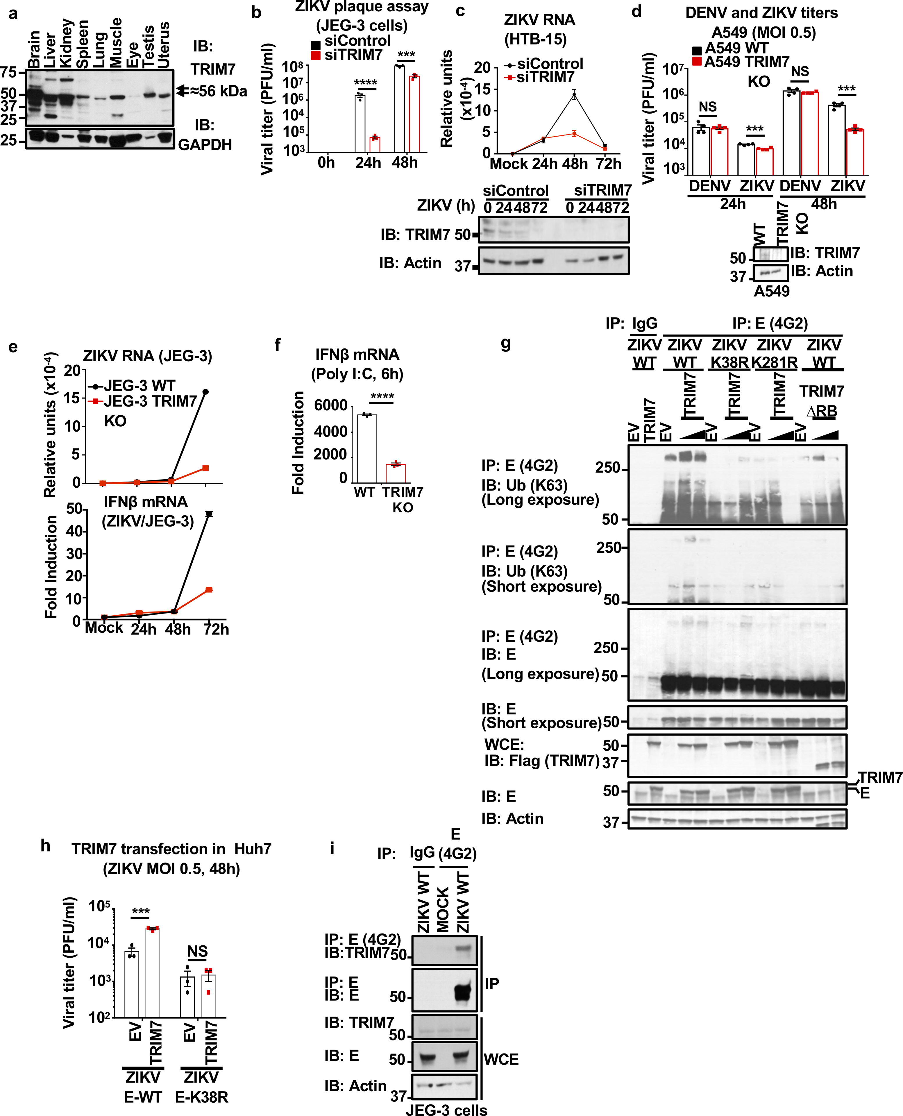
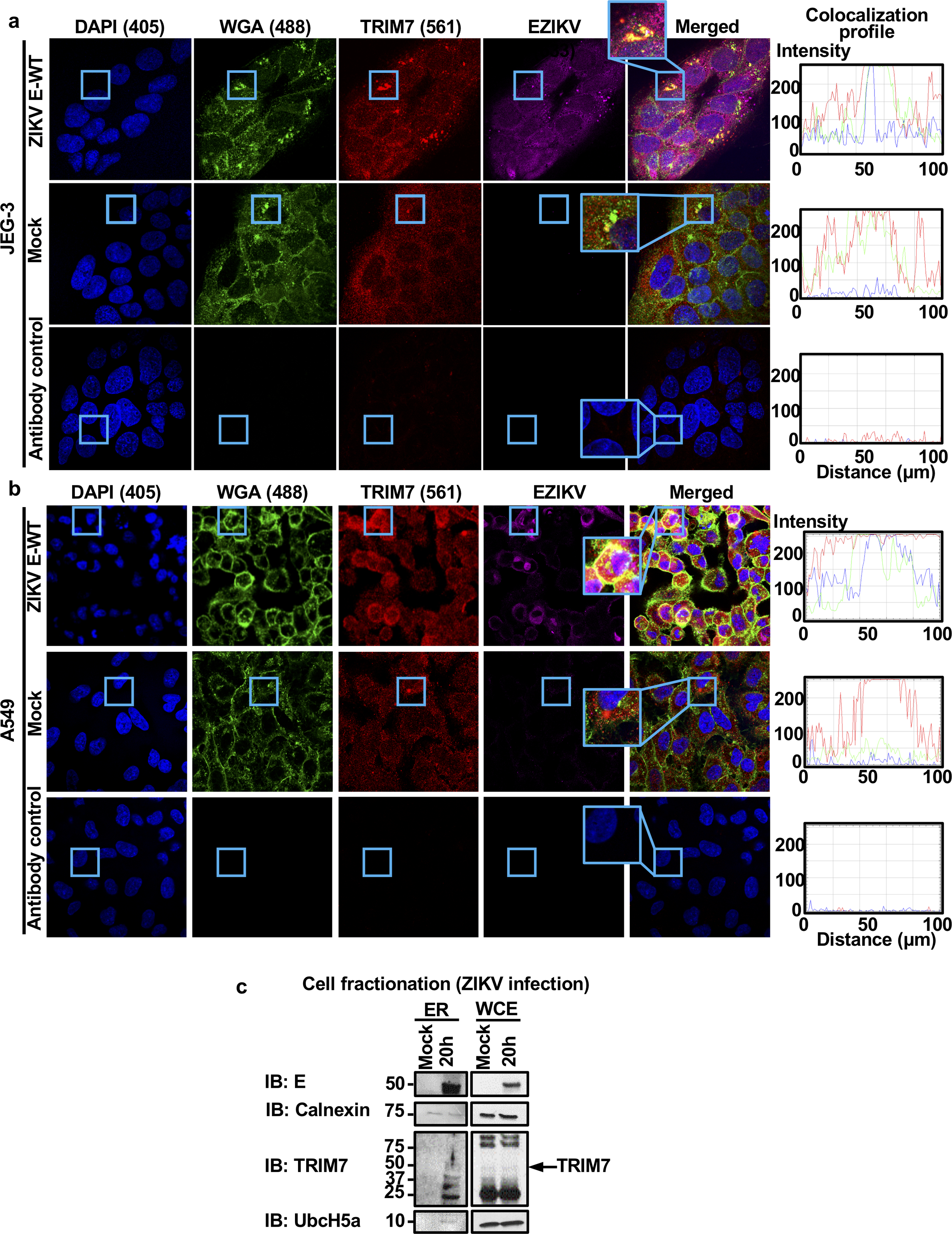
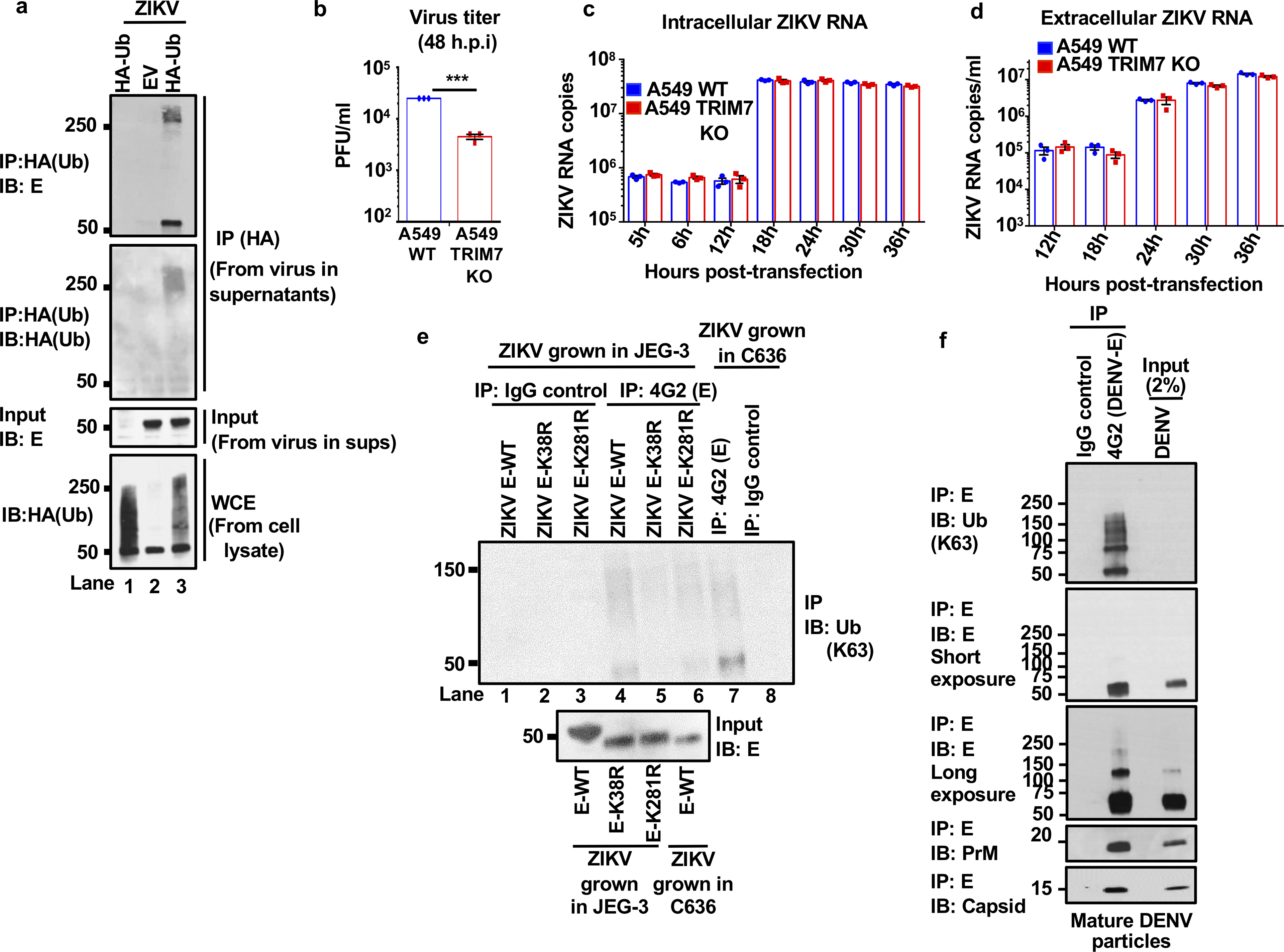
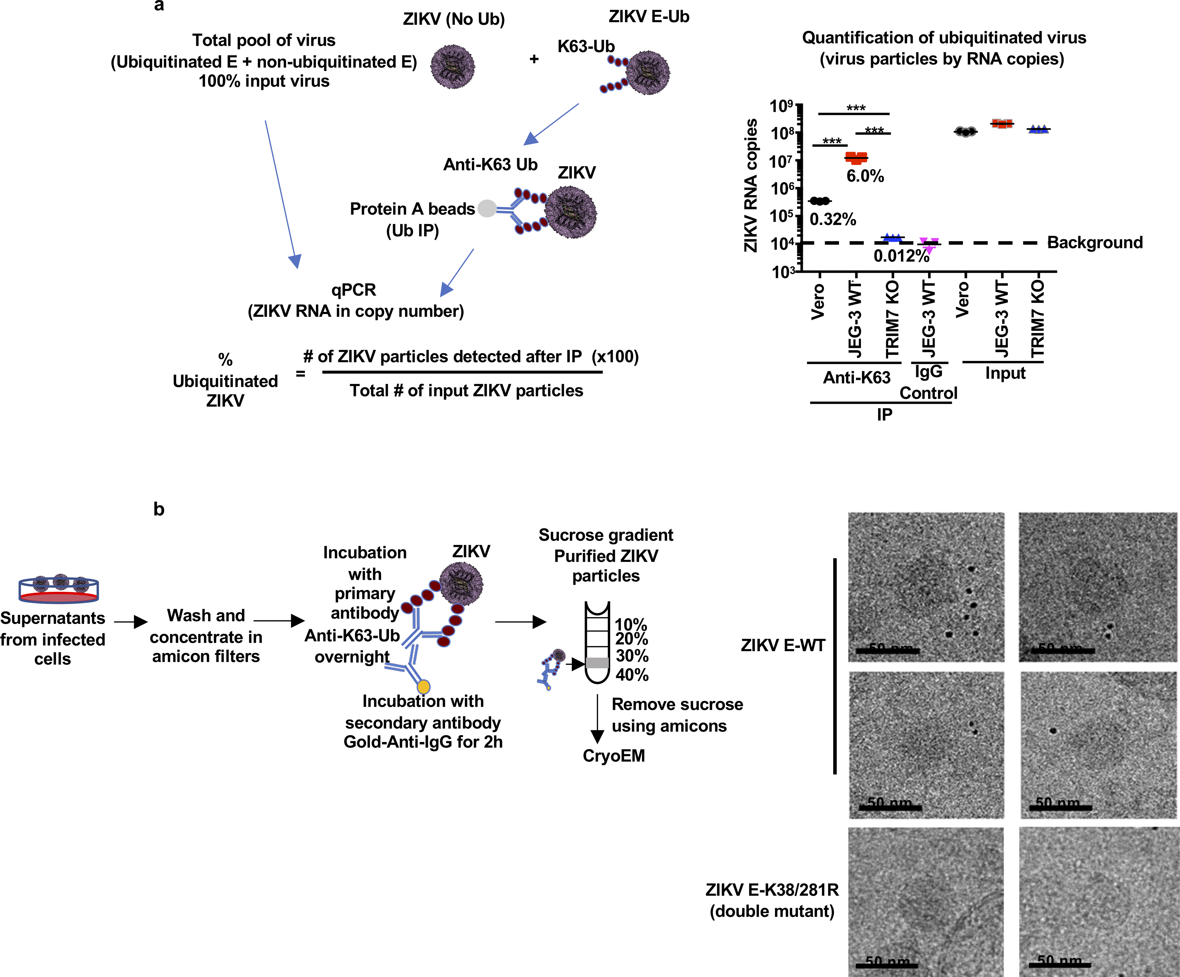
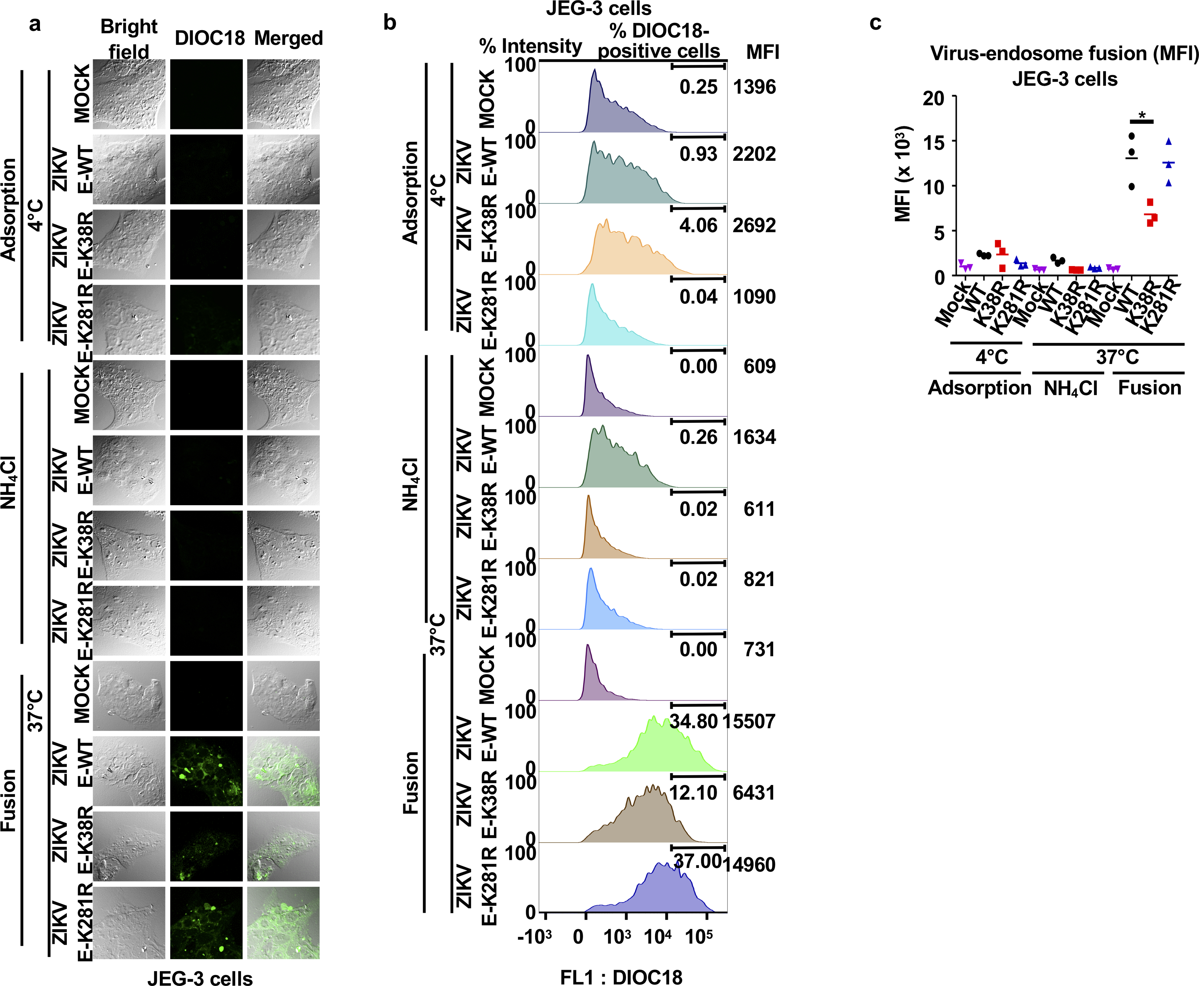
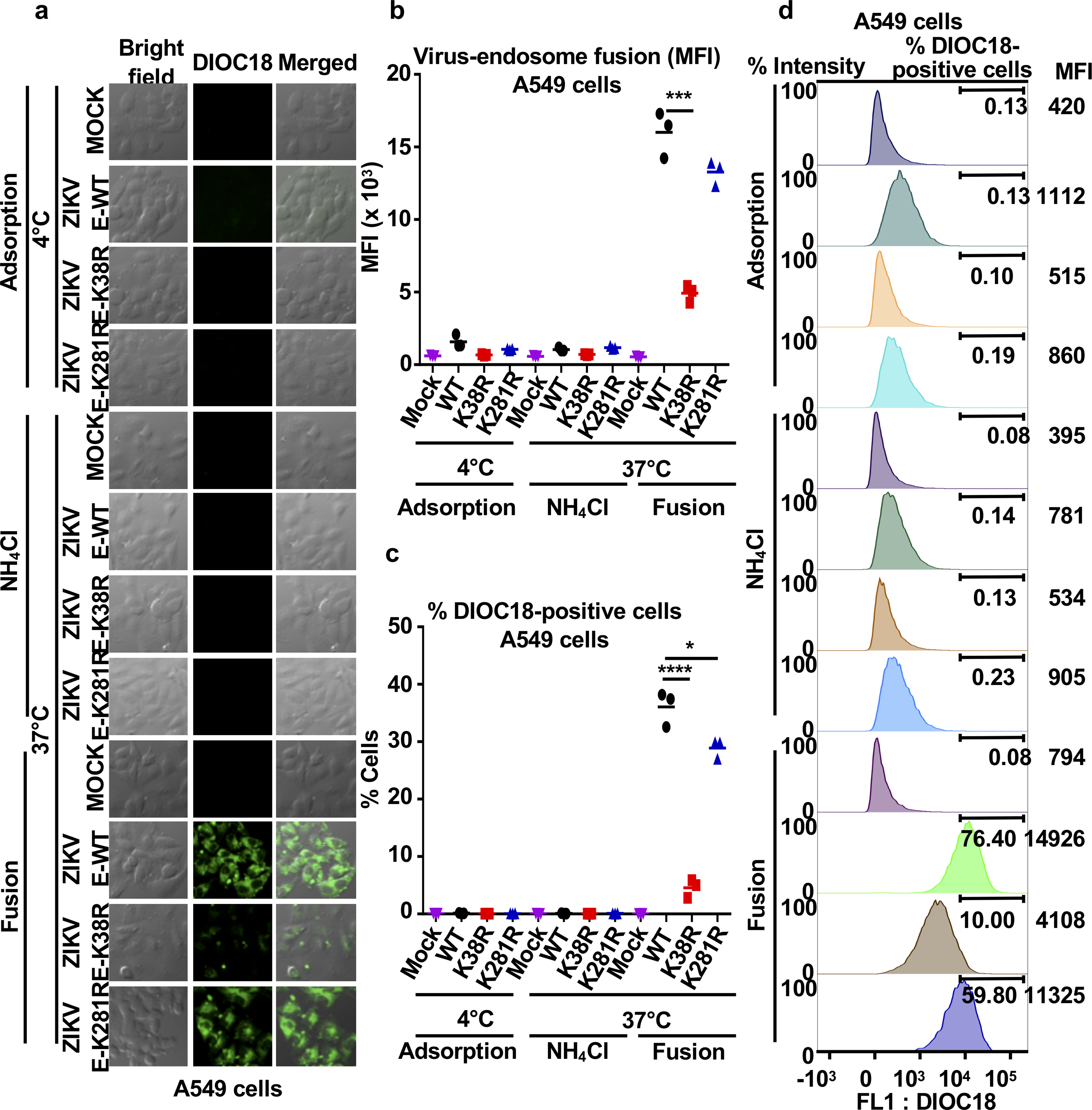
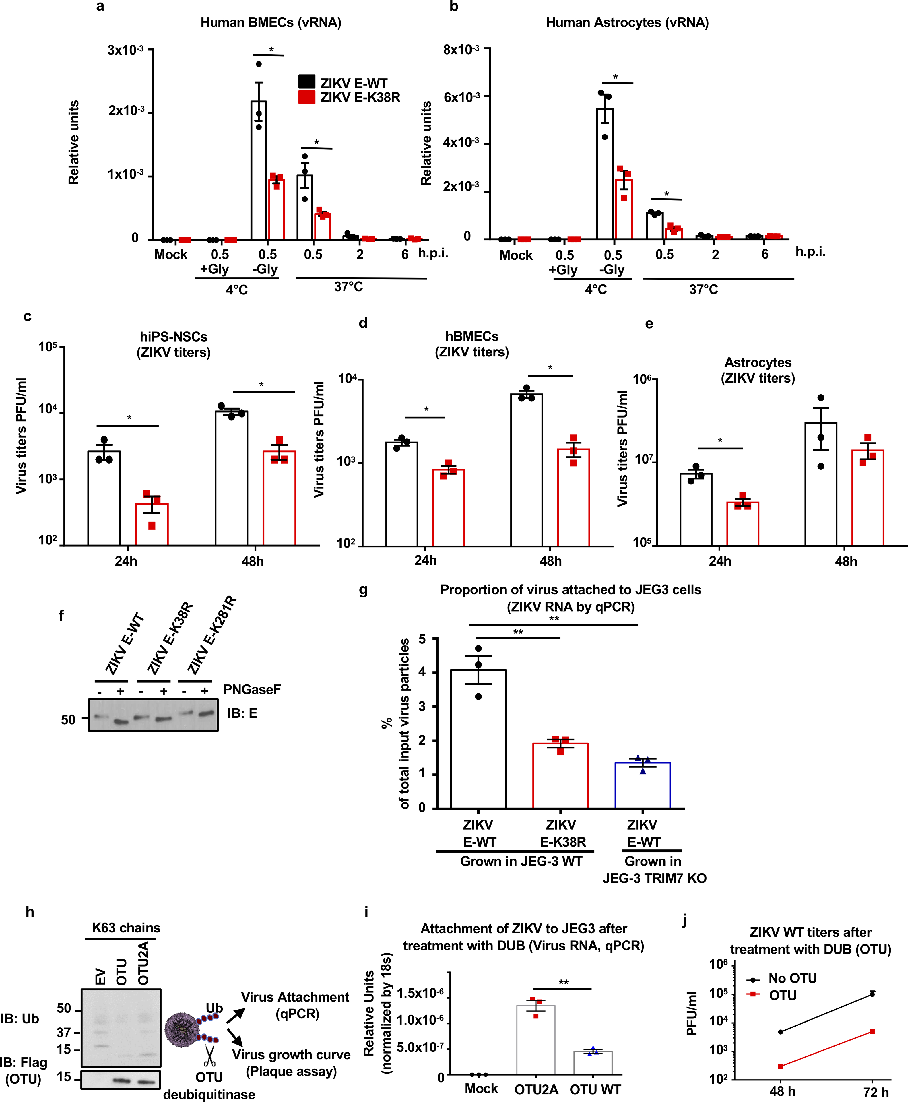
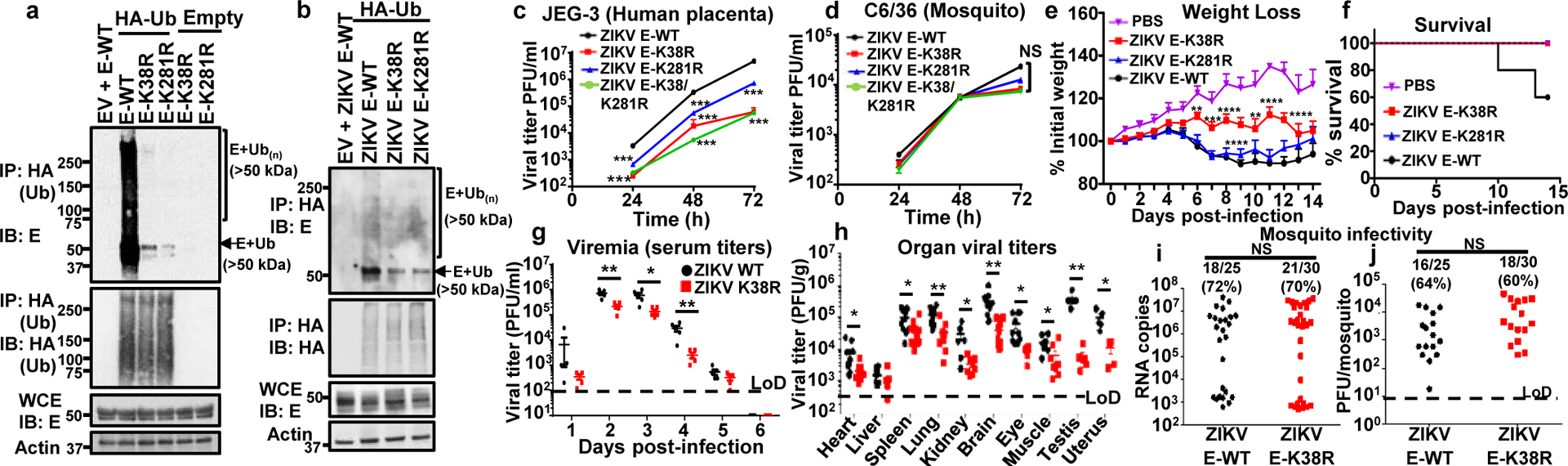
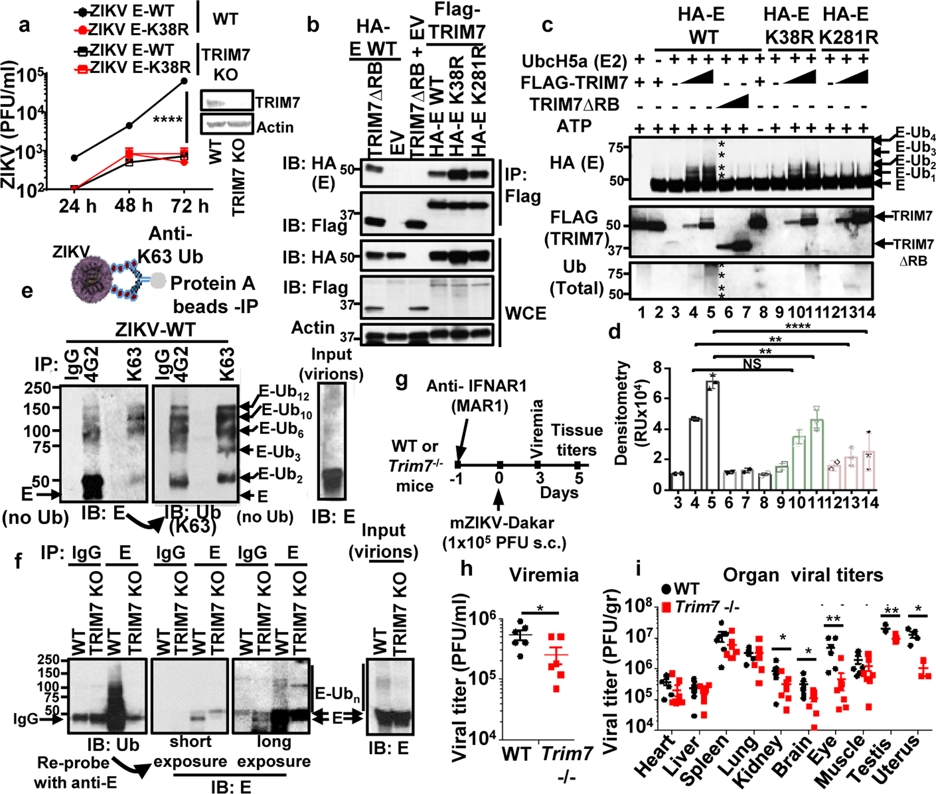
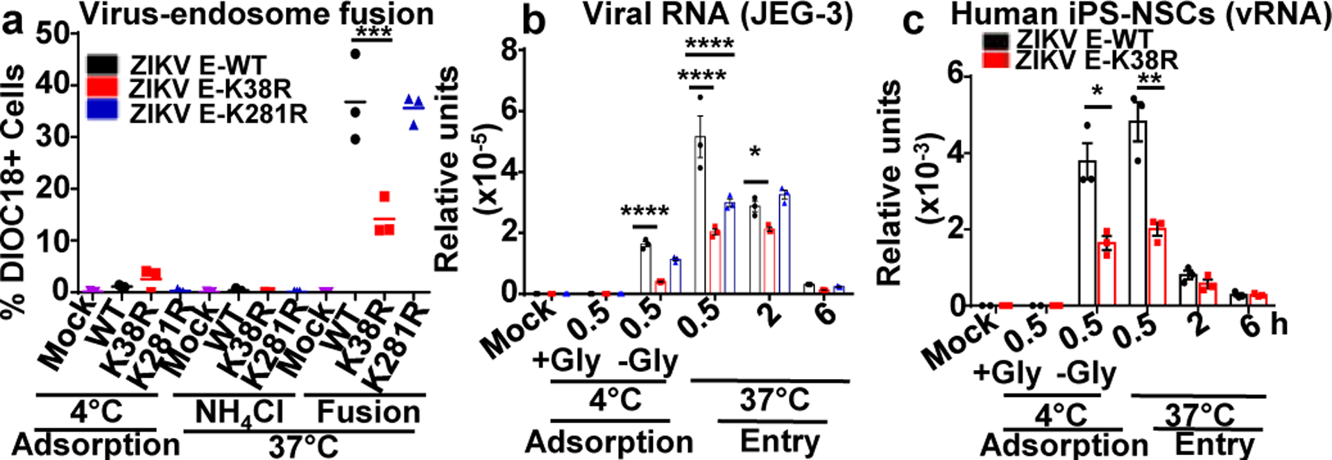
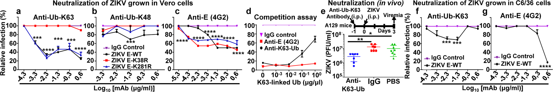
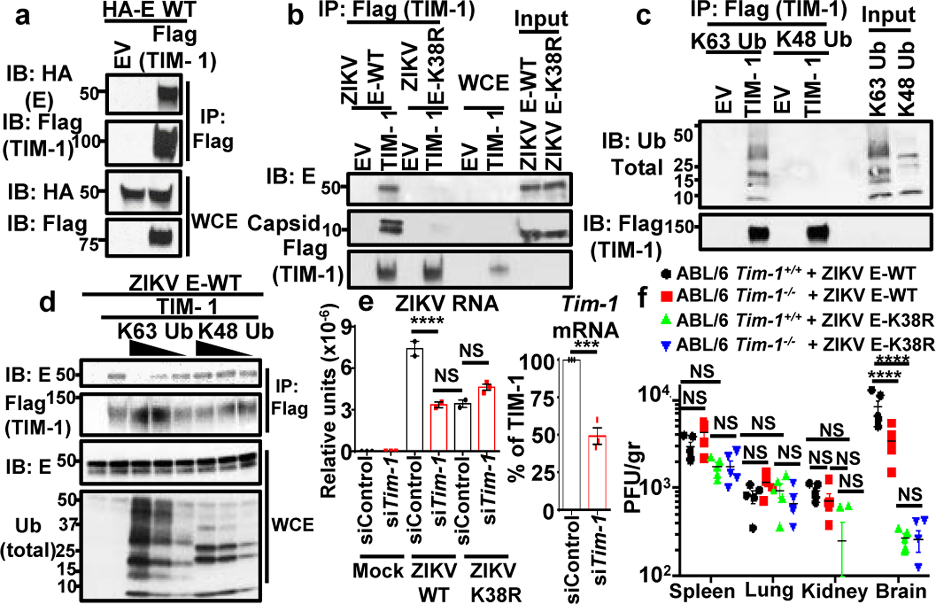
Zika virus (ZIKV) belongs to the family Flaviviridae, and is related to other viruses that cause human diseases. Unlike other flaviviruses, ZIKV infection can cause congenital neurological disorders and replicates efficiently in reproductive tissues1-3. Here we show that the envelope protein (E) of ZIKV is polyubiquitinated by the E3 ubiquitin ligase TRIM7 through Lys63 (K63)-linked polyubiquitination. Accordingly, ZIKV replicates less efficiently in the brain and reproductive tissues of Trim7-/- mice. Ubiquitinated E is present on infectious virions of ZIKV when they are released from specific cell types, and enhances virus attachment and entry into cells. Specifically, K63-linked polyubiquitin chains directly interact with the TIM1 (also known as HAVCR1) receptor of host cells, which enhances virus entry in cells as well as in brain tissue in vivo. Recombinant ZIKV mutants that lack ubiquitination are attenuated in human cells and in wild-type mice, but not in live mosquitoes. Monoclonal antibodies against K63-linked polyubiquitin specifically neutralize ZIKV and reduce viraemia in mice. Our results demonstrate that the ubiquitination of ZIKV E is an important determinant of virus entry, tropism and pathogenesis.


Conflict of interest statement

Competing interests: Authors declare no competing interests

Figures

Extended Data Fig. 1:
Extended Data Fig. 1:. Ubiquitination of flavivirus E protein.
a, Proteasome inhibition blocks ZIKV replication. JEG-3 were pre-treated with DMSO or MG132 (2 h) followed by ZIKV infection (MOI 2, 24 h, visualized by immunofluorescence with anti-E 4G2). b, Ubiquitinated peptides from flavivirus-infected cells identified by mass spectrometry (peptides highlighted in yellow, diglycine residues indicating ubiquitination in red, conserved residues in green). Sequences for strains ZIKV FSS13025 (GenBank: KU955593.1), DENV-2 Y98P (JF327392.1), WNV NY99 (DQ211652) and YFV (ANC33490.1). JEG-3 cells were used for ZIKV infections, Huh7 for DENV, and A549 for WNV (repeated in U2OS cells with identical results, two independent experiments). Representative Mass Spectra for ubiquitinated peptides found for WNV are shown. b and y ions are indicated in blue and red, respectively. c, Whole cell extracts (WCE) from DENV or ZIKV HuH7 transfected with HA-Ub and infected (MOI 2, 20h), followed by DMSO or MG132 treatment (6 h) were used for HA immunoprecipitation (IP). Immunoblots (IB) are shown. NT: non-treated d, WCE from cells transfected with vectors expressing E and Ub-WT or all K-to-R mutants except for K63 or K48 (only), or K48R and K63R, followed by IP. e, JEG-3 pre-treated with MG132 or DMSO. Cells were incubated with ZIKV at 4°C for 30min, followed by wash with or without glycine to test virus adsorption. Additional samples were then switched to 37°C to allow virus internalization. Viral RNA detection by qRT-PCR. Representative of 2 independent experiments, N=3 technical replicates, mean +/− SE, Unpaired t-test, two-sided, * (p < 0.05). NS (Not significant). All experiments are representatives from 2 independent experiments, with similar results.
Extended Data Fig. 2:
Extended Data Fig. 2:. Differences in Zika virus cell tropism are associated with ZIKV E-ubiquitination.
a, JEG-3 cells stably expressing HA-Ub were infected with ZIKV E-WT, or recombinant infectious ZIKV E-K38R and ZIKV E-K281R mutants. WCE were used for IP with HA beads (same experiment as shown in Figure 1b, but without normalizing the input for the IP). Reduced ZIKV E-K38R replication can be seen represented by the levels of E in the WCE. Representative of two independent experiments. b-e, Different cell types were infected with either ZIKV E-WT, E-K38R, or E-K281R (MOI 0.5). Cells were lysed for RNA extraction and virus quantification by qRT-PCR in c, and supernatants were collected for plaque assays at different time-points, for placenta HTR-8 in b, testis 15P-1 in d, liver HuH7 in e. f, Back titration for the virus used on these experiments, and for Fig 1e and 1f. Representatives from 2 independent experiments (n=3 technical replicates, mean +/− SE, multiple t-test, Holm-Sidak correction, *p < 0.05, ***p < 0.001)
Extended Data Fig. 3:
Extended Data Fig. 3:. Ubiquitination of E in tissues from infected mice.
Tissues from testis (a), and brain (b) from mock or ZIKV-WT infected A129 mice were collected at day 8 post-infection. Tissues were homogenized and 200 μg of total input protein was used for immunoprecipitation (IP) of E using 4G2 antibody or an IgG control. Ubiquitination of WT-E was detected with anti-Ub antibody by immunoblot (IB). IPs shown are from mixed tissue lysates from 3 different mice. c-d, A129 mice (male and females) were mock treated (5 mice) or infected with ZIKV E-WT, ZIKV E-K38R or ZIKV E-K281R (1×10⁴ PFU, 9 mice per group, combined from 2 independent experiments). Weight loss and survival is shown in Fig 1e–f. c, serum titers (viremia), were determined at day 2 p.i. by plaque assay, after blood collection from 6 mice for ZIKV E-WT and K38R, and 7 mice for ZIKV E-K38R. d, virus titers (at day 8 p.i.) in brain (14 mice for ZIKV E- WT, and 9 mice for ZIKV E- K38R and ZIKV E- K281R), testis (6 mice/each gorup), and eye (14 mice for ZIKV E-WT, and 9 mice for ZIKV E-K38R and E-K281R). Unpaired, t-test, two-sided, *p < 0.05, **p < 0.01.
Extended Data Fig. 4:
Extended Data Fig. 4:. TRIM7 interacts with and ubiquitinates ZIKV-E and promotes virus replication.
a, Differential expression of TRIM7 in mouse tissues by immunoblot (IB). Predicted molecular weight of full-length TRIM7 is 56 kDa. b-c, TRIM7 knockdown (24h) in JEG-3 (b) or brain HTB-15 cells (c) followed by infection with ZIKV (MOI 1). c, Viral RNA levels were determined by qRT-PCR at different time points (upper panel). TRIM7 knockdown efficiency was confirmed by western blot (lower panel). d, TRIM7 Crispr Knockout A549 and WT parental cells were used for infections with ZIKV or DENV at an MOI of 0.5. Bottom panel shows immunoblot of TRIM7. Plaque assays from supernatants collected at different time-points is shown. e-f, Infections of WT and TRIM7 CRISPR KO JEG-3 cells with ZIKV (MOI 0.5) or poly I:C stimulation (f, transfection of 10 μg/ml with lipofectamine 2000). Quantification of ZIKV RNA (e, top) and IFNβ mRNA expression (e, bottom and f) by qPCR. g, Overexpression of TRIM7 enhances K63-linked polyubiquitination of E-WT but not E-K38R or E-K281R. HEK293T cells were transfected with vectors expressing E-WT, E-K38R, or E-K281R and different amounts of HA-TRIM7 (350ng, 700ng). Thirty hours post-transfection, cells lysates were used for immunoprecipitation (IP) with anti-E 4G2 or isotype control. Immunoblots (IB) with indicated antibodies. h, Transfection of Huh7 cells with empty vector (EV) or vector expressing TRIM7. After 48h cells were infected with ZIKV E-WT or E-K38R. N=3, technical replicates, mean +/−SE. Multiple t-test, two-sided, ***p<0.001, ****p<0.0001, NS (Not significant, p>0.05). i, Endogenous TRIM7 interacts with E in ZIKV placental JEG-3 infected cells. Cells were infected with ZIKV-WT (MOI 2). Thirty hours p.i. cells were lysed and whole cell extracts (WCE) were used for IP with anti-E (4G2) or isotype control. Representatives of 2 independent experiments
Extended Data Fig. 5:
Extended Data Fig. 5:. TRIM7 co-localizes with E in the Golgi and ZIKV-E is detected in supernatants from infected cells.
a-b, JEG-3 (a) or A549 (b) cells were mock treated or infected with ZIKV (MOI 2). Twenty-four hours post-infection, cells were fixed and stained for endogenous TRIM7 (red), Golgi (WGA-FITC, green), and E (4G2, purple) for confocal microscopy. Colocalization is shown in rectangles, and RGB profile graphs are on the right. All images were processed identically using the same conditions with ZEN 2.5.75.0 (Zeiss) and RGB profiles were obtained using ImageJ (NIH) v1.52e. c, cell fractionation of infected JEG-3 cells (20h, MOI 2) for ER was performed following manufacturer instructions (Sigma). Representative of two independent experiments.
Extended Data Fig. 6.
Extended Data Fig. 6.. Zika and dengue virions contain ubiquitinated E, and TRIM7 is important for ZIKV but not DENV replication.
a, Supernatants collected from JEG-3 ZIKV-infected cells stably expressing HA-Ub showed detectable levels of ubiquitinated E. Viruses were immunoprecipitated with anti-HA beads. Immunoblot (IB) for E and HA-Ub. The lower panel shows the total HA-Ub levels expressed in these HA-Ub stable cell lines. b, Infection of A549 WT or TRIM7 KO cells with ZIKV. c-d, Transfection of A549 WT or TRIM7 KO cells with in vitro transcribed ZIKV RNA. Viral RNA was quantified by qPCR from cell lysates (intracellular, c) or from supernatants (extracellular, d). Representatives of 2 independent experiments. N=3, technical replicates, mean +/− SE. e, K63-linked polyubiquitinated E was detected on ZIKV E-WT particles but reduced in ZIKV E-K38R and K281R, after IP anti-E 4G2 antibody. Although reduced, ubiquitination on E from ZIKV grown in mosquito C6/36 can also be detected. f, DENV virus from supernatants from infected BHK-21 was immunoprecipitated with anti-E antibody (4G2) or an IgG control. Immunoblot (IB) for K63-linked polyUb and each viral protein are shown. Representatives of 2 independent experiments.
Extended Data Fig 7.
Extended Data Fig 7.. Proportion of ubiquitinated E in Zika virions and CryoElectron microscopy of ubiquitinated ZIKV.
a-b, ZIKV stocks were grown in Vero cells, JEG-3 WT or TRIM7 KO cells were used for IP using an anti-K63-Ub antibody, or an IgG control to set the background levels. The immunoprecipitated virus as well as a sample of input viruses were lysed in Trizol for virus RNA quantification by qPCR (a). The virus RNA copy number was determined using a standard of purified ZIKV RNA and its known molecular weight. The proportion of ubiquitinated virus was calculated taking as 100% the input virus. N=3 technical replicates, mean, unpaired two-sided t-test, ***p<0.001. b, CryoElectron microscopy of ubiquitinated ZIKV. Experimental approach. Supernatants from Vero cells infected with WT or ZIKV E-K38/281R double mutant were washed and concentrated in Amicon filters followed by labelling with a primary antibody against K63-Ub and secondary nano-gold-labelled antibody. Virus-antibody complexes were then purified by sucrose gradient. A visible band containing these complexes was recovered and passed through Amicon filters to remove sucrose and concentrate the complexes. Samples were flash-frozen in liquid ethane cooled to liquid nitrogen temperatures on holey carbon grids and images were recorded in movie mode at 40K magnification using a 200 KV JEOL 2200FS transmission electron microscope. To facilitate visualization of virus particles, frames were further binned 3X to yield a pixel size of 4.398 Å/pixel. These binned micrographs were manually examined using EMAN2. To identify potentially gold-labelled ubiquitinated particles, we looked for spherical particles corresponding to the known ~500 Å (50nm) size of mature ZIKV and which were within 200 Å of the easily recognizable nano-gold clusters. Approximately 15% of visible ~500 Å ZIKV E-WT particles satisfied these criteria. None of the ZIKV E-K38/281R double mutant viruses were found labelled with gold particles (b). The cryoEM experiments with Gold particle labeling were performed only one time due to the large amount of virus needed.
Extended Data Fig. 8:
Extended Data Fig. 8:. Ubiquitination of ZIKV-E promotes virus-endosome membrane fusion. In JEG-3 cells.
a-c, ZIKV E-WT, E-K38R and E-K281R were labeled with DiOC18. After filtration, viruses were incubated at 4°C with JEG-3 cells at MOI 2 and after 30 min were washed and collected for controls as adsorbed viruses. Additional samples were then incubated at 37°C for 1h, in the presence or absence of NH4Cl to block acidification (as control), washed, fixed and visualized with a confocal microscope (a). The same experiment was repeated for quantification by FACS (mean fluorescence intensity, MFI, shown in b, % of cells infected is shown in main Figure 3a, and c, Representative histograms). N=3, technical replicates, mean, unpaired two-sided t-test , *p < 0.05. Representatives of 2 independent experiments.
Extended Data Fig. 9:
Extended Data Fig. 9:. Ubiquitination of ZIKV-E promotes virus-endosome membrane fusion in A549 cells.
a-d, ZIKV E-WT, E-K38R, and E-K281R were labeled with DiOC18. After filtration, viruses were incubated at 4°C with A549 cells at MOI 2 and after 30min were washed and collected for controls as adsorbed viruses. Additional samples were then incubated at 37°C for 1h, and NH4Cl was used as control to block acidification of the endosome. Cells were washed, fixed, and visualized in a confocal microscope in a. Quantification by FACS is shown (mean fluorescence intensity, MFI) in b-c. d, Representative histograms showing green florescence (FL1: DiOC18) during ZIKV-endosome fusion. N=3, technical replicates, mean, unpaired two-sided t-test, *P < 0.05, ***P < 0.05. Representatives of 2 independent experiments.
Extended data Fig. 10.
Extended data Fig. 10.. Ubiquitination of E on K38 promotes ZIKV attachment and enhanced replication in relevant human cells.
a-e, Human brain microvascular endothelial cells (hBMECs, a, d), human astrocytes (b, e), Human primary induced-pluripotent neural stem cells (hiPS-NSCs, c), were infected with ZIKV E-WT or E- K38R (MOI 2) as described in Fig 3b–c. Viral RNA was quantified by qPCR (a-b) and Virus titers by plaque assay (c-e). One experiment, N=3, technical replicates, mean +/− SE. Unpaired two-sided t-test, *p < 0.05. f, Endoglycosidase analyses of E. Proteins from ZIKV E-WT and E- K38R and E-K281R viruses were analyzed by western blot. Viruses were treated with PNGase F for 1h at 37°C. g, ZIKV E-WT or E-K38R mutant grown in JEG-3 WT or TRIM7 KO were used for attachment assays. Viruses were incubated at 4°C for 30min with JEG-3 cells and attachment was determined by measuring virus RNA by qPCR. The percentage of virus attachment was calculated by taking the input virus as 100%. h-j, The deubiquitinase (DUB) domain of the ovarian tumor (OTU) of the Crimean Congo Hemorrhagic fever (CCHF), which can cleave polyubiquitin chains (shown in h as control for activity), and a mutant (2A) with reduced activity were used to cleave ubiquitinated E of ZIKV. After incubation of ZIKV with purified recombinant OTU, the ability of the deubiquitinated virus to attach to cells and to replicate was tested by incubation with JEG-3 cells at 4°C for 30min and viral RNA quantified by qPCR (i), and replication by plaque assay (j). Representatives of 2 independent experiments, N=3 technical replicates, mean +/−SE, unpaired two-sided t-test, **p < 0.01.
Fig. 1:
Fig. 1:. ZIKV-E ubiquitination on K38 and K281 promotes virus replication in cells and in vivo.
a, Whole cell extracts (WCE) from HEK293T cells transfected with empty vector (EV), E-WT or mutants and HA-Ub were used for IP with anti-HA beads. b, JEG-3 cells stably expressing HA-Ub were infected with ZIKV E-WT, or ZIKV mutants followed by AHA IP. Since the mutant viruses are attenuated, the input E was normalized for immunoprecipitation. c-d, Virus titers in supernatants from infected JEG-3 cells (c); or mosquito C6/36 (d), at MOI 0.5. Representatives from 2 independent experiments (n=3 technical replicates, mean +/−SE, ***P < 0.001). e-h, A129 mice (5 mock) or infected with ZIKV mutants (1×10⁴ PFU, 9 mice/group, from 2 independent experiments). e, Body weight (Two-way ANOVA, Tukey’s test). f, Survival. Virus titers shown in Extended Data Fig 3c–d. g, Same experiment as in e repeated with 5 males, 5 females (Viremia), and h, virus titers (day 6 p.i.). Unpaired two-sided t-test, *p<0.05, **p<0.01. i-j, Mosquito infectivity. Aedes aegypti mosquitoes were fed with bloodmeal (106 PFU/ml) of ZIKV. At day 10, individual mosquitoes were quantified for viral RNA (qPCR, i) and virus (plaque assay, j). LoD, limits of detection. Unpaired, two-sided t-test, NS (Not significant, p>0.05).
Fig. 2.
Fig. 2.. TRIM7 ubiquitinates ZIKV-E and promotes virus replication.
a, Virus titers from TRIM7 JEG-3 Knockout (TRIM7 KO) infected with ZIKVs (MOI 0.5). (n=3, technical replicates, mean +/−SE, multiple t-test, Holm-Sidak correction, ****p<0.0001). b, HEK293Ts transfected with TRIM7 or a short isoform (TRIM7 ΔRB), WT and mutant E, followed by IP. Representative of 2 independent experiments. c, TRIM7 ubiquitinates recombinant ZIKV-E on both K38 and K281 in an in vitro ubiquitination assay. Representative of four independent experiments. Densitometry shown in d (N=3, mean +/− SE, One-way Anova, Tukey’s multiple comparison, **p<0.01, ****p<0.0001, NSp>0.5).). e, K63-linked polyubiquitinated E was detected on ZIKV particles concentrated from supernatants from Vero after IP with anti-K63-linked or anti-E 4G2 antibodies. After immunoblot (IB) with anti-E, the blots were re-probed with anti-K63-Ub. f, IP with anti-E (4G2) of virus stocks from WT or TRIM7 KO JEG-3 cells. Representative from 2 independent experiments. g-i, Trim7−/− and Trim7+/+ littermates from 3 separate CRISPR KO lines (4–5 weeks old) were treated i.p. with anti-IFNAR1 (MAR1–5A3). Next day infections performed with mouse-adapted ZIKV Dakar (footpad, 105 PFU, n=7 Trim7+/+, 8 Trim7−/− mice). Serum (h) and tissue titers (i). Unpaired, t-test, two-sided *p<0.05, **p<0.01.
Fig. 3.
Fig. 3.. Ubiquitination of ZIKV-E promotes virus attachment and virus-endosome membrane fusion.
a, ZIKVs were labeled with DiOC18. After filtration, viruses were incubated at 4°C with JEG-3 at MOI 2. After 30 min were washed and collected as adsorbed viruses. Additional samples were then incubated at 37°C for 1h, +/−NH4Cl to block acidification (as control), washed, fixed and quantification by FACS. b-c, Ubiquitination of E promotes virus attachment. JEG-3 (b) or human primary induced-pluripotent neural stem cells (hiPS-NSCs, c), were incubated with viruses as described in a. Viral RNA (qPCR). Incubation at 4°C without Glycine (-Gly) represents attached viruses. Each are representative of 2 independent experiments (N=3, technical replicates, mean +/−SE. Unpaired t-test, two-sided, *p<0.05, **p<0.01 ***p<0.001, ****p<0.0001.
Fig. 4.
Fig. 4.. Specific anti-K63-linked polyUb antibody neutralizes ZIKV in vitro and in vivo.
a-d, WT and mutant viruses grown in Vero cells or mosquito C6/36 cells (f-g) were incubated at 37°C with dilutions of indicated antibodies for 1h, followed by plaque assay. d, Purified K63-Ub chains were incubated together with ZIKV WT and anti-K63-Ub antibody, IgG control or 4G2. Relative infection was calculated as a percentage of antibody effects on each virus relative to its own IgG control (n=3, technical replicates, mean +/−SE, Two-Way ANOVA, with Tukey correction.). e, In vivo neutralization assay, A129 mice were inoculated i.p. with anti-K63-Ub or an IgG control. Next day mice were infected with ZIKV (1×10⁴ PFU). Viremia (day 3). N=7, mean +/−SE, Unpaired t-test, two-sided, *p<0.05, **p<0.01, ***p<0.001, ****p<0.0001.
Fig. 5.
Fig. 5.. Ubiquitination of ZIKV E promotes binding to the Tim-1 receptor.
a, WCE from HEK293Ts transfected with empty vector (EV), HA-E WT and Flag-TIM-1 were used for IP with anti-Flag beads. b, Interaction of ZIKV E-WT but not ZIKV-E K38R viral particles with TIM-1. Ectopically expressed TIM-1 was isolated using anti-Flag beads. After washes, viruses were incubated with the TIM-1. c, Interaction of K63-, but not K48-linked polyUb, with TIM-1 in the absence of E, coIP assay. d, WT was bound to TIM-1 containing beads (as in b). Increasing amounts of purified K63-Ub or K48-Ub chains were added. Representatives of 2 independent experiments (a-d). e, Virus attachment (viral RNA, qPCR) after Tim-1 knockdown in JEG-3 (n=2, mean +/−SE, One-Way ANOVA, multiple comparison, Tukey correction, ****p<0.0001, *p<0.05, NS p>0.05). f, Titers in infected Tim-1−/− mice, n=5/group, mean +/−SE, Two-Way ANOVA multiple comparison, Tukey correction. ****p<0.0001, NS p>0.05.

References

    1. Musso D et al. Potential sexual transmission of Zika virus. Emerg Infect Dis 21, 359–361, doi:10.3201/eid2102.141363 (2015). - PMC - PubMed
    1. Hills Susan L., Morgan Hennessey KR, Williams Charnetta, Oster Alexandra M., Fischer Marc, Mead Paul. Transmission of Zika Virus Through Sexual Contact with Travelers to Areas of Ongoing Transmission — Continental United States, 2016. CDC MMWR Morb Mortal Wkly Rep 65, 215–216 (2016). - PubMed
    1. Driggers RW et al. Zika Virus Infection with Prolonged Maternal Viremia and Fetal Brain Abnormalities. N Engl J Med 374, 2142–2151, doi:10.1056/NEJMoa1601824 (2016). - PubMed
    1. van Tol S, Hage A, Giraldo MI, Bharaj P & Rajsbaum R The TRIMendous Role of TRIMs in Virus-Host Interactions. Vaccines (Basel) 5, doi:10.3390/vaccines5030023 (2017). - DOI - PMC - PubMed
    1. Byk LA et al. Dengue Virus Genome Uncoating Requires Ubiquitination. MBio 7, doi:10.1128/mBio.00804–16 (2016). - PMC - PubMed

Publication types

MeSH terms