. 2020 Jul;29(7):1133-1146.
doi: 10.1007/s00044-020-02582-9. Epub 2020 Jun 7.

Rheostat positions: A new classification of protein positions relevant to pharmacogenomics

Affiliations

Rheostat positions: A new classification of protein positions relevant to pharmacogenomics

Aron W Fenton et al. Med Chem Res. 2020 Jul.

Abstract

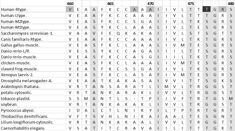
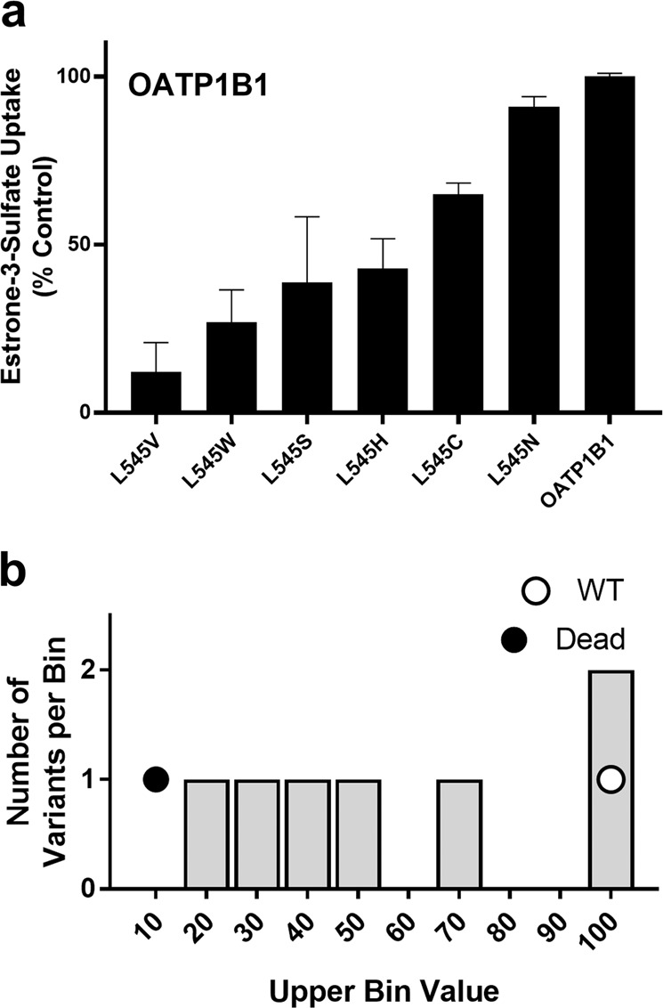
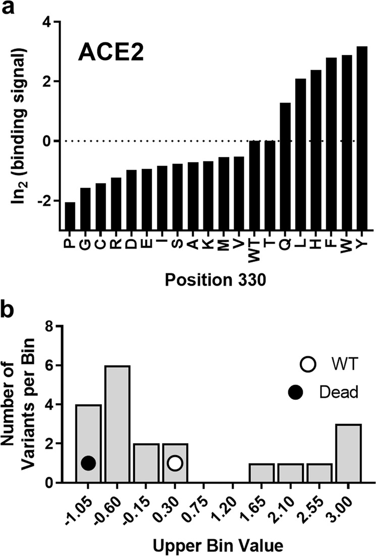
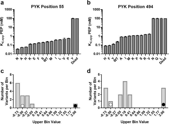
To achieve the full potential of pharmacogenomics, one must accurately predict the functional out comes that arise from amino acid substitutions in proteins. Classically, researchers have focused on understanding the consequences of individual substitutions. However, literature surveys have shown that most substitutions were created at evolutionarily conserved positions. Awareness of this bias leads to a shift in perspective, from considering the outcomes of individual substitutions to understanding the roles of individual protein positions. Conserved positions tend to act as "toggle" switches, with most substitutions abolishing function. However, nonconserved positions have been found equally capable of affecting protein function. Indeed, many nonconserved positions act like functional dimmer switches ("rheostat" positions): This is revealed when multiple substitutions are made at a single position. Each substitution has a different functional outcome; the set of substitutions spans arange of outcomes. Finally, some nonconserved positions appear neutral, capable of accommodating all amino acid types without modifying function. This manuscript reviews the currently-known properties of rheost at positions, with examples shown for pyruvate kinase, organic anion transporting polypeptide 1B1, the beta-lactamase inhibitory protein, and angiotensin-converting enzyme 2. Outcomes observed for rheostat positions have implications for the rational design of drug analogs and allosteric drugs. Furthermore, this new framework - comprising three types of protein positions - provides a new approach to interpreting disease and population-based databases of amino acid changes. In conclusion, although a full understanding of substitution out comes at rheostat positions poses a challenge, utilization of this new frame of reference will further advance the application of pharmacogenomics.

Keywords: Rheostat position; protein evolution; saturating mutagenesis; specificity.


Conflict of interest statement

Conflict of Interest. The authors declare that they have no conflict of interest.

Figures

Fig. 1
Fig. 1
An example sequence alignment. Sequence alignments are represented with related protein (homologs) in horizontal rows, aligned so that equivalent positions fall into vertical columns. Amino acids are represented with the one letter code. When the same amino acid is present in many homologs, that position is considered to be conserved. Conservation is interpreted to indicated that other amino acids are not tolerated at that position, which in corollary indicates the importance of that particular side chain. This example shows part of the pyruvate kinase sequence alignment. The four human isozymes are the top rows and position numbering corresponds to RPYK; note that more sequences and more amino acid positions are in the alignment than shown. Light gray columns indicate positions with conserved ConSurf scores in the full alignment (Glaser et al. ; Landau et al. ; Ashkenazy et al. 2010). Black (conserved) and dark gray (not conserved) cells indicate positions of RPYK disease mutations (Pendergrass et al. 2006)
Fig. 2
Fig. 2
Examples of toggle, neutral, and rheostat substitution outcomes for individual positions, using three of the functional parameters measured for human liver pyruvate kinase (LPYK). “Kd,app PEP” is the apparent affinity for substrate phosphoenol pyruvate (corresponding to either (i) Km if the Hill number is 1 or (ii) K1/2 if the Hill number is < >1); “Kd,ala” and “Kd,FBP” are affinities for the two allosteric ligands. Amino acid substitutions are listed on the x-axis in panels (ac). a At a toggle position, substitutions are either like wild-type or dead enzyme (i.e., no detectable function). b At a neutral position, most substitutions are like wild-type. c At a rheostat position, substitutions range from better than wild-type, to wild-type, to dead. To quantitatively summarize the overall substitution outcomes, data were binned for further analyses (Hodges et al. 2018). Histograms for each type of position in (ac) are represented in (df). A white dot is used to indicate the bin that includes wild-type data and a black dot is used to indicate the bin corresponding to no detectable function. Note especially the binning pattern of a rheostat position (f), which has entries in multiple bins. Data are from (Wu et al. ; Hodges et al. ; Tang et al. ; Ishwar et al. ; Martin et al. 2020). The RheoScale scores determined from these histograms are as follows. d Position 483: neutral 0.00, rheostat 0.16, and toggle 0.91. The toggle score is above the significance threshold of 0.7. e Position 138: neutral 1.00, rheostat 0.03, and toggle 0.00. The neutral score is above the significance threshold of 0.7. f Position 446: neutral 0.00, rheostat 0.58, and toggle 0.00. The rheostat score is above the significance threshold of 0.5
Fig. 3
Fig. 3
Rheostat behavior in the drug uptake transport protein, OATP1B1. a Relative to wild-type OATP1B1, substitutions at position L545 show a range of diminished function depending on the amino acid substitution. Amino acid substitutions are listed on the x-axis. The measure of function, esterone-3-sulfate uptake, is on the y-axis. b Histogram of OATP1B1 functional data. The upper bin limit is shown along the x-axis. The number of amino acid substitutions that occupy each bin is shown along the y-axis. A white and black dot are used to denote the bin that contains the wild-type and dead values, respectively. Data are from Ohnishi et al. (2014). The RheoScale scores determined from these histograms are neutral 0.00, rheostat 0.63, and toggle 0.00. The rheostat score falls above the significance threshold of 0.5
Fig. 4
Fig. 4
Example rheostat position from human ACE2; data were taken from Procko (2020). a Functional data for position 330 of ACE2. Amino acid substitutions are listed on the x-axis. A measure of the binding of ACE2 and the SARS-CoV-2 spike protein is shown on the y-axis. Negative values indicate that binding is weaker than wild-type, whereas positive values indicate “better” function. b Histogram analyses of the functional data for ACE2 position 330. The upper bin value for each bin is shown along the x-axis. Bins that contain the “dead” and “wild-type” values are shown with a black and white dot, respectively. All data with scores above 3.00 were reset to this limit, following the example of Procko (2020). The nonfunctional “dead” protein value of −1.5 was calculated from (average + standard deviation) derived from all nonsense mutations (all replicates) plus the standard deviation. The Rheoscale scores calculated from this histogram were: neutral 0.05, rheostat 0.78, and toggle 0.21. The rheostat score is well above the significance threshold of 0.5
Fig. 5
Fig. 5
BLIP position 50 is a rheostat position. In this study, BLIP position 50 was substituted with 18 amino acids (the 19th could not be purified) and binding was assessed for the three different beta-lactamases noted on the plot. The amino acid rank order for the left-most data is preserved in the middle and right datasets; the jagged patterns show that rank order changes for these two binding partners. Note that even the tightest and weakest substitutions differ among the datasets. Data were taken from Adamski and Palzkill (2017)
Fig. 6
Fig. 6
a Protein monomer extracted from a structure of the LPYK homo-tetramer. This figure was rendered with UCSF chimera (Pettersen et al. 2004) using the pdb 4ip7 (Holyoak et al. 2013). Black spheres at the top of the structure highlight positions in the catalytic site; dark gray spheres indicate the allosteric site for alanine inhibitor binding; black spheres at the bottom of the structure indicate the allosteric site for fructose-1-6-bisphosphate activator binding. Binding affinities for the allosteric effectors are denoted with “Kd,ala” and “Kd,FBP”, respectively. Arrows indicate the allosteric coupling (“Q”) that occurs between the catalytic site and each of the allosteric sites. Both allosteric effectors alter the apparent affinity for substrate phosphoenol pyruvate binding (“Kd,app PEP”; see the legend to Fig. 2). bd LPYK functional parameters altered by substitutions at rheostat positions do not correlate. For the example rheostat position 514, the values of three functional parameters with rheostat scores ≥0.5 were compared for each amino acid substitution (individual dots). (Note that two substitutions have estimates for Kd,ala, but binding was too weak to estimate Qala; this leads to a different number of data points on (b) and (c)). No correlation was observed among the parameters for this or other LPYK rheostat positions, which indicates that each substitution has independent effects on the different functional parameters. Data were taken from Wu et al. (2019)
Fig. 7
Fig. 7
Example of a near-neutral position and a part-toggle-part-rheostat position in LPYK. Amino acid substitutions are listed on the x-axis in panels a, b. a Position 55 shows a substitution pattern that is between neutral and rheostatic. Most of the substitutions have activities close to that of wild-type, but some substitutions result in “dead” protein variants. b Position 494 shows a substitution pattern that is partially that of a rheostat position and partially that of a toggle position. Some of the amino acid substitutions improve the function to different degrees whereas other substitutions result in wild-type-like or “dead” values. c Binned data further demonstrates the near-neutral data for position 55, with the wild-type like data shown in the bin with the white dot. Substitutions that result in no detectable function are denoted by the black dot as a “dead” bin. The RheoScale scores determined from this histogram are as follows: neutral 0.08, rheostat 0.28, and toggle 0.08. None of these scores fall above the relevant significance thresholds; this position is best described as “weakly rheostatic”. d Binned data further demonstrates the range of behaviors exhibited by substitutions at position 494. The RheoScale scores determined from this histogram are as follows: neutral 0.00, rheostat 0.41, and toggle 0.21. None of these scores fall above the relevant significance thresholds; this position is best described as “weakly rheostatic”. Substitutions show both (i) toggle behavior, acting closely to the wild-type behavior (white circle) or showing no detectable function (black circle), and (ii) rheostat behavior, falling into a range of bins between wild-type and dead. Data were taken from (Tang et al. ; Ishwar et al. ; Wu et al. ; Hodges et al. 2018)
Fig. 8
Fig. 8
Toggle and conservation scores agree whereas rheostat scores yield a different view of substitution data. Experimental data were obtained from Procko (2020) and comprised saturating mutagenesis for 117 positions in and near the binding site for the spike SARS-CoV-2 protein (for a total of 2223 variants plus wild-type). Conservation scores are shown along the x-axis. According to Procko, “Conservation scores are calculated from the average of the log2 enrichment ratios for all amino acid substitutions at each residue position.” Positive conservation scores show that most substitutions at a position lead to enriched binding of the SARS-CoV-2 spike protein, whereas negative values indicate that most substitutions at a position diminished binding. a Comparison between toggle scores and conservation scores. Toggle scores were calculated from the substitution data for each of the 117 positions, using the RheoScale calculator and the “nCov-S-High sorts” log2 enrichment ratios of replicate 1. Further details to the calculation are in the legend to Fig. 4. The dashed line at y = 0.7 indicates the empirical significance threshold for toggle scores that was previously determined (Wu et al. 2019). b Comparison between rheostat scores and conservation scores. Rheostat scores were calculated simultaneously with toggle scores. The dashed line at y = 0.5 indicates the empirical significance threshold for rheostat scores that was previously determined (Hodges et al. 2018). As seen by the low Pearson correlation coefficient, rheostat and conservation scores reveal different aspects of the aggregate substitution data for each position. This can be understood by considering two hypothetical examples, one with the set of functional values [−2, −1, 0, 1, 2] and one with the set of functional values [−1, −1, 0, 1, 1]. Both positions have a conservation score of 0, but the first position has a stronger rheostat score than the second position. In the ACE2 data, positions with common conservation scores have a wide variety of rheostat scores

References

    1. Acuna-Hidalgo R, Veltman JA, Hoischen A. New insights into the generation and role of de novo mutations in health and disease. Genome Biol. 2016;17:241. doi: 10.1186/s13059-016-1110-1. - DOI - PMC - PubMed
    1. Adamski CJ, Palzkill T. Systematic substitutions at BLIP position 50 result in changes in binding specificity for class A β-lactamases. BMC Biochem. 2017;18:2. doi: 10.1186/s12858-017-0077-1. - DOI - PMC - PubMed
    1. Andreoletti G, Pal LR, Moult J, Brenner SE. Reports from the fifth edition of CAGI: the critical assessment of genome interpretation. Hum Mutat. 2019;40:1197–1201. doi: 10.1002/humu.23876. - DOI - PMC - PubMed
    1. Ashkenazy H, Erez E, Martz E, Pupko T, Ben-Tal N. ConSurf 2010: calculating evolutionary conservation in sequence and structure of proteins and nucleic acids. Nucl Acids Res. 2010;38(Suppl):W529–W533. doi: 10.1093/nar/gkq399. - DOI - PMC - PubMed
    1. Bubp J, Jen M, Matuszewski K. Caring for glucose-6-phosphate dehydrogenase (G6PD)-deficient patients: implications for pharmacy. P T. 2015;40:572–574. - PMC - PubMed

LinkOut - more resources