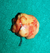
A Clinical and Radiological Investigation of the Use of Dermal Fat Graft as an Interpositional Material in Temporomandibular Joint Ankylosis Surgery
- PMID: 32642033
- PMCID: PMC7311854
- DOI: 10.1177/1943387520903876
A Clinical and Radiological Investigation of the Use of Dermal Fat Graft as an Interpositional Material in Temporomandibular Joint Ankylosis Surgery
Abstract
Management of temporomandibular joint (TMJ) ankylosis is mainly through surgical intervention. Interpositional materials are a necessity when it comes to prevention of TMJ re-ankylosis after arthroplasty. Early aggressive postoperative physiotherapy is essential for the prevention or treatment of TMJ hypomobility or ankyloses. Recently, it has been shown that abdominal dermis fat helps promote smooth, pain-free joint function and it is stable after interposition and less prone to fragmentation. The purpose of this study was to assess that whether dermal fat is a good choice of interpositional material when it comes to decreased pain perception during aggressive physiotherapy after release of ankyloses thus ensuring good compliance by the patient. We also assessed the fate of the graft material on computed tomography to evaluate any volume changes if occurred after interposition.
Keywords: ankylosis; dermal fat; reconstruction; temporomandibular joint; trauma.
© The Author(s) 2020.
Conflict of interest statement
Declaration of Conflicting Interests: The author(s) declared no potential conflicts of interest with respect to the research, authorship, and/or publication of this article.
Figures


References
-
- Long X, Li X, Cheng Y, et al. Preservation of disc for treatment of traumatic temporomandibular joint ankylosis. J Oral Maxillofac Surg. 2005;63(7):897–902. - PubMed
-
- Buchbinder D, Currivan RB, Kaplan AJ, Urken ML. Mobilization regimens for the prevention of jaw hypomobility in the radiated patient: a comparison of three techniques. J Oral Maxillofac Surg. 1993;51(8):863–867. - PubMed
-
- Khalifa GA. Monitoring of incremental changes in maximum interincisal opening after gap arthroplasty omits the risk of re-ankylosis. J Craniomaxillofac Surg. 2018;46(1):75–81. - PubMed
-
- Chuong R, Piper MA. Cerebrospinal fluid leak associated with proplast implant removal from the temporomandibular joint. Oral Surg Oral Med Oral Pathol. 1992;74(4):422–425. - PubMed
-
- Risdon F. Ankylosis of the temporo-mandibular joint. J Am Dent Assoc. 1934;21:1933.
LinkOut - more resources
Full Text Sources
