Review
. 2020 May;12(5):2945-2954.
doi: 10.21037/jtd.2020.02.42.

The tricuspid valve in review: anatomy, pathophysiology and echocardiographic assessment with focus on functional tricuspid regurgitation

Affiliations
Review

The tricuspid valve in review: anatomy, pathophysiology and echocardiographic assessment with focus on functional tricuspid regurgitation

Evin Yucel et al. J Thorac Dis. 2020 May.

Abstract

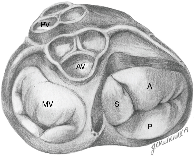
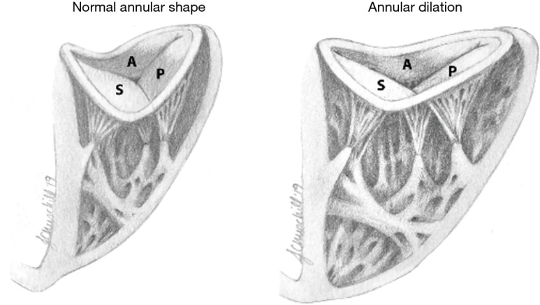
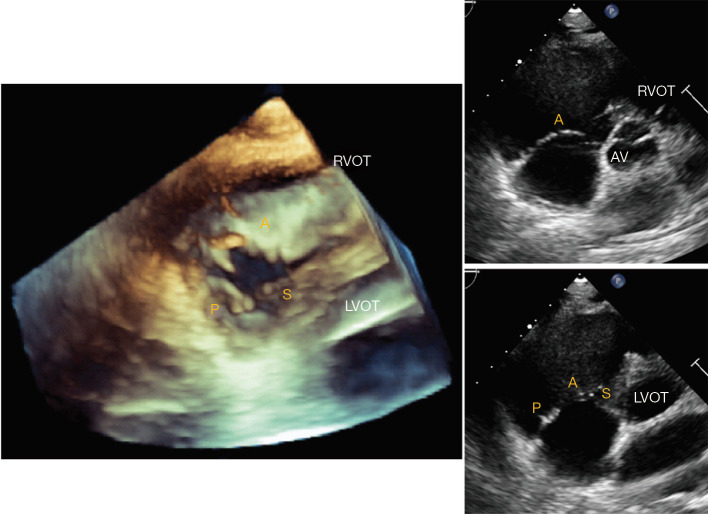
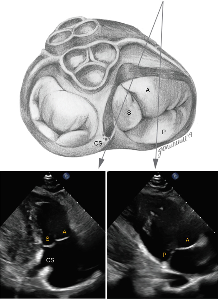
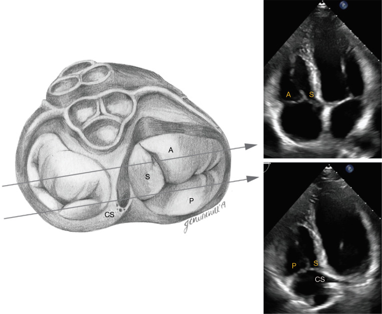
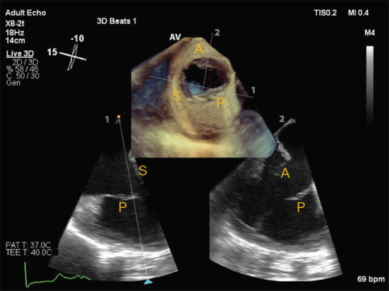
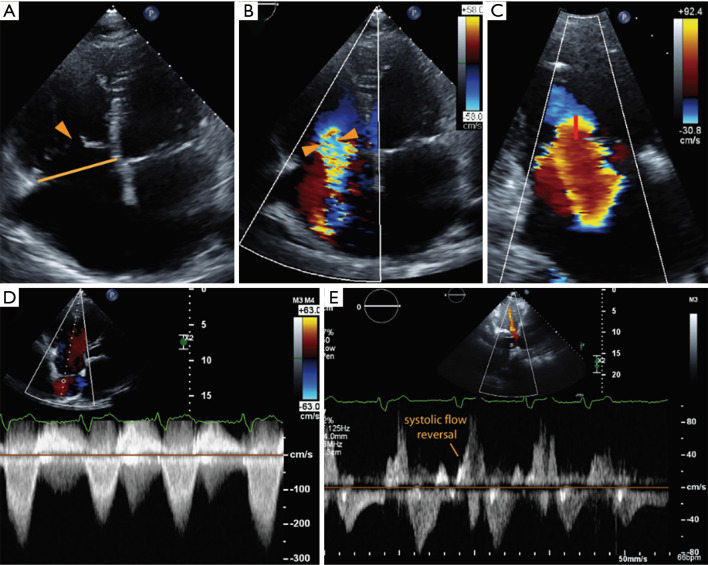
The tricuspid valve (TV) is a complex anatomical structure that incorporates a saddle-shaped annulus, asymmetric leaflets, the subvalvular apparatus and the right ventricle and its loading conditions. In this paper, an appreciation of the normal anatomy and physiology of the TV is reviewed before discussing functional tricuspid regurgitation (TR), a disease that has garnered renewed interest due to increased awareness of adverse outcomes and novel transcatheter therapeutic options. Two and three-dimensional echocardiographic imaging of the TV using transthoracic and transesophageal windows are subsequently discussed. The future of cardiovascular medicine will have more to offer the "forgotten" right-sided chambers and valves, and this review aims to refresh knowledge and enthusiasm around the forgotten but crucially important TV.

Keywords: Tricuspid valve (TV); echocardiography; functional tricuspid regurgitation; three-dimensional echocardiography; tricuspid regurgitation (TR).


Conflict of interest statement

Conflicts of Interest: All authors have completed the ICMJE uniform disclosure form (available at http://dx.doi.org/10.21037/jtd.2020.02.42). The series “Novel Concepts in Cardiopulmonary and Structural Heart Disease” was commissioned by the editorial office without any funding or sponsorship. The authors have no other conflicts of interest to declare.

Figures

Figure 1
Figure 1
Tricuspid valve anatomy at the base of the heart. Of the three leaflets (S, septal; A, anterior; P, posterior), the anterior leaflet is typically the largest. Note the position of the tricuspid valve relative to the MV, AV, coronary arteries, and the PV. MV, mitral valve; AV, aortic valve; PV, pulmonic valve.
Figure 2
Figure 2
Tricuspid annular shape in normal versus dilated valves. The tricuspid valve has a three-dimensional, saddle-shaped annulus, with most atrial (superior) portions of the annulus in anteroseptal (A-S) and posterior (P) position, as opposed to the more ventricular (apical) portions of the annulus in septo-posterior and anterior (A) position. Tricuspid annular dilation secondary to a dilated RA and/or RV is associated with a flattening of the saddle-shape. Dilation occurs in the direction of the right ventricular free wall (anterior-posterior).
Figure 3
Figure 3
Transthoracic echocardiography of the tricuspid valve in the parasternal short axis view. Three-dimensional imaging of the tricuspid valve from short axis view (A) shows the relative position of the three leaflets with respect to the right ventricular outflow tract (RVOT) and left ventricular outflow tract (LVOT). Angulation towards the RVOT (B) predominantly visualizes the anterior leaflet (A), while angulation towards the LVOT (C) enables visualizing the septal (S) and posterior (P) leaflets.
Figure 4
Figure 4
Transthoracic imaging of the tricuspid valve in the modified parasternal long axis view (“right ventricular inflow view”). The anterior leaflet (A) is usually visualized from this view along with either the posterior leaflet (P) or septal leaflet (S). The septal leaflet appears when angulating towards the ostium of the coronary sinus (CS).
Figure 5
Figure 5
Transthoracic imaging of the tricuspid valve in apical 4-chamber view. In apical 4-chamber views, the septal leaflet (S) is visualized in the septal position. The lateral leaflet can be the anterior (A) or posterior (P) leaflet, depending on angulation. Visualization of the coronary sinus (CS) indicates a posterior angulation, in which case the posterior leaflet is demonstrated.
Figure 6
Figure 6
Transesophageal echocardiography of the tricuspid valve, using biplane two-dimensional assessment of the tricuspid valve as well as en face three-dimensional visualization of the three leaflets. The latter is particularly useful for guidance of percutaneous interventions. In this example, a deep transesophageal view of the tricuspid valve is obtained at 15 degrees, showing septal (S) and posterior (P) leaflets. In the orthogonal biplane view, the anterior (A) and posterior (P) leaflets can be appreciated.
Figure 7
Figure 7
The echocardiographic assessment of severe tricuspid regurgitation (TR). (A) The right heart chambers are both dilated, with apical tethering of the tricuspid leaflets and central coaptation gap (arrowhead). Vena contracta width (B) and proximal isovelocity surface area (PISA) radius (C) are indicative of severe TR; (D) the continuous wave Doppler demonstrates a dense, triangular waveform, and there is evidence of systolic flow reversal in the hepatic veins (E).

References

    1. Hung J. The pathogenesis of functional tricuspid regurgitation. Semin Thorac Cardiovasc Surg 2010;22:76-8. 10.1053/j.semtcvs.2010.05.004 - DOI - PubMed
    1. Badano LP, Muraru D, Enriquez-Sarano M. Assessment of functional tricuspid regurgitation. Eur Heart J 2013;34:1875-85. 10.1093/eurheartj/ehs474 - DOI - PubMed
    1. Friedman G, Kronzon I, Nobile J, et al. Echocardiographic findings after tricuspid valvectomy. Chest 1985;87:668-70. 10.1378/chest.87.5.668 - DOI - PubMed
    1. Koelling TM, Aaronson KD, Cody RJ, et al. Prognostic significance of mitral regurgitation and tricuspid regurgitation in patients with left ventricular systolic dysfunction. Am Heart J 2002;144:524-9. 10.1067/mhj.2002.123575 - DOI - PubMed
    1. Hung J, Koelling T, Semigran MJ, et al. Usefulness of echocardiographic determined tricuspid regurgitation in predicting event-free survival in severe heart failure secondary to idiopathic-dilated cardiomyopathy or to ischemic cardiomyopathy. Am J Cardiol 1998;82:1301-3, A10. - PubMed