Jejunal Enterolith: A Rare Case of Small Bowel Obstruction
- PMID: 32642343
- PMCID: PMC7336684
- DOI: 10.7759/cureus.8427
Jejunal Enterolith: A Rare Case of Small Bowel Obstruction
Abstract
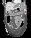
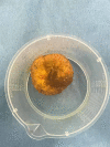
Small bowel obstruction is a common operative finding following an acute surgical admission. However, small bowel obstruction due to an enterolith is a rarer finding. Enteroliths are formed in conditions contributing to hypomotility and stasis within the gastrointestinal tract. These include Crohn's disease, strictures, and intestinal diverticulae. We present a case of small bowel obstruction due to an enterolith in an 89-year-old female. In our case, CT identified an inflamed jejunal diverticulum pre-operatively.
Keywords: enterolith; jejunal diverticulosis; small bowel obstruction.
Copyright © 2020, Patel et al.
Conflict of interest statement
The authors have declared that no competing interests exist.
Figures


References
-
- Fecalith in the ileum causing intestinal obstruction. Ain QU, Azhar SA, Baloch S, Khan SA, Salim A. https://www.ncbi.nlm.nih.gov/pubmed/27323592. J Ayub Med Coll Abbottabad. 2016;28:189–190. - PubMed
-
- Enteroliths masquerading as urinary bladder stone. Karim T, Dey S. JCR. 2015;5:467–469.
-
- Subacute intestinal obstruction by an enterolith: a rare case. Hiremath SC, Ahmed Z, Iqbal T. http://www.ijars.net/articles/PDF/2425/36750_CE[VSU]_F(SHU)_PF1(A_OM)_PF... Int J Anat Radiol Surg. 2018;7:1–3.
-
- Enterolithiasis: an unusual cause of small intestinal obstruction. Singal BM, Kaval S, Kumar P, Singh CP. Arch Int Surg. 2013;3:137–141.
Publication types
LinkOut - more resources
Full Text Sources
