Central Mirror Foot: Treatment and Review of the Literature
- PMID: 32642361
- PMCID: PMC7336690
- DOI: 10.7759/cureus.8448
Central Mirror Foot: Treatment and Review of the Literature
Abstract
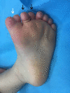
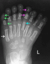
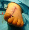
Mirror foot is a rare abnormality which presents as a preaxial, postaxial, or central polydactyly of the foot. The latter is encountered infrequently. We describe the case of a central mirror foot. Our patient had eight digits of a central ray pattern type with fully developed metatarsal, proximal, middle, and distal phalanges, as well as a medial toe syndactyly. He had no tarsal bone duplications. He was treated by central ray resection via double V-shaped incisions on the dorsal and plantar aspects of the foot, while preserving the medial and lateral rays. The results were satisfactory. We describe the technique and attempt a review of the literature.
Keywords: central ray; mirror foor; polydactyly.
Copyright © 2020, Papamerkouriou et al.
Conflict of interest statement
The authors have declared that no competing interests exist.
Figures


References
-
- Classification of postaxial polydactyly of the foot. Lee HS, Park SS, Yoon JO, Kim JS, Youm YS. Foot Ankle Int. 2006;27:356–362. - PubMed
-
- The spectrum of preaxial polydactyly of the foot. Belthur MV, Linton JL, Barnes DA. J Pediatr Orthop. 2011;31:435–447. - PubMed
-
- Congenital diplopodia with hypoplasia or aplasia of the tibia. A report of six cases. Karchinov K. J Bone Joint Surg Br. 1973;55:604–611. - PubMed
-
- Fibula dimelia in association with ipsilateral proximal focal femoral deficiency, tibial deficiency, and polydactyly. A case report. Igou RA Jr, Kruger LM. https://pubmed.ncbi.nlm.nih.gov/2394051/ Clin Orthop Relat Res. 1990;258:237–241. - PubMed
-
- Mirror foot. Skoll PJ, Silfen R, Hudson DA, Bloch CE. Plast Reconstr Surg. 2000;105:2086–2088. - PubMed
Publication types
LinkOut - more resources
Full Text Sources
